EISSN: 2086-4094 DOI: 10.4308/hjb.21.4.173
Correlation Between Akt and p53 Protein Expression and
Chemoradiotherapy Response in Cervical Cancer Patients
IIN KURNIA1*, BUDININGSIH SIREGAR2, SETIAWAN SOETOPO3, IRWAN RAMLI4,
TJAHYA KURJANA3, ANDRIONO5, MARINGAN DIAPARI LUMBAN TOBING6,
BETHY SURYAWATHI7, TEJA KISNANTO1,DEVITA TETRIANA1
1Department of Nuclear Medicine and Biology Radiation, Center for Technology of Radiation Safety and Metrology,
National Nuclear Energy Agency, Jalan Lebak Bulus Raya 49, Jakarta 12070, Indonesia
2Department of Pathology Anatomy, Cipto Mangun Kusumo Hospital, Jalan Diponegoro 71, Jakarta 10310, Indonesia 3Department of Radiation Oncology, Hasan Sadikin Hospital, Jalan Pasteur 38, Bandung 40161, Indonesia
4Department of Radiotherapy, Cipto Mangun Kusumo Hospital, Jalan Diponegoro 71, Jakarta 10310, Indonesia 5Department of Obstetric Ginaecology, Cipto Mangun Kusumo Hospital, Jalan Diponegoro 71, Jakarta 10310 Indonesia
6Department of Obstetric Ginaecology, Hasan Sadikin Hospital, Jalan Pasteur 38, Bandung 40161, Indonesia 7Department of Pathology Anatomy, Hasan Sadikin Hospital, Jalan Pasteur 38, Bandung 40161, Indonesia
Received March 6, 2014/Accepted November 13, 2014
Akt is a protein that is associated with cell proliferation and is expressed at high levels in cancer cells. Some research indicates it may play a role in increasing the resistance of cancer cells to chemotherapy treatment. P53 is a tumor suppressor protein that influences the cell cycle and apoptosis. The purpose of this study was to examine the relationship between the expression of Akt and p53 in cancerous tissue before chemoradiation treatment, and the clinical response to treatment of cervical cancer patients. Twenty microscopic tissue samples were taken from cervical cancer biopsies obtained from patients before cancer treatment. The tissue samples were stained with p53 and Akt antibodies via immunohistochemistry technique, to measure expression of both proteins. After completion of chemoradiotherapy, patients’ clinical response to treatment was determined using the pelvic control method. Our results revealed no correlation between expression of Akt and p53 index (P = 0.74) as well as between p53 Index and chemoradiotherapy clinical response (P=0.29). There was significant correlation between expression of Akt and cervical cancer chemoradiotherapy response (P = 0.03). There was no correlation found between p53 index and chemoradiotherapy clinical response (P = 0.29). High expression of Akt may related with high cell proliferation and resistance to chemoradiotherapy.
Key words: Akt, p53, cervical cancer, chemoradiotherapy
___________________________________________________________________________
_________________
∗Corresponding author. Phone: +62-21-7513906 axt 201, Fax: +62-21-7657950, E-mail: [email protected]
INTRODUCTION
Cervical cancer is the most common type of malignancy diagnosed in Indonesia. In general, patients seek treatment when the disease is not yet metastatized, but already at an advanced stage in its original location. In Indonesia, the treatment of choice for this type and stage of cancer is radiotherapy combined with chemotherapy or concurrent chemoradiotherapy (Didit & Rukmini 2002; Mufyala & Wofson 2008).
Radiotherapy provides significant curative and palliative benefits and is used in about 40-50% of all
cases of cervical cancer (Delaney et al. 2005). The
efficacy of radiotherapy is influenced by two factors
i.e. physical and biological factors. Two important biological factors are the patient’s intrinsic rate of cell proliferation, as well as the extent of hypoxia in
the cancer cells (Wilson et al. 2006). Physical factors include cell kinetics: the rate of proliferation in cancer cells correlates positively with its radiosensitivity and death by irradiation. The risk of cancer cell repopulation is greater after a failure of radiation treatment (Wilson 2003; Eriksen et al. 2004; Pedicini
et al. 2012).
Akt [protein kinase B (PKB)] is a serine threonine protein kinase with oncogenic and anti apoptotic activities (Gasinska et al. 2004). This protein consists of Akt1, Akt2, and Akt3, all of which have been found to play a role in the regulation of fundamental cellular functions such as cell proliferation, survival and programmed cell death; glucose metabolism, ribosomal function, genetic transcription, and cell migration via phosphorylation of a multitude of substrates. Dysregulation of Akt signaling pathways featured in many human malignancies (Song et al.
transformation of normal cervical to neoplasm cell (Crowell et al. 2007).
The p53 gene acts as a tumor suppressor. It works to regulate cell proliferation by stimulating
transcription of other genes specific to cell cycle
control (Brainwaithe & Prives 2006) and the process of apoptosis (Bai & Zhou 2006). Cells with a wild-type p53 gene have normal cell cycle function, including the ability to shut down replication of abnormal DNA during the G1 to S phase. Cancer cells with an inactivated, or mutant p53 protein cannot function properly and so replication of abnormal DNA is not prevented. In fact, inactivation of wild-type p53 gene represents the most common genetic mutation implicated in human carcinogenesis (Fei & El Deiry 2003; Brenna et al. 2004), and some researchers maintain that its over-expression, as detected by immunohistochemistry, indicates a worse prognosis in some malignancies (Wootipoom et al. 2004; Kocak et al. 2013; Min et al. 2014).
Cancer cell death in radiotherapy is caused in part by apoptosis, a process which is also regulated by p53. A proposed association between Akt expression and the effectiveness of radiotherapy and chemoradiotherapy on cervical cancer is still controversial. Some studies report a positive correlation between Akt expression and prognosis/response, while others have found a negative correlation. In the majority of studies, the expression of Akt is observed qualitatively using immunohistochemistry method (Kim et al. 2006; Faried et al. 2006; Zhang & Zhang 2008; Kocak et al. 2013). The aim of the present research was to detect and evaluate any relationship between the expression of Akt in cervical cancer tissues measured by the IRS
(Immno Reactive Score) method and p53 index with cervical cancer chemoradiotherapy response.
MATERIALS AND METHODS
Patients. Twenty consecutive subjects, were selected from among a whole series of 60 patients suffering from non-metastatic localized cervical carcinoma (stage IIB-IIIB). The subjects received a complete course of treatment, and were studied prospectively from July 2010 to March 2011 (Table 1). All patients were diagnosed and treated by definitive and concurrent chemoradiotherapy at Cipto Mangunkusumo Hospital (Jakarta) and Hasan Sadikin Hospital (Bandung) and provided written informed consent. Our research protocol was approved by the Research and Ethical Committee from Faculty of Medicine, University of Indonesia. Clinical staging of the patients was conducted by speculoscopy, bimanual examination and cystoscopy or rectoscopy, if necessary. Abdominal pelvic CT scans and chest X-rays were performed
on all patients. Histological grade was classified
according to the guidelines published by the Union for International Cancer Control as follows: G1 well differentiated; G2 moderately differentiated; and G3 poorly differentiated or undifferentiated. All patients had squamous cell carcinoma tumors with 13 patients diagnosed at clinical stage IIB, 1 patient at stage IIIA and 6 patients at stage IIIB.
Treatment. Patients were treated with a combination of External Beam Radiotherapy (EBRT) with 60Co gamma rays and 192Ir High Dose-Rate
Intracavitary Brachytherapy (HDR-ICBT). EBRT
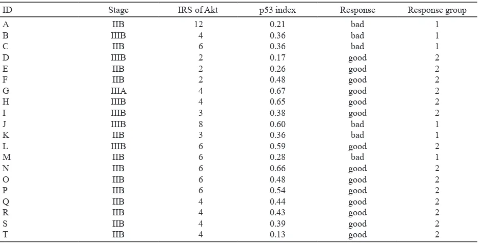
Table 1. Expression of Akt, p53 index, and chemoradiotherapy clinical response in cervical cancer treated with chemoradiotherapy
ID Stage IRS of Akt p53 index Response Response group
was administered to the whole pelvis with a clinical target volume that included the primary cancer, uterus, internal iliac artery, pre sacral, external iliac artery, and lower common iliac lymph nodes. This
was usually achieved by a “four-field box technique,”
or sometimes by antero-posterior and postero-anterior
parallel opposed portals. The usual field borders for anterior and posterior fields were superior at the
L4-L5 inter space, inferior at the bottom of the obturator foramen or 2 cm distal from the tumor, and 1.5 to
2.0 cm lateral to the bony pelvic wall. Lateral fields
had their anterior border at the symphysis pubis and the posterior border encompassed the entire sacral silhouette. A total dose of 50 Gy was prescribed in 25 equal fractions to the isocenter. HDR-ICBT using a Microselectron (Nucletron International, Amsterdam, Netherlands) followed by EBRT in two fractions (850 cGy/fraction) at point A. Cisplatin was administered at dose of 40 mg/m2 on day 1, 8, 15,
22, and 29; 2 hours or less prior to EBRT treatment given concurrently on the same days (Pearcey et al. 2002; Elizabeth et al. 2006; Palmer et al. 2012).
Clinical Chemoradiotherapy Response.
Clinical response to radiation treatment was evaluated
by a radiotherapist and classified according to the
Hong Criteria (Gonzalez et al. 2002) as follows:(i)
NRT (no gross residual tumor); complete or nearly
complete regression of pelvic tumor; non specific fibrosis; or granulation over the cervix. These results
are designated in our research as a positive (good) clinical response to treatment. (ii) GT response (gross residual tumor): gross tumor or palpable nodularity on cervix, and/or palpable in duration on the parametrium. These results are designated as a negative (bad) clinical response to treatment.
Immunohistochemistry. The p53 index and expression of Akt were analyzed by performing immunohistochemistry of pre-treatment biopsy samples. These samples of tumor tissue were embedded in paraffin, then incubated with Akt antibody (Abcam, USA) applied at a 1:100 dilution and with anti p53 monoclonal antibody (Leica,
Novocastra, ready to used-p53-D07), over night at 5
oC, in a moist chamber, followed by post primary, post
protein, and Novolink HRP system (Novolink) and revealed with DAB (Novolink) and counterstained with Mayer Hematoxylin. The primary antibody was omitted in one of slide as a negative control. Staining for p53, observed in the nucleus, was scored as percent of stained cells (Kurnia et al. 2009). Up to 1000 cells were counted in each slide. Expression of Akt found in nucleus, cell cytoplasm and membrane, was observed in zones of maximum expression of the marker in at least 10 high power
fields (400×). Expression of Akt was described using
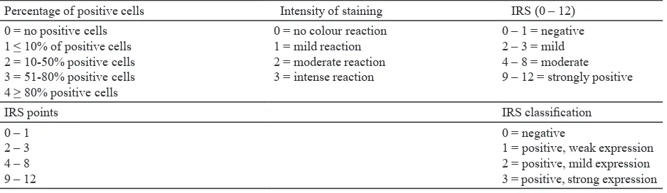
semi-quantitative scoring with the Immuno Reactive Score (IRS). The semi-quantitative analysis of the stained sections was carried out by light-microscopy and scored according to the IRS system introduced (Kaemerer et al. 2012) (Table 2).
Statistical Analysis. The p53 index was analyzed by using the Kolmogorov test for normal distribution data. Statistical analysis was conducted to assess any correlation between the Akt IRS score and the p53 index. Analysis of variance (ANOVA) test was used to analyze the relationship between the Akt IRS score and the clinical response of subjects to chemoradiotherapy. All statistical analyses were performed using Medcalc Software Version 9.2.0.1 (Harris & Taylor 2008).
RESULTS
In terms of clinical response to radiation treatment,
14 patients (70%) showed a positive clinical response and 6 patients (30%) showed a negative clinical
response after completion of treatment (Table 2). Our goal was to determine whether there exists any correlation between these outcomes with Akt and p53 expression.
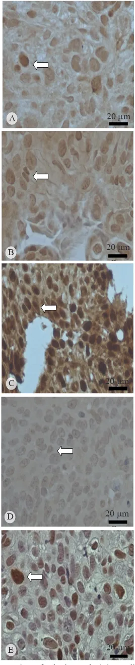
Measures of both Akt and p53 expression were obtained from tumor samples from 20 subjects. Expression of Akt was observed as brown coloration in the cancer cell cytoplasm (Figure 1A,B,C) and
Table 2. Akt and IRS classification scoring systems
Percentage of positive cells Intensity of staining IRS (0 – 12)
0 = no positive cells 1 < 10% of positive cells 2 = 10-50% positive cells 3 = 51-80% positive cells 4 > 80% positive cells
0 = no colour reaction 1 = mild reaction 2 = moderate reaction 3 = intense reaction
0 – 1 = negative 2 – 3 = mild 4 – 8 = moderate 9 – 12 = strongly positive
IRS points IRS classification
0 – 1 2 – 3 4 – 8 9 – 12
0 = negative
Figure 1D as negative control. We observed strong
expression of Akt in one patient (5%), mild-positive expression in 14 patients (70%), and low or negative expression of Akt in 5 patients (25%).
Patients who showed a positive clinical response
to radiation treatment were #4 (28%) showed weak expression of Akt, and #7 (50%) showed mild
expressions of Akt. Patients who had a negative
clinical response to radiation treatment, #1 (14%) showed strong Akt expression, and #6 (85 %) showed
mild expression of Akt.
The expression of p53 protein can be observed as brown coloration in the nucleus (Figure 1E) and is measured as percentage of nucleus affected. Among our research subjects, the p53 index varied from
13 to 67% (mean 41%). As shown in Figure 2, we
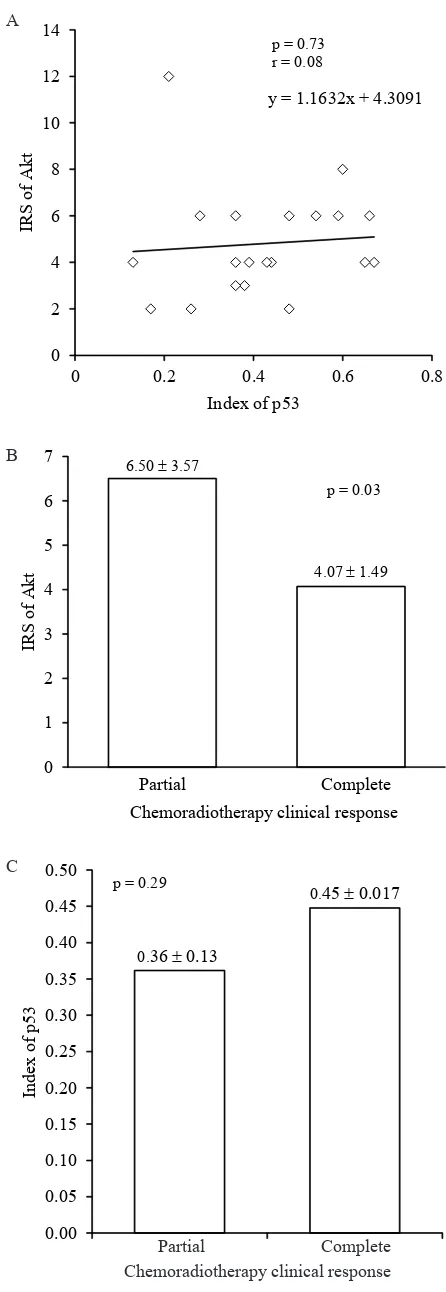
found no statistical correlation between expression of Akt and expression of p53 (P = 0.73, r = 0.08).
We did find a significant correlation between higher
expression of Akt and negative clinical response (P = 0.03) and between higher expression of p53 and positive clinical response (P = 0.29) after completion of chemoradiotherapy. Our results indicate that high expression of Akt before treatment correlates to negative clinical responses in chemoradiotherapy treatment.
DISCUSSION
Expression of Akt in our patient samples can also be categorized as either positive, or weak/
negative expression, with a “positive” defined as ≥ 75% of cervical cancer tissue testing positive for Akt expression; and “weak/negative” defined as ≤ 25% of
cancer tissue testing with low or no Akt expression. Other studies have analyzed pre-treatment tissues of patients with locally advanced cervical cancer and
found that 86% (Kocak et al. 2013), and 74% (Kim et al. 2006), classify as “positive” for Akt. These studies
used a different method to analyze the expression of
Akt, which classified according to the intensity and
quality of staining from Akt inside the cancer cell (Kim et al. 2006; Suzuki et al. 2010). In a study of glioblastoma (Suzuki et al. 2010) Akt was found in
the nucleus as well in cytoplasm, with 29.7% of the subjects categorized as 50% high positive.
Our results showed only a weak, non-significant
negative correlation between Akt and p53 expression. Prior research suggests that Akt will suppress expression of p53 related with mitochondria function. In wild-type p53, Akt clearly functions to modulate the direct action of p53 on the caspase-dependent mitochondrial function in death pathway (Yang et al. 2006). The relationship between Akt and p53 activity
20 mm
20 mm
20 mm
20 mm
20 mm A
B
C
D
E
also influenced by MDM2 function by degradation of
p53 protein (Zhou et al. 2001; Yoko et al. 2002; Milne
et al. 2004). Although almost all p53 mutations can be detected using immunohistochemical techniques, sequencing analysis is still necessary to ensure the type of DNA mutation.
Expression of p53 and Akt each showed a different association with chemoradiotherapy clinical response. Higher expression of p53 tended to correlate with positive clinical response; it may be that mutated forms of p53 promote uncontrolled proliferation of cancer cells, which in turn increases radiosensitivity during the cell cycle phase, making the cells more susceptible to radiation than wild– type p53. Researchers have observed resistance to chemotherapy and radiotherapy in NSCLC (non small cell lung carcinoma) cell lines, when Akt is highly expressed (Brognard et al. 2001). Other studies have sought to understand and clarify the relationship between Akt and clinical response, in head and neck cancer treated with radiotherapy, and breast cancer treated with chemotherapy. Clinical
studies have confirmed an association between Akt
expression and resistance to radiotherapy in head and neck tumors (Gupta et al. 2002) and to chemotherapy in breast cancer (Stall et al. 2003). In this study, we surmise that Akt may disrupt the apoptosis process induced by irradiation that should lead to cancer cell death. One study of Non Small Cell Lung Carcinoma (NSCLCs) also showed a correlation between high expression of Akt and indicators of poor prognosis, in NSCLCs with lymph-node involvement, but it can’t be considered to indicate distant of metastasis (Hirami et al. 2004).
Radiation exposure produces highly reactive free radicals, such as reactive oxygen species, that can interact with and damage DNA interfering with the cell’s ability to reproduce normally. Reactive molecules may also affect intracellular functions such as cytokine production, growth factors, gene transcription, and apoptosis (Benz & Yau 2008; Flohe & Flohe 2011). Akt may contribute to treatment resistance by stimulating Non Homolog End Joining (NHEJ) repair and promoting cancer cell survival (Toulany et al. 2008) or by associating with DNA-PK in response to genotoxic stresses (Bozulic et al. 2008). Thus Akt can inhibit imminent cell death or apoptosis that would normally occur as a result of radiation treatment. PI3K/AKT signaling may
influence clinical response to radiation in various
ways, by altering regulation of mitochondrial proteins,
0.36 ±0.13
0.45 ±0.017
0.00 0.05 0.10 0.15 0.20 0.25 0.30 0.35 0.40 0.45 0.50
1 2
In
d
ex
o
f
p
5
3
p = 0.29 6.50 ±3.57
4.07 ±1.49
0 1 2 3 4 5 6 7
IRS
o
f
A
k
t
Partial Complete Chemoradiotherapy clinical response
p = 0.03 y = 1.1632x + 4.3091
0 2 4 6 8 10 12 14
0 0.2 0.4 0.6 0.8
IRS
o
f
A
k
t
Index of p53 p = 0.73 r = 0.08
Partial Complete Chemoradiotherapy clinical response
Figure 2. Corellation between expression of Akt, and p53 (A), expression of Akt (B), and p53 (C) in complete and partial response of chemoradiotherapy.
A
B
transcription factors, translation machinery, and cell-cycle progression (Zhan & Han 2004; Cheung & Testa 2013). Our research clearly supports the idea that the level of Akt expression prior to treatment can serve as a parameter to predict positive or negative clinical response to chemoradiotherapy in cervical cancer cases. High expression of Akt correlates to increased resistance of cervical cancer cells to chemoradiotherapy. Pre-treatment measurements of Akt expression can be used as a biomarker of patient response to chemoradiotherapy for cervical cancer.
ACKNOWLEDGEMENT
This research was supported by a Research Grant from Center for Technology of Radiation Safety and Metrology, National Nuclear Energy Agency, Fiscal Year 2011. The authors thank to Soehartati Gondhowiardjo, Endy Moegni, Endang SR Harjolukito (Cipto Mangunkusumo Hospital)
and Firman (Hasan Sadikin, Hospital) for scientific
advice.
REFERENCES
Bai L, Zhou WG. 2006. p53: Structure, function and therapeutic applications. Cancer Mol 2:141-153.
Benz CC, Yau C. 2008. Ageing, oxidative stress and cancer: paradigms in parallax. Nat Rev Cancer 8:875-879. http:// dx.doi.org/10.1038/nrc2522
Bozulic LB, Surucu DH, Hemmings BA. 2008. PKBα/Akt1 acts downstream of DNA-PK in the DNA double-strand break response and promotes survival. Mol Cell 30:203-213. http:// dx.doi.org/10.1016/j.molcel.2008.02.024
Braithwaite AW, Prives CL. 2006. p53: more research and more questions. Cell Death Differ 13:877-880. http://dx.doi. org/10.1038/sj.cdd.4401938
Brenna SMF, Silva IDCG, Zefrino LC, Pereira JS, Martinez EZ, Syijanen KJ. 2004. Prognostic value of P53 codon 72 polymorphism in invasive cervical cancer in Brazil.
Gynecol Oncol 93:374-380. http://dx.doi.org/10.1016/j. ygyno.2004.03.004
Brognard J, Clark AS, Ni Y, Dennis PA. 2001. Akt/protein kinase B is constitutively active in non-small cell lung cancer cells and promotes cellular survival and resistance to chemotherapy and radiation. Cancer Res 61:3986-3997. Cheung M, Testa JR. 2013. Diverse mechanisms of AKT pathway
activation in humanmalignancy. Curr Cancer Drug Targets
1:234-244. http://dx.doi.org/10.2174/1568009611313030002 Crowell JA, Steele VE, Fay JR. 2007. Targeting the AKT protein
kinase for cancer chemoprevention. Mol Cancer Theraupetic
6:2139-2147. http://dx.doi.org/10.1158/1535-7163.MCT-07-0120
Delaney G, Jacob S, Featherstone C, Barton M. 2005. The role of radiotherapy in cancer treatment: estimating optimal utilization from a review of evidence-based clinical guidelines. Cancer 104:1129-1137. http://dx.doi. org/10.1002/cncr.21324
Didit T, Rukmini M. 2002. Cancer in Indonesia, present and future. Jpn J Clin Oncol 32:(Supplement1) S17-S21. http:// dx.doi.org/10.1093/jjco/hye123
Elizabeth F, Robert YK, Sreelatha M, Jennifer DLS, Sharon S. 2006.Low-dose-rate vs. high-dose-rate intracavitary brachytherapy for carcinoma of the cervix: The University of Alabama at Birmingham (UAB) experience. Brachyihterapy
5:49-55.
Eriksen JG, Steiniche T, Askaa J. 2004. The prognostic value of epidermal growth factor receptor is related to tumour differentiation and the overall treatment time of radiotherapy in squamous cell carcinomas of the head and neck. Int J Radiat Oncol Biol Phys 58:561-566. http://dx.doi. org/10.1016/j.ijrobp.2003.09.043
Faried LS, Faried A, Kanuma T. Sano T, Nakazato T, Tamura T, Kuwano H, Minegishi T. 2006. Predictive and prognostic role of activated mammalian target of rapamycin in cervical cancer treated with cisplatin based neoadjuvant chemotherapy. Oncol Reports 16:57-63.
Fei P, El-Deiry WS. 2003. P53 and radiation responses. Oncogene
22:5774-5783. http://dx.doi.org/10.1038/sj.onc.1206677 Flohe RB, Flohe L. 2011. Basic principles and emerging concepts
in the redox control of transcription factors. Antioxidants & Redox Signaling 15:2335-2381. http://dx.doi.org/10.1089/ ars.2010.3534
Gasinska A, Fowler JF, Lind BK. 2004. Influence of overall treatment time and radiobiological parameters on biologically effective doses in cervical cancer patients treated with radiation therapy alone. Acta Oncol 43:657-666. http:// dx.doi.org/10.1080/02841860410018511
Gonzales AD, Graniel CL, Ensico AG, Mohar A, Rivera L, Motta A, Guaddarrama R, Chanona G, Garza JDL. 2002. Concomitant chemoradiation versus neoadjuvant chemotherapy in locally advanced cervical carcinoma: result from II concecutive studies. Annals Oncology 13:1212-1219. http://dx.doi.org/10.1093/annonc/mdf196
Gupta AK, Mckenna MG, Weber CN, Feldman MD, Goldsmith JD, Mick R. 2002. Local recurrence in head and neck cancer: relationship to radiation resistance and signal transduction.
Clin Cancer Res 8:885-892.
Harris M, Taylor G. 2008. Medical Statistics Made Easy, Oxfordshire: Scion Publishing Ltd.
Hirami Y, Aoe M, Tsukuda K, Hara F, Otani Y, Koshimune R. 2004. Relation of epidermal growth factor receptor, phosphorylated-Akt, and hypoxia-inducible factor-1a in non-small cell lung cancers. Cancer Lett 14:157-164. http:// dx.doi.org/10.1016/j.canlet.2004.04.028
Kaemerer D, Peter L, Lupp A, Schulz S, Sanger J, Baum RP, Prasad V, Homman M. 2012. Comparing of IRS and Her2 as immunohistochemical scoring schemes in gastro enteropancreatic neuroendocrine tumors. Int J Clin Exp Pathol 5:187-194.
Kim TJ, Lee JW, Song SY. 2006. Increased expression of pAKT is associated with radiation resistance in cervical cancer. Brit J Cancer 94:1678-1682.
Kocak ME, Barisik NO, Mayadagli A, Gemici C, Eren M, Naki M, Ozseker N, Aksu A. 2013. The prognostic and predictive value of expression of P-AKT in patients with FIGO IIB locally advanced cervical cancer under chemotherapy. Int J Hematol Oncol 3:153-159. http://dx.doi.org/10.4999/ uhod.11070
Kurnia I, Suzuki Y, Budiningsih S, Andrijono A, Ramli I, Badri C, Yoshida Y, Nakano T. 2009. Nucleolar organizer regions in squamous cell carcinomas of the uterine cervix treated with chemoradiotherapy. Aust Asian J Cancer 8:93-102. Milne D, Kampanis P, Nicol S, Dias S, Campbell DG, Pace FF,
Min Q, Jianglin Z, Weiqi Z, Xiang C. 2014. DNAJB1 stabilizes MDM2 and contributes to cancer cell proliferation in ap53-dependent manner. Biochim et Biophys Acta 1839:62-69. http://dx.doi.org/10.1016/j.bbagrm.2013.12.003
Mufyala S, Wofson AH. 2008. Cervical cancer. In: Lu JJ, Brady LW (eds). Radiation Oncology. An Evidence Base Approach. Berlin: Springer Verlag. p 357-369.
Palmer A, Hayman O, Muscat S. 2012. Treatment planning study of the 3D dosimetric differences between Co-60 and Ir-192 sources in high dose rate (HDR) brachytherapy for cervix cancer. J Contemp Brachyther 4:52-59. http://dx.doi. org/10.5114/jcb.2012.27952
Pearcey R, Brundage M, Drouin P, Jeffrey J, Johnston D, Lukka H, Maclean G, Souhami L, Stuart G, Tu D. 2002. Phase III trial comparing radical radiotherapy with and without Cisplatin chemotherapy in patients with advanced squamous cell cancer of the cervix. J Clin Oncol 20:966-972. http:// dx.doi.org/10.1200/JCO.20.4.966
Pedicini P, Antonio N, Lidia S, Barbara AJF, Daniela A, Marta C, Francesca B, Barbara V, Rocchina C, Alba F. 2012. Correlation between EGFR expression and accelerated proliferation during radiotherapy of head and neck squamous cell carcinoma. Radiat Oncol 7:143-154. http://dx.doi. org/10.1186/1748-717X-7-143
Ree AH. 2008. Highly proliferative neuroendocrine carcinoma influence of radiotherapy fractionation on tumor response.
Radiat Oncol 3:13-17. http://dx.doi.org/10.1186/1748-717X-3-13
Song G, Ouyang G, Bao S. 2005. The activation of Akt/PKB signaling pathway and cell survival. J Cell Mol Meds 8:59-71. http://dx.doi.org/10.1111/j.1582-4934.2005.tb00337.x Stall O, Perez T, Akerberg GL, Olsson B, Nordenskjold B,
Skoog L. 2003. Akt kinase in breast cancer and the results of adjuvant therapy. Breast Cancer Res 5:34-37. http://dx.doi. org/10.1186/bcr693
Suzuki Y, Shirai K, Oka K, Mobaraki A, Yoshiha Y, Noda S, Okamoto M, Suzuki Y, Itoh J, Itoh H, Ishiuci S, Nakano T. 2010. Higher pAkt expression predicts a significant worse prognosis in glioblastomas. J Radiat Res 51:343-348. http:// dx.doi.org/10.1269/jrr.09109
Taylor RC, Cullen SP, Martin SJ. 2008. Apoptosis: controlled demolition at the cellular level. Nat Rev Mol Cell Biol 9:231-241. http://dx.doi.org/10.1038/nrm2312
Toulany M, Kehlbach R, Florczak U. 2008. Targeting of AKT1 enhances radiation toxicity of human tumor cells by inhibiting DNA-PKcs-dependent DNA double-strand break repair. Mol Cancer Ther 7:1772-1781. http://dx.doi. org/10.1158/1535-7163.MCT-07-2200
Wilson GD. 2003. Proliferation models in tumours. Int J R a d i a t O n c o l B i o l 7 9 : 5 2 5 - 5 3 0 . h t t p : / / d x . d o i . org/10.1080/0955300031000114710
Wilson GD, Saunders M, Stanley D, Frances MD, Francesca MB, Paul IR, Søren MB. 2006. Pre-treatment proliferation and the outcome of conventional and accelerated radiotherapy.
Eur J Cancer 42:363-371. http://dx.doi.org/10.1016/j. ejca.2005.10.022
Wootipoom V, Lekhyananda N, Phongrassan T, Boopnyapiphat P, Tongsuksai P. 2004. Prognostic significance of Bax, Bcl-2, and p53 expressions in cervical squamous cell carcinoma treated by radiotherapy. Gynecol Oncol 94:636-642. http:// dx.doi.org/10.1016/j.ygyno.2004.03.012
Yang X, Fraser M, Moll UT, Basak A, Benjamin KT. 2006. Akt-mediated cisplatin resistance in ovarian cancer: Modulation of p53 action on caspase-dependent mitochondrial death pathway. Cancer Res 66:3126-3136. http://dx.doi. org/10.1158/0008-5472.CAN-05-0425
Yoko O, Shohei K, Toshiyuki O, Yuko I, Toshiaki S, Keiji T, Norihisa M, Yukiko G. 2002. Akt enhances Mdm2-mediated ubiquitination and degradation of p53. J Biol Chem
24:21843-21850.
Zhan M, Han ZC. 2004. Phosphatidylinositide 3-kinase/AKT in radiation responses. Histol Histopathol 19:915-923. Zhang XY, Zhang HY. 2008. Elevated phosphatidylinositol
3-kinase activation and its clinicopathological significance in cervical cancer. Eur J Obstet Gynecol Reprod Biol 139:237-244. http://dx.doi.org/10.1016/j.ejogrb.2007.12.021 Zhou BP, Liao Y, Xia W, Zou Y, Spohn B, Hung MC. 2001.