Identiication of pathogenesis pathway in basal-like breast cancer
based on mutant p53 protein and topoisomerase-IIα expression
Abstrak
Latar belakang: Keganasan payudara jenis basal-like
sulit untuk diterapi dengan regimen terapi standar karena tidak mengekspresikan reseptor hormon atau reseptor faktor pertumbuhan epidermis yang menjadi target terapi.
Diperlukan upaya identiikasi molekul yang berperan
pada karsinoma payudara jenis basal-like, terutama molekul yang dapat dijadikan target pengobatan, salah
satunya topoisomerase IIα yang diregulasi oleh protein
p53. Penelitian bertujuan untuk membandingkan ekspresi
protein p53 mutan dan topoisomerase-IIα pada karsinoma
payudara basal-like dan non basal-like, serta menentukan hubungan antara ekspresi protein p53 mutan dan
topoisomerase-IIα pada kelompok basal-like.
Metode: Sampel 40 blok parain karsinoma payudara yang sudah terveriikasi triple negative, dibagi menjadi 2 kelompok berdasarkan ekspresi cytokeratin - 5 (CK-5) yaitu kelompok karsinoma payudara jenis basal-like dan
non basal-like. Ekspresi p53 mutan dan Toposiomerase-IIα
diperiksa dengan imunohistokimia, kemudian dihitung dan dibandingkan pada kedua kelompok. Analisis statistik untuk analisis deskriptif, kappa test, uji normalitas, perbandingan dua mean dan kategori menggunakan SPSS versi 16.
Hasil: Median (min-max) ekspresi p53 mutan pada kelompok basal-like adalah 21 (0-100), kelompok
non basal-like 2 (0-80), p = 0,061. Min-max ekspresi
topoisomerase IIα pada kelompok basal-like 263 (15-492), kelompok non basal-like 262 (0-481), p = 0,409.
Ditemukan hubungan bermakna antara positiitas p53
mutan dengan subtipe karsinoma payudara (p = 0,027). Terdapat pula koekspresi yang lebih sering p53 mutan -
topoisomerase IIα pada basal-like dibandingkan dengan
non basal-like (p = 0,018).
Kesimpulan: p53 mutan dan topoisomerase-IIα secara
bersama-sama dapat berperan dalam salah satu jalur patogenesis karsinoma payudara jenis basal-like.
Abstract
Background: Basal-like breast cancer is dificult to treat
with standard regimen therapy, because it doesn’t express hormone receptors or epidermal growth factor receptors.
Identiication of oncogenesis pathway is expected to ind
molecules which can be used as target for therapy. One
candidate molecule is topoisomerase-IIα whose expression
is regulated by p53. This study aimed to compare the expression of mutant p53 proteins and topoisomerase
IIα in basal-like and non basal-like breast cancer, and to
determine the association between mutant p53 proteins and
topoisomerase IIα in basal-like group.
Methods: The samples were 40 formalin ixed parafin embedded tissues from veriied triple negative
breast cancer tissue. The samples were divided into 2 groups, basal-like and non basal-like breast cancer, based on cytokeratin - 5 (CK-5) expression. Mutant
p53 proteins and topoisomerase IIα were stained
using immunohistochemistry method, scored and compared. Statistical test used SPSS software version 16 for descriptive statistics, kappa test, normality test, comparison of two mean, and categorical comparison. Results: Median (min-max) of mutant p53 protein expression in basal-like group was 21 (0-100), the non basal-like group was 2 (0-80), p = 0.061. Min-max of
topoisomerase IIα in basal-like group was 263 (15-492),
non basal-like group was 262 (0-481), p = 0.409. There was an association between mutant p53 positivity with breast cancer subtype (p = 0.027) and between mutant
p53-topoisomerase IIα coexpression with breast cancer
subtype (p = 0.018).
Conclusion: Co-expression of mutant p53 with
topoisomerase IIα has the role in one of the pathway of
basal-like breast cancer pathogenesis.
Keywords: basal-like breast cancer, mutant p53, topoisomerase-IIα
pISSN: 0853-1773 • eISSN: 2252-8083 • http://dx.doi.org/10.13181/mji.v23i4.995 • Med J Indones. 2014;23:197-202
Correspondence author: Yayi Dwina, [email protected]
B a s i c M e d i c a l R e s e a r c h
Copyright @ 2014 Authors. This is an open access article distributed under the terms of the Creative Commons Attribution-NonCommercial-ShareAlike 4.0 International License (http://creativecommons.org/licenses/by-nc-sa/4.0/), which permits unrestricted non-commercial use, distribution, and reproduction in any medium, provided the original author and source are properly cited.
Yayi Dwina, Ria Kodariah, Endang S.R. Hardjolukito
Breast cancer is the most common malignancy in women worldwide. In developing country the prevalence continues to increase and often diagnosed at an advanced stage.1 Breast cancer has heterogeneous clinical, pathological, histological, molecular, and response to treatment characteristics. In order to better understand and to ind the appropriate treatment strategy for this disease, various attempts have been made to classify breast cancer.2
Based on the expression of its mRNA, breast cancer is divided into 5 types: luminal A, luminal B, normal breast like, human epidermal growth factor receptor 2 (HER2) positive, and basal-like.3,4 Basal-like type has the most aggressive clinical behavior and poor prognosis. Basal-like breast cancer often do not express estrogen (ER), progesterone (PR) and HER2 receptors, hence triple negative breast cancer terminology.5 This type of breast cancer also frequently express high molecular weight cytokeratins, such as cytokeratin - 5 (CK-5). In other word, basal-like breast cancer is triple negative breast cancer that express high molecular weight cytokeratins.5 The non-expression of ER, PR, and HER2 in basal-like breast cancer is link to dificulties in therapy, because targeted therapy using standard hormone receptor with HER2 as its target, will not work.5 Therefore it is necessary to explain the characteristic of basal-like breast cancer, so that we will be able to explain the aggressiveness, and to ind the potential molecule involved and hence design the right treatment.
In basal-like breast cancer, there are several known genetic defects, such as decreased expression of breast cancer genes 1 (BRCA1),4,5 a genes responsible for repair of deoxyribose-nucleic acid (DNA) double stranded break.5 The high amount of DNA double stranded breaks in normal cells causes cell cycle stalled; but basal-like breast cancer seems to have high tolerance for BRCA1 defect, and cell cycle keep going on despite the high accumulation of DNA damage. This resistance is hypothetically caused by mutations in the p53 gene, a tumor suppressor gene. In a p53-gene-mutated-cell the cell cycle will not be interrupted even if there has been a signiicant defect in DNA.5,6
One protein in which transcription is regulated by p53 is topoisomerase IIα.7,8 Topoisomerase IIα is an ubiquitous enzyme in the cell nucleus, which plays a role in the regulation of DNA topology.7,9
It also regulate many biological processes such as replication, segregation, transcription, regulation of chromatin structure and general gene expression.10,11 Wang, et al7 and Sandri, et al8 in in vitro study has proved that topoisomerase IIα gene expression can be inhibited by p53 gene, even though the mechanisms is not completely understood.
These studies raised possibilities that pathogenesis of basal-like breast cancer could be mediated by pathway that involve the p53 gene and topoisomerase IIα, in which p53 tumor suppressor gene dysfunction causing increase expression of topoisomerase IIα.
Topoisomerase IIα is one of the molecular targets for chemotherapy, but the prognosis has not improved signiicantly.12 There are also several side effects caused by anti-topoisomerase IIα regimen such as secondary malignancies as well as cardiotoxicity Therefore, ideally the regiment should only be given to cancer patients with positive topoisomerase IIα.13 It is important to study oncogenesis pathways of p53-topoisomerase IIα. If this pathway proves important in the pathogenesis of basal-like breast cancer then it could serve as the basis/justiication for the treatment of breast cancer with a regimen targeting topoisomerase IIα.
METHODS
Cell was categorized as positive for CK-5 if demonstrated cytoplasmic and membrane staining.14 The semi-quantitative assessments was as follows: negative if < 10% of tumor cells show positive results, positive if ≥ 10% of tumor cells show positive results. Mutant p53 positive cell was nuclear stained cell and scored using proportion of 100 cells count, and categorized as positive if more than 10% showed positive result, and negative if less than 10%.15
Topoisomerase IIα was positive if showed nuclear staining and scored by counting the positivity based on 500 cells count using 400x magniication.11 It was grouped as negative if < 25% tumor cells show positive results, positive if ≥ 25% tumor cells show positive results.
The assessment of immunohistochemistry staining was done by two independent pathologists and the results were analyzed by Kappa test. Statistical test was using SPSS software version 16 for descriptive statistics, kappa test, normality test, comparison of two mean, and categorical comparison.
RESULTS
Forty two conirmed triple negative cases were obtained, among which 2 cases were excluded due to minimal amount of tumor tissue. Of the 40 cases, 20 cases were positive for CK-5 so it was grouped as the basal-like breast cancer, and the other 20 cases were negatively stained with CK-5 and were grouped as the non basal-like breast cancer. Assessment of mutant p53, topoisomerase II and CK-5 was performed by two independent pathologists, the kappa values were as followed: kappa values for mutant p53 = 0.866, p < 0.001; kappa values for topoisomerase IIα = 1, p = 0.002; kappa values for CK-5 = 0.879, p < 0.001; all showed good concordance.
Marker expression
Type of breast cancer
p* Non
basal-like (n = 20)
Basal-like (n = 20)
Topoisomerase IIα
median (min - max) 262 (0-481) 263 (15-492) 0.409
Mutant p53
median (min - max) 2 (0-80) 21 (0-100) 0.061 *Mann-Whitney
Table 1. Median score of topoisomerase IIα and mutant p53
expression in basal-like breast cancer and non basal-like breast cancer
Table 1. is about the median score of Topoisomerase IIα and mutant p53 expression in basal-like breast cancer and non basal-like breast cancer, it showed there was no difference in the expression of Topoisomerase IIα and mutant p53 between two groups.
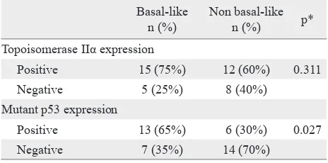
Table 2. showed positivity of Topoisomerase IIα and mutant p53 in basal-like breast cancer and non basal-like breast cancer, in this table we can see there was an association between mutant p53 expression and the type of breast cancer, in the basal-like group mutant p53 expressed more often compared to non basal-like group.
Table 3. is about the correlation between expression of mutant p53 and Topoisomerase IIα in basal-like breast cancer, it showed that there was co-expression of mutant p53 and topoisomerase IIα in half of
Basal-like n (%)
Non basal-like
n (%) p*
Topoisomerase IIα expression
Positive 15 (75%) 12 (60%) 0.311
Negative 5 (25%) 8 (40%)
Mutant p53 expression
Positive 13 (65%) 6 (30%) 0.027
Negative 7 (35%) 14 (70%)
*Chi-square
Table 2. Positivity of topoisomerase IIα and mutant p53 in
basal-like breast cancer and non basal-like breast cancer
Mutant p53 Topoisomerase IIα p*
Positive n (%) Negative n (%)
Positive 10 (50%) 3 (15%) 0.727
Negative 5 (25%) 2 (10%)
Table 3. The correlation between expression of mutant p53 and
topoisomerase IIα in basal-like breast cancer
*Mc Nemar
Mutant p
53-topoisomerase IIα
co-expression n (%)
Mutant p
53-topoisomerase IIα
non co-expression n (%) p*
Basal-like 10 (50%) 10 (50%) 0.018
Non
basal-like 3 (15%) 17 (85%)
Table 4. Proportion of p53 mutant-topoisomerase IIα co-expres -sion in basal-like breast cancer and non basal-like breast cancer
the basal-like cases, but there was no signiicant difference between mutant p53 and topoisomerase IIα expression in the whole cases of basal-like breast cancer.
Table 4. showed proportion of p53 mutan-Topoisomerase IIα co-expression in basal-like breast cancer and non basal-like breast cancer, in this table we can see there was signiicant difference in co-expression mutant p53-topoisomerase IIα between basal-like breast cancer (50%) and the non basal-like breast cancer (15%).
Figure 1 showed the positivity of CK-5, mutant p53, and topoisomerase IIα, immunohistochemistry in breast cancer tissues.
DISCUSSION
In this study the positive expression of mutant p53 in basal-like breast cancer (65%) was higher than that of the non-basal-like (30%). This is in line with Conforti, et al16 who found 51% positivity of mutant p53 in basal-like breast cancer, while in the non-basal like the positivity was only 11%. The explanation for this phenomenon was that as much as 80% - 90% basal-like breast cancer were associated with reduced expression of BRCA1.17 BRCA1 is a protein that has a role in various cellular processes such as response to DNA damage repair, control check points in the cell cycle, transcription modulation, and ubiquitinisation.5,6,18 In normal condition cells with reduced levels BRCA1, either due to mutation or promoter’s methylation should undergo cessation of the cell cycle and eventually death. In basal-like breast cancer it seems that the tumor cells are not responding to the lack of BRCA1 levels so that the cell cycle continue. This phenomenon might be caused by mutation of the tumor suppressor gene p53, allowing genetically defective cells to continue replicating and eventually transforming into cancer
cells.2,5,6 This explanation is consistent with the results of the study by Holstege, et al6 who found that all breast cancers with BRCA1 expression defects were also contained p53 mutations, while other studies found 60%-77% p53 mutations in breast carcinoma with BRCA1 defects.6
No difference was found in topoisomerase IIα expression between basal-like breast cancer and the non like, although the basal-like group has slightly higher positivity (75%), compared to non basal-like (60%). This inding is actually consistent with the results reported by Romero, et al19 They stated that topoisomerase IIα protein expression which was measured by immunohistochemistry method, was higher in the basal-like breast carcinoma (72.72%), compared with the other subtypes: luminal type B (57.14%), HER2 positive (62.5%), luminal A (9%), and normal-like (0%).
This study also assessed the relationship between mutant p53 expressions with topoisomerase IIα expression in basal-like breast cancer; although we didn’t ind any signiicant association, but it showed that mutant p53-topoisomerase IIα co-expressed in half of cases. This didn’t explain the hypothesis of mutant p53 and topoisomerase IIα role in basal-like breast canacer. There might be other carcinogenesis pathway involved in the pathogenesis of basal-like breast cancer. They were: epidermal growth factor receptor (EGFR), proto-oncogene c-kit (c-KIT), and BRCA1.5 Another possibility was that the biological trait of topoisomerase IIα doesn’t expressed continuously in the cell cycle. The highest expression is in late S phase and peaked in growth2 - mitosis (G2-M) phase.20 Because of this feature topoisomerase IIα considered as cell proliferation surrogate marker, and doesn’t expressed in the cell constantly,19 instead it depends on the cell cycle status.21
A B C
The non association between mutant p53 and topoisomerase IIα may be caused by the method used to assess p53 status. In this study we used immunohistochemistry to detect the presence of mutant p53, because mutant p53 have longer half-life than the wild type variant, according to literature. Half-life of wild-type p53 is very short and it’s basically expressed in a very small amount below the detection threshold of immunohistochemistry.22,23 Sjogren, et al24 stated that mutant p53 detection using immunohistochemistry produce false positive results as much as 30% and false-negative results around 33% when compared to measurements using copy dna (cDNA) sequencing. It was mentioned that increased expression of p53 by immunohistochemistry standard methods can represent mutations in the p53; veriication using cDNA sequencing are considered as gold standard.22 False positive staining by immunohistochemistry method can be due to cellular stress induced p53 wild-type stabilization, Whereas the incidence of false negatives may occur due to mutation causing stop codons, deletions, or mutation causing destabilization of the protein.25 Truncation at the carboxyl end of p53 could cause false negative result, since the truncated mutant p53 protein will be degraded due to lost of several important function, for example the function for DNA binding domain, nuclear localization signals, and oligomerization domain24
We also assess the relationship between type of breast carcinoma with the incident of topoisomerase IIα-mutant p53 co-expression, and it revealed signiicant results with p = 0.018. Half of basal-like breast cancer showed topoisomerase IIα-mutant p53 co-expression, which is higher than the co-expression in non basal-like group which only expressed in 3 samples (15%). This supports the hypothesis of mutant p53 and topoisomerase IIα involvement in basal-like breast cancer pathogenesis. According to Wang, et al7 and Sandri, et al8 p53 has the ability to regulate topoisomerase IIα expression, in in-vitro
studies they found that p53 has the ability to regulate topoisomerase IIα transcription, by interacting with topoisomerase promoter (-32 to +90) in dna sequence cytosine cytosine adenine adenine tyrosine (CCAAT) segments. Wild-type p53 can act as a trans-acting elements capable of suppressing transcription of topoisomerase IIα, while mutant p53 has less ability to suppressed topoisomerase IIα expression. This inding can also serve as the consideration for treatment planning using anti topoisomerase IIα drug in part of basal like breast cancer.
In conclusion, this study showed a signiicant association between mutant p53 expression and basal like subtype. There was no association between p53 and topoisomerase IIα expression in basal like group. Co-expression of mutant p53-topoisomerase IIα occurred signiicantly more often in basal like, which indicates the role of mutant p53-topoisomerase IIα pathway in basal like pathogenesis in part of cases.
Conlicts of interest
The authors afirm no conlict of interest in this study.
Acknowledgments
We would like to thank Professor Santoso Cornain, and Mr. Kusmardi for their invaluable contribution in statistical analysis of this study, and also dr. Vinesia Lestari Riddi for allowing authors to continue using her samples and her work in the basal like breast cancer.
REFERENCES
1. World Health Organization [Internet]. Breast cancer:
prevention and control. Breast cancer. [updated 2008; cited
2010 Dec 20]. Available from: http://www.who.int/cancer/ detection/breastcancer/en/
2. Rakha E, Reis-Filho JS. Basal like breast carcinoma from
expression proiling to routine practice. Arch Pathol Lab Med. 2009;133(6):860-8.
3. Lester SC. The breast. In: Kumar V, Abbas AK, Fausto N, Aster JC, editors. Robbins and Cotran pathologic basis of disease. 8th ed. Philadelphia, PA: Sauders Elseviers; 2010.
p. 1065-96.
4. Perou CM, Sørlie T, Eisen MB, van de Rijn M, Jeffrey SS, Rees CA, et al. Molecular portraits of human breast
tumours. Nature. 2000;406(6797):747-52.
5. Rakha E, Reis-Filho JS, Ellis IO. Basal-like breast cancer:
a critical review. J Clin Oncol. 2008;26(15):2568-81.
6. Holstege H, Joosse SA, van Oostrom CT, Nederlof PM, de Vries A, Jonkers J. High Incidence of protein truncating
8. Sandri MI, Isaacs RJ, Ongkeko WM, Harris AL, Hickson ID, Broggini M, et al. p53 regulates the minimal promoter
of the human topoisomerase IIα gene. Nucleic Acids Res. 1996;24(22):4464-70.
9. Roca J. Survey and summary topoisomerase II: a itted
mechanism for the chromatin landscape. Nucleic Acid Res.
2009;37(3):721-30.
10. Nitiss JL. DNA topoisomerase II and its growing repertoire of biological function. Nat Rev Cancer.
11. Hellemans P, van Dam PA, Geyskens M, van Oosterom AT, Buytaert P, Van Marck E. Immunohistochemical
study of topoisomerase II-α ekspression in primary
ductal carcinoma of the breast. J Clin Pathol.
1995;48(2):147-50.
12. Sørlie T, Perou CM, Tibshirani R, Aas T, Geisler S, Johnsen H, et al. Gene expression patterns of breast carcinomas distinguish tumor subclasses with clinical implications.
Proc Natl Acad Sci USA. 2001;98(19):10869-74.
13. Nitiss JL. Targeting DNA topoisomerase II in cancer
chemotherapy. Nat Rev Cancer. 2009;9(5):338-50.
14. Dogu GG, Ozkan M, Ozturk F, Dikilitas M, Er O, Ozturk A. Triple-negative breast cancer: immunohistochemical correlation with basaloid markers and prognostic value of
survivin. Med Oncol. 2010;27(1):34-9.
15. Lukas J, Niu N, Press MF. p53 mutations and expression in breast carcinoma in situ. Am J Pathol.
2000;156(1):183-91.
16. Conforti R, Boulet T, Tomasic G, Spielmann M, Delaloge S, Arriagada R, et al. Predictive value of MRP2, p53, bcl2
and topoisomerase II immunostainings for the eficacy
of anthracyclines-based adjuvant chemotherapy in breast cancer: Results from two randomized trials. J Clin Oncol (Meeting Abstracts). 2008;26(15 Suppl):616.
17. Turner NC, Reis-Filho JS, Russell AM, Springall RJ, Ryder K, Steele D, et al. BRCA1 dysfunction in sporadic basal-like breast cancer. Oncogene. 2007;26(14):2126-32. 18. Ahmed M, Lalloo F, Evans DG. Update on genetic
predisposition to breast cancer. Expert Rev Anticancer
Ther. 2009;9(8):1103-13.
19. Romero A, Martín M, Cheang MC, López García-Asenjo
JA, Oliva B, He X, et al. Assessment of topoisomerase II α
status in breast cancer by quantitative PCR, gene expression
microarrays, immunohistochemistry, and luorescence in
situ hybridization. Am J Pathol. 2011:178(4):1453-60. 20. Woessner RD, Mattern MR, Mirabelli CK, Jhonson
RK, Drake FH. Proliferation- and cell cyle-dependent differences in expression of the 170 kilodalton and 180 kilodalton forms of topoisomerase II in NIH-3T3 cells. Cell
Growth Differ. 1991;2(4):209-14.
21. Moelans CB, de Weger RA, van Blokland MT, van der Wall E, van Diest PJ. Simultaneous detection of TOP2A
and HER2 gene ampliication by multiplex ligation-dependent probe ampliication in breast cancer. Mod Pathol. 2010;23(1):62-70.
22. Vojtĕsek B, Bártek J, Midgley CA, Lane DP. An
immunochemical analysis of the human nuclear phosphoprotein p53 new monoclonal antibodies and epitope mapping using recombinant p53. J Immunol Methods.1992:151(1-2):237-44.
23. Cooper K, Haffajee Z. bcl-2 and p53 protein expression in
follicular lymphoma. J Pathol. 1997;182(3):307-10.
24. Sjögren S1, Inganäs M, Norberg T, Lindgren A, Nordgren H, Holmberg L, et al. The p53 gene in breast cancer: prognostic value of complementary DNA sequencing versus immunohistochemistry. J Natl Cancer Inst.
1996;88(3-4):173-82.
25. Varna M, Bousquet G, Plassa LF, Bertheau P, Janin A. TP53 status and response to treatment in breast cancers. J Biomed