April 2017 1 *Corresponding author:
E-mail: [email protected]; iman [email protected]
Strengthening No: 36b/E/KPT/2016 Available online at http://medpet.journal.ipb.ac.id/
Isolation and Number of Gonadal Primordial Germ Cells (Gonadal PGCs) on the
Stages of Early Embryonic Development of KUB Chicken
S. Sopiyanaa,*, M. A. Setiadib, M. Fahrudinc, & I. Supriatnab,* aIndonesian Research Institute for Animal Production
Jalan Veteran III, Desa Banjarwaru, Ciawi-Bogor 16002, Indonesia
bDepartment of Veterinary Clinic, Reproduction, and Pathology, Faculty of Veterinary Medicine, Bogor Agricultural University,
cDepartment of Anatomy, Physiology, and Pharmacology, Faculty of Veterinary Medicine, Bogor Agricultural University,
Jalan Agatis, Kampus IPB Darmaga, Bogor 16680, Indonesia (Received 24-01-2017; Reviewed 08-03-2017; Accepted 22-03-2017)
ABSTRACT
Primordial germ cells (PGCs) are cells that will differentiate themselves into spermatogonia in the testis or oogonia in the ovary. Primordial germ cells arise from epiblast and circulate through the bloodstream and finally entering gonadal anlage. The aim of this study was to determine the number of gonadal PGCs of KUB chicken at different development stages. Sixty KUB chicken fertile eggs were divided into four groups (6, 7, 8, and 9 days incubation periods), and incubated at 38 oC with a humidity of 60%. Harvesting was synchronized to the embryonic development at 6-9 d. Gonads were collected using sharp tweezers, and were placed in Eppendorf tube 1.5 mL containing 500 µL PBS [-]. Gonadal PGCs were purified using PBS [-]. The results showed that the average number of gonadal PGCs at 6, 7, 8, and 9 d were 113.7; 143.5; 92.9; and 85.7 cells per embryo, respectively. Number of gonadal PGCs per embryo of KUB chicken were significantly affected by stage of embryonic devel-opment (P<0.05), which reached a peak at day 7 of incubation, so that the isolation and collection of PGCs from the gonads were recommended at day 7 of incubation. This information is useful in production of germline chimera of other Indonesian local chickens.
Keywords: KUB chicken, embryo development, gonad, PGCs
ABSTRAK
Primordial germ cell (PGC) adalah sel yang akan berdiferensiasi menjadi spermatogonia dalam
testis atau oogonia dalam ovarium. Primordial germ cell berasal dari epiblast di daerah germinal cres-cent dan beredar melalui aliran darah dalam waktu singkat sebelum memasuki bakal gonad. Setelah migrasi ke gonad, PGC disebut primordial germ cell gonad (PGC gonad). Penelitian ini bertujuan mengetahui jumlah PGC gonad ayam KUB pada tahap perkembangan embrio yang berbeda. Dalam penelitian ini digunakan 60 butir telur fertil ayam KUB yang dibagi menjadi empat kelompok per-lakuan (6, 7, 8, dan 9 hari periode inkubasi) yang diinkubasi pada suhu 38oC dengan kelembapan 60%. Pemanenan disesuaikan dengan tahap perkembangan embrio pada 6-9 hari. Gonad diisolasi menggunakan pinset yang tajam, dan dikumpulkan pada tabung Eppendorf 1,5 mL yang berisi 500 µL larutan PBS [-]. PGC dimurnikan dengan menggunakan PBS [-]. Hasil penelitian menunjukkan bahwa rataan jumlah PGC gonad pada umur embrio 6, 7, 8, dan 9 hari pada ayam KUB berturut-turut adalah 113,7; 143,5; 92,9; dan 85,7 sel per embrio. Jumlah PGC gonad per embrio ayam KUB dipenga-ruhi oleh tahap perkembangan embrio (P<0,05), yang mencapai puncaknya pada hari ke-7 inkubasi sehingga isolasi dan koleksi PGC yang berasal dari gonad disarankan dipanen pada hari ke-7 inkubasi. Hasil penelitian berguna untuk produksi germline chimera untuk preservasi PGC ayam lokal Indonesia lainnya.
2 April 2017
INTRODUCTION
Indonesia has around 43 breeds of local chickens (Sartika et al., 2016). Some of the local chicken popula-tions are very limited in number and even many of them are in the status of endangered species. Some of
them have potentially high benefit that have to be pre
-served in-situ or ex-situ. Kampung Unggul Badan Litbang
Pertanian (KUB) chicken is one of the genetic resources
of Indonesian Kampung chicken breeds, selected for improved egg production conducted at the Indonesian Research Institute for Animal Production. KUB chickens
are able to produce eggs about 50% hen-day production, reaching peak productions of 65%-70% (Sartika et al.,
2016). As a new breed, KUB chicken may be categorized as a new genetic resource.
Preservation can be divided into in-situ preserva-tion and ex-situ preservation. The primary objective of preservation is to ensure the survival of threatened species and the maintenance of associated genetic diversity. In-situ or ex-situ preservation is a form of the living individual or population that needs high costs
and risks associated with the disease. To solve the prob -lem, it is necessary to develop a preservation strategy through the cryopreservation of genetic material, such
as the storage of sperm, ova, and embryos in the frozen form (cryopreserved). However, the method of genetic
resource preservation cannot be applied to poultry
spe-cies primarily because of the technical difficulties on the cryopreservation of poultry eggs. The approach was
developed to overcome the current limitations of in-situ
and ex-situ preservations of genetic resources by collect-ing and transferrcollect-ing avian primordial germ cells (PGCs), collected from developing embryos.
Primordial germ cells are the progenitor cells for
gametes that colonize the developing gonad and sub
-sequently differentiate into spermatogonia in the testis
or into oogonia in the ovary (Kohara et al., 2008; Qian
et al., 2010; Glover & McGrew, 2012). Chicken PGCs are
located at the center of the pellucida area (Yamamoto et al., 2007). At the primitive streak stage, PGCs locates at the extra embryonic region that called germinal crescent region (Nakamura et al., 2007). As soon as the blood vessel is formed at stage 10 of Hamburger & Hamilton
(HH) (1951), The PGCs from the germinal crescent start
to circulate temporarily through the embryonic blood-stream on day 2 of incubation. The cells leave the capil-lary system and migrate to the form of genital ridge,
where they colonize the developing gonad (Minematsu et al., 2004; Glover & McGrew, 2012). After migrating to
the gonadal anlage, the PGCs are called gonadal germ
cells (GGCs) (Nakajima et al., 2011). Gonadal germ cells
or gonadal primordial germ cells (gonadal PGCs) in
the gonad, ultimately differentiate into spermatogonia, which takes place in the testes or into oogonia, which
takes place in the ovaries. Primordial germ cells are cells
with pluripotent characteristic making the PGCs as a
good model to study embryonic development in-vitro
(Wang et al., 2010).
Primordial germ cells have a unique pathway
of migration activity in birds (Song et al., 2010). The
unique migration pathway of PGCs provides a possibil
-ity to isolate subsequent PGCs based on their locations. Primordial germ cells can be isolated from X-stage
blastoderm, blood at 2.5-3-day-old embryos (stages 13-17 HH), or gonads at 5.5-6-day-old embryos (stages 26-28 HH) (Chojbacka-Puchta et al., 2012; Tajima, 2013).
Numerous studies had reported the isolation and col-lection of PGCs from gonadal embryos (Nakamura et al., 2011; Nakajima et al., 2011; Nakajima et al., 2016). Moreover, germline chimeras had been produced in chicken by transferring PGCs or GGCs into the blood-stream at 2-day-old recipient embryos (Furuta et al., 2007, 2008) so that PGCs could be used as a genetic resource to produce germ line chimera (Furuta, 2012).
Nakajima et al. (2012) reported that gonadal PGCs
col-lected from 7-day-old chick embryos and incubated in PBS [-] can be used to produce germline chimeras. It has been reported that gonadal PGCs are used to generate interspecies germline chimeras by transferring pheasant gonadal PGCs into chicken embryos (Kang et al., 2008) and by transferring chicken gonadal PGCs into duck embryos (Liu et al.,2012).
One of the problems with the PGCs collection of embryonic blood at the age of 2.5 days is the low
number of cells found in Indonesian local chicken (Kostaman et al., 2013). Therefore, to obtain more har-vested PGCs, collection of PGCs from embryonic gonad can be used as an alternative method. There are several methods used to isolate avian gonadal PGCs, one is by using proteinases, such as trypsin solution (Park et al., 2008; Kohara et al., 2008) and the other is by using a
phosphate-buffered saline without Ca2+ and Mg2+ solu-tion (PBS [-]) (Nakajima et al., 2011). The present study
was conducted to apply this method to isolate gonadal
PGCs from KUB chicken embryos by using
phosphate-buffered saline without Ca2+ and Mg2+ solution (PBS [-]) (Nakajima et al., 2011). The main objective of this study was to determine the number of gonadal PGCs of KUB chicken at different developmental stages of embryos.
The results of the research are expected to be basic in-formation for determining the appropriate stage for the isolation of gonadal PGCs.
MATERIALS AND METHODS
Preparation of Fertilized Eggs
Sixty fertilized eggs of KUB chickens, produced at
the chicken research laboratory of Indonesian Research
Institute for Animal Production, Bogor-Indonesia, were
used in this research for gonads collection. Those eggs
were divided into 4 treatment groups based on embry
-onic development on days 6, 7, 8, and 9 of incubation to obtain embryos at stage 29-35 of HH. The collected
eggs were incubated at 38oC with humidity of 60% and
rotated 90o every 30 min in a portable incubator (P-008B Biotype; Furanki Showa, Saitama, Japan). The harvest
April 2017 3
Isolation and Collection of Gonadal PGCs
Gonadal PGCs were isolated on days 5.5-6 or more
than 6 days of embryonic gonads (stage 26-28 HH)
(Chojbacka-Puchta et al., 2012). In this study, gonadal
PGCs were collected from the right and left gonads of KUB chicken embryos at the age of 6-9 d. Each embryo was separated from yolk and placed in a petri dish of 90 x 15 mm of LBS60001PT, BIOLAB. Thereafter, the em
-bryos were rinsed with PBS [-]. The abdomen of the em
-bryos and their contents were carefully dissected under microscope and the gonads were collected using sharp tweezers (Dumont No. 5L4 ¼ in., Inox alloy, Sigma Aldrich/F6521-1EA). The collected gonads were placed in an Eppendorf tube of 1.5 mL containing 500 µL PBS [-]. The primordial germ cells were then collected using PBS [-] solution. The gonads were incubated using PBS [-] for one hour (Nakajima et al., 2011). The morphologi-cal characteristics and the total number of gonadal PGCs
of KUB chicken were observed microscopically.
Statistical Analysis
The total number of gonadal PGCs of each
embry-onic development at the ages of 6, 7, 8, and 9 d were analyzed statistically using Completely Randomized Design with 15 replications.
Yij= µ + ti + eij
Note:
Yij = any observation for which X1= i
µ = general location parameter
ti = effect of having treatment level i
eij = random error
Data were analyzed by ANOVA after their normal distribution was analyzed by Kolmogorof-Smirnov Z. The differences among groups were examined by
Duncan's multiple-range test.
RESULTS
Isolation and Identification of Gonadal PGCs from KUB Chicken
Embryonic gonad is generally attached to the
mesonephros (Figure 1). For the PGCs isolation, em-bryonic gonads should be separated from mesonephros
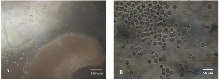
attachment. Results under microscopic observations showed that gonadal PGCs that were released from the
gonads reached a high number in 1 h after incubation
of the gonads (Figure 2). This results show that gonadal
PGCs can be isolated by incubating embryonic gonads of chicks in PBS [-] at 38oC for about 1 h (Nakajima & Tajima, 2013).
349
350 351 352 353
356 357
358
359
360 361 362
365 366 367
368
a
A B
A m B
349
350
351 352 353
356 357
358
359
360 361 362
365 366 367
368
A 200 µm B 50 µm
Figure 2. Gonadal PGCs cells from embryonic gonad of KUB chicken after 1 hour incubation in PBS [-] at 38oC. A) PGCs released from gonad, B) harvested isolated PGCs.
Figure 1. A) A pair of gonads (a) attached to the mesonephros chicken embryo. B) A pair of gonads that were removed from the me -sonephros and ready to be isolated.
4 April 2017
The results were in line with the results reported by Nakajima et al. (2011; 2013) in White Leghorn (WL) and
Rhode Island Red (RIR) chickens. The result also
con-firmed that PGCs isolation from gonad was easier and better with larger number of collected PGCs compared
to the isolation of circulated PGCs.
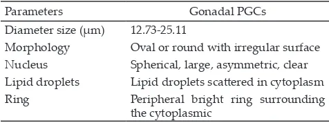
Gonadal PGCs of KUB chicken had morphological
characteristics: (1) large cell size, (2) bright lipid droplet
distributed in the cytoplasm (3) spherical and large nu-cleus and located asymmetrically, (4) peripheral bright ring surrounding the cytoplasmic membrane. More details about KUB chicken PGCs characteristics are
summarized in Table 1.
Number of Gonadal PGCs
The number of gonadal PGCs per embryo isolated
from embryonic development on days 6, 7, 8, and 9 of incubation period (stage 29-35 of HH) are presented in Table 2. Chojbacka-Puchta et al. (2012) reported that PGCs could be collected from embryonic gonad on days
5.5 to 6. Further result of Minematsu et al. (2004) showed
that PGCs began to migrate to the embryonic gonad
starting on day 5 after incubation and subsequent PGCs began to play a part in the gonad between days 6 and 7 (at the onset of sexual differentiation) and sexual dif
-ferentiation of the gonads were occurred after the arrival of germ cells. Meanwhile, germ cells multiplications are occurred at a later stage. Germ cells differentiation phase in female embryos took place between days 8 and 11 of incubation, whereas in male embryos, germ cells differentiation took place between days 13 and 15 of the
incubation period.
In this study, gonads sampling of embryos was
conducted on day 6 of incubation period, since most of PGCs had left the blood vessels and had begun to arrive
in the gonadal anlage (Han, 2009). Collection of embryo gonads was terminated on day 9 of incubation because
the PGCs remained stayed in the gonads. After that
pe-riod, PGCs gradually differentiated into sex cells.
DISCUSSION
Isolation and Identification of Gonadal PGCs from KUB Chicken
The results of morphological characteristics of
PGCs were consistent with the results reported by
Kostaman et al. (2013) in Gaok chicken, Minematsu et al.
(2004) in RIR chicken, Nakajima et al. (2016) in Oriental stork and Northern Bald ibis, and Sopiyana et al. (2016)
in KUB chicken using the lysis buffer ACK method.
Number of Gonadal PGCs
Based on the results of previous studies, one of the problems in the PGCs collection of embryonic blood on
day 2.5 was the low numbers of cells in local chickens
that could be collected (Kostaman et al., 2013). On the contrary to those studies, our results revealed that the
PGCs collection from days 6-9 yielded more PGCs
compared to the collection method conducted on day
2.5. The highest number of gonadal PGCs (P<0.05) was
obtained in the embryonic development at the stage of day 7 among the number of gonadal PGCs collected on
days 6, 8, and 9. This result showed that the number of gonadal PGCs was apparently influenced by the age of
embryonic development.
Starting on day 6 of embryonic development,
the number of gonadal PGCs significantly increased (P<0.05) and reached the peak number of gonadal PGCs on the embryonic development on day 7, which was probably that all PGCs might already reach the
embryonic gonad as reported by Hong et al. (1995).
Furthermore, this opinion was supported by Nakamura et al. (1988) that the PGCs had mostly settled down
in the gonadal ridge on the day 7th of the incubation
period.
The number of PGCs slowly decreased (P<0.05)
on days 8th to 9th incubation period. The reason of the
decreased number of PGCs on the day 8th of
incuba-tion might be due to the start of PGCs to differentiate
sexually to be a female embryo (Hong et al., 1995). Furthermore, PGCs began the meiotic division actively
forming oogonia on days 8-9 of the incubation period (Van Krey, 1990).
Collection of PGCs from avian embryonic gonad, especially in Indonesian chickens, had not been done so far, so this preliminary study can be applied for genetic material preservation technology of embryonic gonad PGCs on Indonesian local chicken, especially for KUB chicken. The advantage of the collection of PGCs using
PBS [-] has also been reported by Nakajima (2011) who
incubated embryos for 30 min and obtained 402 cells per
Parameters Gonadal PGCs
Diameter size (µm) 12.73-25.11
Morphology Oval or round with irregular surface Nucleus Spherical, large, asymmetric, clear Lipid droplets Lipid droplets scattered in cytoplasm Ring Peripheral bright ring surrounding
the cytoplasmic
Incubation period (days)
Average number of gonadal PGCs per embryo (cells)
Table 1. Morphology characteristics of gonadal primordial germ cells (gonadal PGCs) in Kampung Unggul Badan Litbang Pertanian (KUB) chicken
Note: Means in the same column with different superscript differ sig
-nificantly (P<0.05).
Table 2. Number of harvested gonadal primordial germ cells (gonadal PGCs) per embryo of Kampung Unggul Badan Litbang Pertanian (KUB) chicken at incubation period
April 2017 5
embryo at embryonic age of 7 d. Nakamura et al. (2011) harvested PGCs from gonads of WL chicken at the age of 7 d and obtained 237 cells per embryo.
This is the first report about gonadal PGCs in
Indonesian local chicken, particularly in KUB chicken,
although has lower number of PGCs compared to
the number of PGCs resulted from exotic chicken as reported previously by Atsumi et al. (2008), Nakajima et
al. (2011), and Nakamura et al. (2011). However results
of this study were still comparable with the results
reported by Nakajima et al. (2016) on Oriental stork and
Northern Bald ibis with the averages number PGCs as much as 130 and 160 cells. The lower number of gonadal
PGCs was influenced by the type of chicken (Zhao et al.,
2003). The results of this present study indicated gener-ally that KUB chicken as one type of Indonesian local
chicken had lower number of PGCs compared to exotic chicken. This lower number of PGCs seems likely to be correlated with the lower productivity of local chicken,
as was also stated by Zhao et al. (2003) that the number
of chicken PGCs correlated with the number of eggs.
CONCLUSION
The highest number of gonadal PGCs of KUB
chicken was obtained on day 7 of incubation period
and it can be recommended that the best period time to isolate gonadal PGCs is on day 7.
ACKNOWLEDGEMENT
Authors thanked to Dr. Tatan Kostaman, S.Si., MP.,
Dr. Ir. Tike Sartika, M.Si., and Prof. (R) Dr. Ir. Sofjan Iskandar, M.Rur.Sc., who helped both material and non-material until this research and article finished.
REFERENCES
Atsumi, Y., T. Tagami, H. Kagami, & T. Ono. 2008. Restriction of germline proliferation by soft X-ray of chicken embryos and its application to chimera production. J. Poult. Sci.
45:292-297. https://doi.org/10.2141/jpsa.45.292
Chojnacka-Puchta, L., K. Kasperczyk, G. Plucienniczak, D. Sawicka, & Bednarczyk. 2012. Primordial germ cells (PGCs) as a tool for creating transgenic chickens.
Polish. J. Vet. Sci. 15: 181-188. https://doi.org/10.2478/
v10181-011-0132-6
Furuta, H. 2012. Establishing germline chimeric chickens
us-ing primordial germ cells. J. Poult. Sci. 49:1-4. https://doi. org/10.2141/jpsa.011053
Furuta, H., S. Marumiya, I. Nakano, T. Yoshida, H. Mukouyama, & M. Tanaka. 2007. Effect of transfer primordial germ cells
(PGCs) into chick gonad. J. Poult. Sci. 44: 335-338. https:// doi.org/10.2141/jpsa.44.335
Furuta, H., T. Sawada, K. Nishikawa, I. Yamamoto, T. Yoshida, & M. Tanaka. 2008. Transfer of blood containing
primor-dial germ cells between chicken eggs development of embryonic reproductive tract. Cytotechnology 56:27-32. https://doi.org/10.1007/s10616-007-9096-x
Glover, J. D. & M. J. McGrew. 2012. Primordial germ cells tech-nologies for avian germplasm cryopreservation and
inves-tigating germ cell development. J. Poult. Sci. 49:155-162. https://doi.org/10.2141/jpsa.011161
Hamburger, V. & H. L Hamilton. 1951. A series of normal stages
in development of the chick embryo. J. Morphol. 88:49-92.
https://doi.org/10.1002/jmor.1050880104
Han, J. Y. 2009. Germ cells and transgenesis in chickens. CIMID.
32:61-80. https://doi.org/10.1016/j.cimid.2007.11.010
tion. Biol. Reprod. 79:931–937. https://doi.org/10.1095/ biolreprod.108.069989
Kohara, Y., Y. Kanai, & A. Tajima. 2008. Cryopreservation of go-nadal germ cells (GGCs) from the domestic chicken using
vitrification. J. Poult. Sci. 5:57-61. https://doi.org/10.2141/ jpsa.45.57
Kostaman, T., T. L. Yusuf, M. Fahrudin, & M. A. Setiadi. 2013. Isolation and number of circulated primordial germ cells (circulated-PGCs) on stages of embryonic development of
Gaok chicken (In bahasa: Isolasi dan jumlah primordial
germ cell sirkulasi (PGC-sirkulasi) pada stadium perkem-bangan embrio ayam Gaok). Indonesian Journal of Animal and Veterinary Sciences. 18:27-33.
Liu, C., K. A. Khazanehdari, V. Baskar, S. Saleem, J. Kinne, U. Wernery, & I. K. Chang. 2012. Production of chicken prog-eny (Gallus gallus domesticus) from interspecies germline chimeric duck (Anas domesticus) by primordial germ cell
transfer. Biol. Reprod. 86: Article 101, 1–8.
Minematsu T., A. Tajima, & Y. Kanai. 2004. The migratory abil-ity of gonadal germ cells in the domestic chicken. J. Poult.
Sci. 41:178-185. https://doi.org/10.2141/jpsa.41.178
Nakajima Y., H. Fukuda, M. Onuma, K. Murata, M. Ueda, E. Sunaga, T. Shiraishi, & A. Tajima. 2016. Migratory ability of gonadal germ cells (GGCs) isolated from Ciconia boyci-ana and Geronticus eremita embryos into the gonad of
de-veloping chicken embryos. J. Vet. Med. Sci. 78:1055-1058. https://doi.org/10.1292/jvms.15-0664
Nakajima, Y. & A. Tajima. 2013. Development of a novel meth-od for isolating gonadal germ cells from early chick
em-bryos. J. Dev. Sustain. Agri. 8:75-78.
Nakajima, Y., M. Naito, & A. Tajima. 2012. Production of germline chimeras by the transfer of gonadal germ cells (GGCs) recovered from 7-day-old chick embryos by using
newly developed PBS [−] method. World Poult. Sci. J. 68,
Supplement 1.
Nakajima, Y., T. Minematsu, M. Naito, & A. Tajima. 2011. A
new method for isolation viable gonadal germ cells from 7-day-old chick embrios. J. Poult. Sci. 48:106-111. https:// doi.org/10.2141/jpsa.010094
Nakamura, M., T. Kuwana, Y. Miyayama, & T. Fujimoto. 1988. Extragonadal distribution of primordial germ cells in
the early chick embryo. Anat. Rec. 222:90-94. https://doi. org/10.1002/ar.1092220113
Nakamura, Y., F. Usui, D. Miyahara, T. Mori, H. Watanabe, T. Ono, K. Takeda, K. Nirasawa, H. Kagami H, & T. Tagami. 2011. Viability and functionality of primordial germ cells
after freeze-thaw in chicken. J. Poult. Sci. 48:57-63. https:// doi.org/10.2141/jpsa.010085
Nakamura, Y., Y. Yamamoto, F. Usui, T. Mushita, T. Ono, A. R. Setioko, K. Takeda, K. Nirasawa, H. Kagami, & T. Tagami. 2007. Migration and proliferation of primor-dial germ cells in the early chicken embryo. J. Poult. Sci.
86:2182-2193. https://doi.org/10.1093/ps/86.10.2182
Park, T. S., M. A. Kim, J. M. Lim, & J. Y. Han. 2008. Production
of quail (Coturnix japonica) germline chimeras derived
from in-vitro-cultured gonadal primordial germ cells.
Mol. Rep. and Dev. 75:274-281. https://doi.org/10.1002/
mrd.20821
6 April 2017
W. Chen, N. Yang, & Z. Li. 2010. Influence of microgravity on the concentration of circulating primordial germ cells
in Silky chicken offspring. J. Poult. Sci. 47:65-70. https:// doi.org/10.2141/jpsa.009036
Sartika, T., S. Iskandar, & B. Tiesnamurti. 2016. Sumberdaya Genetik Ayam Lokal Indonesia dan Prospek Pengembangannya. Indonesian Agency for Agricultural Research and Development Press, Jakarta, Indonesia. Song, G., T. S. Park, T. M. Kim, & J. Y. Han. 2010. Avian
bio-technology: Insight from germ cells-mediated transgenic
system. J. Poult. Sci. 47:197-207. https://doi.org/10.2141/ jpsa.009108
Sopiyana, S., I. Supriatna, M. A. Setiadi, & M. Fahrudin. 2016. Determination of production capacity of circulated primordial germ cells (circulated-PGCs) of KUB chicken
using lysis buffer ammonium chloride potassium (ACK).
Indonesian Journal of Animal and Veterinary Sciences.
21:55-61. https://doi.org/10.14334/jitv.v21i1.1315
Tajima, A. 2013. Conservation of avian genetic resources. J.
Poult. Sci. 50:1-8. https://doi.org/10.2141/jpsa.0120083
Van Krey, H. P. 1990. Reproductive biology in relation to breeding and genetics. In: Poultry Breeding and Genetics.
Edited by R. D. Crawford. Elsevier Science Publisher, Amsterdam. Pp. 61-90.
Wang Y., L. Hou, C. Li, W. Guan, L. Chen, X. Li, W. Yue, & Y. H. Ma. 2010. Isolation, culture, and biological
charac-teristics of primordial germ cells from Beijing Fatty chick
-en. J. Reprod. Dev. 56:303-308. https://doi.org/10.1262/ jrd.09-126N
Yamamoto, Y., T. Ono, & H. Kagami. 2007. Dynamic analysis of the developmental fate of cells in the center of the area
pel-lucida of the blastoderm in chicken. J. Poult. Sci. 44:85-91. https://doi.org/10.2141/jpsa.44.85
Zhao, D. F., H. Yamashita, M. Matsuzaki, T. Takano, S. Abe, M. Naito, & T. Kuwana. 2003. Genetic factors affect the number of circulating primordial germ cells in early chick