Determination of Production Capacity of Circulated Primordial Germ Cells
(Circulated-PGCs) of
KUB Chicken using Lysis Buffer Ammonium Chloride
Potassium (ACK)
Sopiyana S1, Supriatna I2, Setiadi MA2, Fahrudin M3
1Indonesian Research Institute for Animal Production, Bogor PO BOX 221, Bogor 16002 2 Department of Reproduction and Pathology Clinic, Faculty of Veterinary Sciences
Bogor Agricultural University, Jl. Agatis Kampus IPB Darmaga, Bogor 16680
3 Department of Anatomy, Physiology, and Farmacology, Faculty of Veterinary Sciences
Bogor Agricultural University, Jl. Agatis Kampus IPB Darmaga, Bogor 16680 E-mail: [email protected]
(received 03-02-2016; revised 27-02-2016; 09-03-2016)
ABSTRAK
Sopiyana S, Supriatna I, Setiadi MA, Fahrudin M. 2016. Penentuan kapasitas produksi primordial germ cell sirkulasi (PGC-sirkulasi) pada ayam KUB menggunakan metode buffer lisis ammonium chloride potassium (ACK). JITV 21(1): 55-61. DOI: http://dx.doi.org/10.14334/jitv.v21i1.1315
Pada embrio unggas, primordial germ cell (PGC) merupakan sel progenitor untuk gamet yang memiliki jalur migrasi yang unik. Primordial germ cell pertama kali muncul dari epiblast di daerah germinal crescent, kemudian bersirkulasi melalui pembuluh darah dalam periode waktu yang singkat, dan bermigrasi menuju gonad. Penelitian ini bertujuan menentukan kapasitas potensi produksi PGC-sirkulasi embrio ayam KUB pada stadia perkembangan embrio yang berbeda untuk mengisolasi dan mengoleksi PGC-sirkulasi menggunakan metode yang cepat dan sederhana. Dalam penelitian ini digunakan 75 butir telur fertil ayam KUB yang dibagi menjadi lima kelompok perlakuan, dan diinkubasi pada suhu 38,5 0C dengan kelembaban 60%. Pemanennan embrio diatur sesuai dengan tahapan perkembangan embrio 14-18. Pengambilan darah embrio dilakukan melalui aorta dorsalis dengan menggunakan mikropipet di bawah mikroskop. Darah yang terkumpul ditempatkan pada tabung eppendorf 1,5 ml yang telah diisi dengan 100 µl larutan phosphate buffered saline tanpa Ca2+ dan Mg2+ (PBS-) yang dicampur dengan fetal bovine serum (FBS) dengan perbandingan 90%:10%. PGC dimurnikan dengan menggunakan proses buffer lisis ammonium chloride potassium (ACK). Hasil penelitian menunjukkan bahwa rataan produksi PGC-sirkulasi per embrio dipengaruhi oleh stadia perkembangan embrio (P<0,05). Rataan produksi PGC-sirkulasi dari stadia perkembangan embrio dari 14, 15, 16, 17, dan 18 pada ayam KUB berturut-turut adalah 37,9; 53,5; 49,8; 38,3; dan 33,5 sel per embrio. Produksi PGC-sirkulasi antara stadia 14, 17, dan 18 tidak berbeda nyata (P>0,05). Produksi PGC-sirkulasi terbanyak didapatkan pada stadia 15, sehingga isolasi dan koleksi PGC melalui sirkulasi darah disarankan pada stadia 15.
Kata Kunci: Ayam KUB, PGC, Stadia Perkembangan Embrio, Ammonium Chloride Potassium
ABSTRACT
Sopiyana S, Supriatna I, Setiadi MA, Fahrudin M. 2016. Determination of production capacity of circulated primordial germ cells (circulated-PGCs) of KUB chicken using lysis buffer ammonium chloride potassium (ACK). JITV 21(1): 55-61. DOI: http://dx.doi.org/10.14334/jitv.v21i1.1315
In poultry embryos, primordial germ cells (PGCs) are progenitor cells for gametes, which have unique migration pathway. Primordial germ cells arise from epiblast in germinal crescent and circulate through the bloodstream for a short period of time, then leave blood vessel to migrate toward gonads. The aim of this study was to determine the potential production capacity of circulated-PGCs of KUB chicken at different developmental stages of embryo using a rapid and simple method. Seventy five KUB chicken fertile eggs were divided into five groups and incubated at 38.5 0C with a humidity of 60%. Hatching was set to the embryonic development stage of 14-18. The blood was collected through dorsal aorta using micropipette under microscope. The collected blood was placed in a 1.5 ml eppendorf tube which was previously filled with 100 µl phosphate buffered saline without Ca2+ and Mg2+ (PBS-) mixed with fetal bovine serum (FBS) with a ratio of 90%:10%. The PGCs were purified using lysis buffer ammonium chloride potassium method. The results showed that average production of circulated-PGCs per embryo of KUB chicken were significantly affected by stage of embryonic development (P <0.05). The average production of circulated-PGCs at stage 14, 15, 16, 17, and 18 were 37.9; 53.5; 49.8; 38.3; and 33.5 respectively. The number of circulated-circulated-PGCs was not different among stages 14, 17 nor 18. The highest number of circulated-PGCs of KUB chicken was obtained at stage 15, so that the isolation and collection of PGCs through the blood circulation was recommended in stage 15.
INTRODUCTION
Indonesia is known as one of world megabiodiversities. Indonesian native poultry is one of animal genetic resources which potential for egg and meat, and fancy animal. Sartika & Iskandar (2007) reported that there were 43 recognized native chicken breeds in Indonesia, which can be further developed and preserved in-situ and ex-situ. Since 2012 Indonesian Research Institute for Animal Production has developed a strain derived from native Kampung chicken breed as improved egg producers strain. KUB chicken is the name of the strain which was able to produce 44.33% henday production, reaching peak production of 60% (Iskandar & Sartika 2014). As a new strain, KUB chicken may be categorized as new genetic resources that is needed to be conservated.
Conservation activity was generally done through in-situ method where the maintenance was conducted in its natural habitat and ex-situ method by maintaining live animals outside its natural habitat. Somehow, in-situ and ex-in-situ conservations of live animals require high maintainance cost and disease risks.
Conservation technique on semen, ovum even embryo in bird did not come to satisfy than when applied in mammal. Blesbois & Labbe (2003) reported that percentage of live sperm after thawing was only 60%. Ovum freezing in bird still was not succeeded regarding its big size and complexity and high fat content in bird (Blesbois & Labbe 2003). Bird’s ovum had some issues causing cryopreservation might not be done (Hagedorn et al. 2004). An approach currently developed to solve limitation on in-situ and ex-situ conservations especially in bird is primordial germ cells (PGCs) cryopreservation. Cryopreserved primordial germ cells might be preserved for unlimited time and might be used everytime needed.
Primordial germ cells were gamet progenitor cells for or spermatozoa and ovum progenitor and were early development of gamet cells (Qian et al. 2010; Glover & McGrew 2012; Tajima 2012). Qian et al. (2010) reported that PGCs was formed out of gonad and migrated toward gonad during embryogenesis which will be developed into spermatozoa and ovum. Primordial germ cells might be used as genetic source and for establishing transgenic chicken (Furuta 2012). Primordial germ cells were cells with plurypotent characteristic making the PGCs as a good model to study embryonic development by in-vitro (Wang et al. 2010).
Primordial germ cells were first detected in chicken embryo in the end of gastrulation (Ginsburg 1997) and first appeared from epiblast and first located in ventral part of pelucyde area of embryo stage X, then it was translocated into dorsal side of hypoblast in stage
XI-XIV or stage 2 to 6 (Hamburger & Hamilton 1951), then was migrated into germinal crescent area in stage 4 through bottom layer. PGCs migration was stoped at the end of gastrulation (Ginsburg 1997). During blood vessel forming, PGCs entered blood vessel between stage 10 and 12 and begun to circulate in bloodstream. During stage 12 to 16, PGCs was stay still in circulation of embryo blood after eggs incubated for 2.5 to 3 days (stage 13-16 HH) known as circulated-PGCs); (3) gonad embryo after eggs incubated for 5.5 to 6 days (stage 26-28 HH) known as gonad-PGCs.
Unique migration path of chicken PGCs easy PGCs collection based on its place. A method to collect PGCs from chicken widely used was by isolating PGCs from 2.5-3 days old embryo blood (Yamamoto et al. 2007; Nakamura et al. 2007). The number of circulated-PGCs would be different in every embryonic development stage (Tajima et al. 1999; Li et al. 2001). Zhao & Kuwana (2003) reported that the number of circulated-PGCs in embryonic development stage would be affected by variated egg production.
The most embryonic development stage with circulated-PGCs of chicken was in stage 13-18 (Tajima et al. 1999), stage 13 in quail (Li et al. 2001), stage 15 in White Leghorn chicken (Kuwana et al. 2006), and stage 14 in Kureko Dori chicken (Qian et al. 2010). Information of embryonic development stage and circulated-PGCs in native chicken was varied among 15-29 cells. The most number of circulated-PGCs was found in Indonesian native Gaok chicken in stage 15 by 51 cells per embryo (Kostaman 2013a).
This study was to determine embryonic development stage with the highest circulated-PGCs production of KUB chicken using buffer lysis ACK. This study result was expected to be basic of proper embryonic development stage determination in native chicken in isolating PGCs through blood vessel using buffer lysis ammonium chloride potassium (ACK) method.
MATERIALS AND METHODS
Time and place
Fertile eggs preparation
Seventy five fertile eggs of KUB chicken from Chicken Research laboratory of IRIAP, Ciawi-Bogor were used in this study for circulated-PGCs collection. Those eggs were divided into 5 treatment groups based on embryonic development stage at 14-18 hours (there were 5 embryonic development stages by 5 eggs in each group). Eggs were incubated in 38.5°C with 60% humidity and stirred as 90° every 30 minutes using portable incubator (P-008B Biotype; Showa Furanki, Saitama, Jepang). Embryo collection from each treatment was set as embryonic development stage, that was in 14-18 HH (Hamburger & Hamilton 1951) or after 50-72 hours incubation.
Isolation and collection of circulated-PGCs using buffer lysis ACK
After egg reached desired embryonic development stage, eggshell was broken and the content was moved into plastic petri dish (90 x 15 mm, LBS60001PT, BIOLAB). Embryo blood was collected through dorsalis aorta using micropipette (50 μm; Drummond Scientific, Broomall, PA USA) under microscop (Olympus SZX7, Japan). Dorsalis aorta was near the heart and providing blood in a large amount. The accumulated blood was divided based on embryonic development stage (14-18 HH) and each was put in
eppendorf 1.5 ml tube filled by 100 µl phosphate
buffered saline solution without Ca2+ and Mg2+ (PBS-) + fetal bovine serum (FBS) with ratio by 90%:10%. PGCs was purified using buffer lysis ACK process (Yamamoto et al. 2007) which was new method to be developed to isolate circulated-PGCs from embryo blood circulation. This was an easy and fast method to
purify circulated-PGCs using ammonium chloride-potassium (ACK).
Variables evaluated in this study were morphology characteristic and production of circulated-PGCs of KUB chicken determined by standard reported by Hamburger & Hamilton (1951) and based on incubation time, embryonic morphology form, the number of somit, and embryonic development. Circulated primordial germ cells might be distinguished from red blood cells apart from cell size and other characteristics such as nucleus, refractive fat, or ring of surrounding cells.
Evaluation and analysis of study results
Circulated-PGCs isolation result as morphology characteristic was descriptively analyzed. Production potential of circulated-PGCs from each embryonic development stage (stage 14, 15, 16, 17, and 18) was statistically analyzed using Completely Randomized Design with 15 replications. Data were analyzed by ANOVA which its normality was analyzed by Kolmogorof-Smirnov Z before. If there was different between those treatments, it was continued by Duncan multiple range test (Steel & Torrie 1995).
RESULT AND DISCUSSION
Issolation and identification of circulated primordial germ cells (circulated-PGCs) using buffer lysis ACK
Circulated PGCs were isolated from embryo blood after egg incubated for 2.5 – 3 days (stage 14-18, Hamburger & Hamilton 1951). In this study, circulated-PGCs of KUB chicken has been successfully isolated and identified using buffer lysis ACK from each blood sample in stage 14-18.
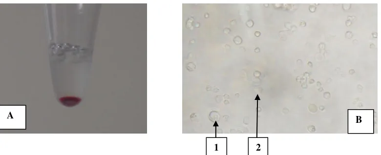
Figure 1. Issolation of circulated-PGCs of KUB chicken using buffer lysis ACK method. A) Blood sample collected from 14-18 stadium of embryonic development. B) First purification of ACK process and centrufigation, circulated-PGCs which is still mixed with red blood cells. 1=circulated-PGCs and 2=red blood cells.
A
1 2
Table 1. Comparation of morphology characteristic between circulated-PGCs and red blood cells
Chriteria Circulated-PGCs Red blood cells (erythrocytes)
Size (µm)1) 14-19 6-8
Cell form Oval or round, irregular contours Round
Core Spherical, large, asymmetrical, clear Exist, not clear
Specific gravity2) 1.029-1.040 1.054-1.060
Fat Droplets There were fat droplets in cytoplasm Not exist
Ring Teher was light ring surrounds the cell Not exist
Source: 1)Yashuda et al. (1992); 2)Qian et al. (2010)
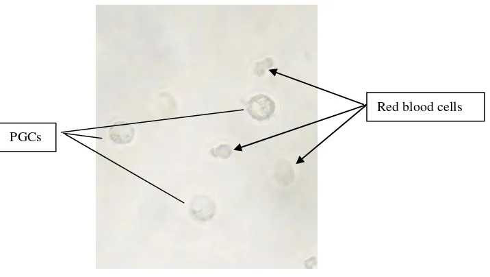
Figure 2. Circulated-PGCs of KUB chicken obtained using buffer lysis ACK (magnification by 400x).
The result from descriptive analysis showed that circulated-PGCs had the same morphological characteristic with the one reported by Ginsburg (1997); Yashuda et al. (1992); Kostaman (2013b) in previous study which showed that PGCs in chicken might be identified morphologically as (1) it had bug cells (14-19 μm), (2) oval or round and irregular contours, (3) there was light fat droplets distributed in sitoplasm, (4) sperical and big core located unsymmetrically and containing prominent nucleous, (5) there was light ring under cell membrane on the outskirts of PGCs.
Other information reported that generally, basic characteristic of PGCs in chicken was about the same with in quail, pheasant, and duck. By those characteristics, PGCs might be distinguished with red blood cells as in Table 1.
In this study, circulated-PGCs was purified using buffer solution and centrifugation with speed developed
by Yamamoto et al. (2007) consisting of two time purifications and two time flushings. Main principle of this purification method was dividing red blood cells with circulated-PGCs by lysising red blood cells where ACK process would dissolve red blood cells and did not affect circulated-PGCs morphology.
Buffer lysis ACK solution consisted of ammonium
chloride, potassium bicarbonate, and EDTA.
Ammonium choride was anorganic compound with as NH4Cl as white crystal salt which was highly water soluble. Potassium bicarbonate was a colorless, odorless and salty substance. EDTA was an anticoalgulant of an odorless aminopolycarboxylic acid and a solid substance dissolved in water. Buffer lysis ACK solution was an odorless substance used to lysis red blood cells and did not affect the circulated-PGCs morphology (Yamamoto et al. 2007).
PGCs
Table 2. Production of circulated-PGCs per embryo using buffer lysis ACK method in KUB chicken
Embryonic development
stage (HH) Incubation time (hour) Average of circulated-PGCs per embryo (cells)
14 50-52 37.9±9.2a
15 53-55 53.5±8.0b
16 56-59 49.8±6.2b
17 60-63 38.3±4.3a
18 64-67 33.5±5.7a
Numerals in the same column followed by the same letter show not significantly different at 5% (Duncan Multiple Range Test)
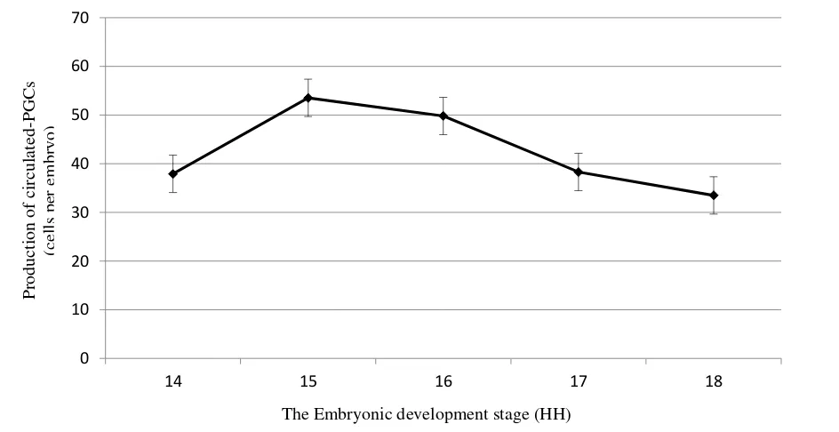
0 Figure 3. Circulated-PGCs of KUB chicken in different incubation time
Circulated primordial germ cells (Circulated-PGCs) of KUB Chicken
Based on isolation obtained and calculation of circulated-PGCs per embryo, it was showed that the number of circulated-PGCs varied between embryonic development stage (Table 2).
The highest average number of circulated-PGCs (P<0.05) was found in stage 15 and 16 compared to stage 14, 17, and 18 where there was varied average circulated-PGCs amount in each embryonic development stage. This showed that circulated-PGCs production was affected by embryonic development stage.
From stage 14, production of circulated-PGCs in embryo blood sample was increase and reached the peak at stage 15. At stage 15, it was possible that all PGCs gathered in embryo circulatory system (Kostaman et al. 2013a). The peak of PGCs production
circulated-PGCs in embryonic development stage 14-18 HH (Figure 3).
This result was not significantly different with previous study in Indonesian native Gaok chicken reporting that the number of circulated-PGCs, when purified by nicodenz density gradient sentrifugation method reached peak amount at stage 15 to of 51.0 cells per embryo (Kostaman 2013a). That result was a considerably lower than previous research result reported that the most circulated-PGCs in Rhode Island Red (RIR) chicken blood circulation system was 67-73 cell at the stage of 15 (Nakamura et al. 2007), and it was different from Silky chicken where the circulated-PGCs peak amount was at stage 14 by 65 cells (Qian et al. 2010). Setioko (2007) reported that average amount of PGCs in White Leghorn chicken was 84.2 cells. Some research results showed that the number of PGCs of native chicken was varied between 15-51 cells (Setioko et al. 2010; Kostaman 2013a). It also showed that appropriate time to harvest the circulated-PGCs was in stage 15 (53.5 cells). At that stage, circulated-PGCs might be obtained in the largest number. This was an appropriate time to preservate PGCs and form germline chimera.
In this study, embryo blood collection did not begin at stage 13, but at stage 14 where the amount of that it would be difficult to inject and would be broken when imposed.
CONCLUSION
The highest number of circulated-PGCs in KUB chicken was in stage 15 and this stage was the best time to isolate PGCs to obtain maximum circulated PGCs.
ACKNOWLEDGEMENT
Authors thanked to Dr. Tatan Kostaman, Dr. Tike Sartika, Prof. (R) Dr. Sofjan Iskandar, and Dr. L. Hardi Prasetyo who helped both material and non-material until this research and article finished.
REFERENCES
Blesbois E, Labbé C. 2003. Main improvements in semen and embryo cryopreservation for fish and fowl. In:
Planchenault D, editor. Cryopreservation of Animal Genetic Resources in Europe, Paris, France. p. 55-65. Chojbacka-Puchta L, Kasperczyk K, Plucienniczak G,
Sawicka D, Bednarczyk. 2012. Primordial germ cells (PGCs) as a tool for creating transgenic chickens. Polish J Vet Sci. 15:181-188.
Furuta H. 2012. Establishing germline chimeric chickens using primordial germ cells. J Poult Sci. 49:1-4. Ginsburg M. 1997. Primordial germ cells development in
avians. J Poult Sci. 76:91-95.
Glover JD, McGrew MJ. 2012. Primordial Germ Cells Technologies for Avian Germplasm Cryopreservation and Investigating Germ Cell Development. J Poult Sci. 49:155-162.
Hagedorn M, Peterson A, Mazur P, and Kleinhans FW. 2004. High ice nucleation temperature of zebrafish embryos: slow-freezing is not an option. Cryobiology. 49:181-189.
HamburgerV,HamiltonHL.1951. A series of normal stages in development of the chick embryo. J Morphol. 88:49-92.
Iskandar S, Sartika S. 2014. KUB Chicken: The first Indonesia Kampung chicken selected for egg production. Proceedings of the 16th AAAP Animal Science Congress Vol II 10-14 November 2014. Yogyakarta (Indones): Gadjah Mada University. p. 157-160.
Kostaman T, Yusuf TL, Fahrudin M, Setiadi MA. 2013a. Isolasi dan jumlah primordial germ cell sirkulasi (PGC-sirkulasi) pada stadium perkembangan embrio ayam Gaok. JITV. 18:27-33.
Kostaman T. 2013b. Isolasi dan kriopreservasi primordial germ cell (PGC) menggunakan kriopotektan DMSO untuk pembentukan germline chimera ayam Gaok (Disertation). [Bogor (Indones)]: Institut Pertanian Bogor.
Kuwana T, Kawashima T, Naito M, Yamashita H, matsuzaki M, Takano T. 2006. Conservation of a threathened indigenous fowl (Kureko Dori) using the germline chimeras transpanted from primordial germ cells. J Poult Sci. 43:60-66.
Li HC, Matsui K, Ono T. 2001. Population of circulating primordial germ cells in early Japanese quail embryos. J Poult Sci. 38:175-180.
Nakamura Y, Yamamoto Y, Usui F, Mushita T, Ono T, Setioko AR, Takeda K, Nirasawa K, Kagami H, Tagami T. 2007. Migration and proliferation of primordial germ cells in the early chicken embryo. J Poult Sci. 86:2182-2193.
Sartika T, Iskandar S. 2007. Mengenal plasma nutfah ayam Indonesia dan pemanfaatannya. Edisi Pertama. Bogor (Indones): Balai Penelitian Ternak.
Setioko AR, Tagami T, Tase H, Nakamura Y, Takeda K, Nirasawa K. 2007. Cryopreservation of primordial germ cells (PGCs) from White Leghorn embryo using commercial cryoprotectants. J Poult Sci. 44:73-77. Setioko AR, Kostaman T, Sopiyana S. 2010. Jumlah
primordial germ cell (PGC) pada beberapa tingkat umur embrio yang berbeda pada ayam buras dan ras. Prosiding Seminar Nasional Biologi. Bandung (Indones): Universitas Padjajaran. hlm. 133-141. Steel RGD, Torrie JH. 1995. Prinsip dan prosedur statistika:
Suatu pendekatan biometrik. Sumantri B, penerjemah. Principles and procedurs of statistics: A biometrical approach. Jakarta (Indones): Gramedia Pustaka Utama. Tajima A, Hayasi H, Kamizumi A, Ogura J, Kuwana T,
Chikamune T. 1999. Study on the concentration of circulating primordial germ cells (cPGCs) in early chick embryos. J Exp Zool. 28:759-764.
Tajima A. 2012. Conservation of avian genetic resources. J Poult Sci. 50:1-8.
Ukeshima A, Yoshinaga K, Fujimoto T. 1991. Scanning and transmission electron microscopic observations of chick primordial germ cells with special reference to the extravasation in their migration course. J Elec Micros. 40:124-128.
Wang Y, Hou L, Li C, Guan W, Chen L, Li X, Yue W, Ma YH. 2010. Isolation, culture, and biological characteristics of primordial germ cells from Beijing Fatty chicken. J Reprod Dev. 56:303-308.
Yamamoto Y, Usui F, Nakamura Y, Ito Y, Tagami T, Nirasawa K, Matsubara Y, Ono T, Kagami H. 2007. A novel method to isolate Primordial Germ Cells and its use for the generation of germline chimeras in chicken. Biol Reprod. 77:115-119
Yashuda Y, Tajima A, Fujimoto T, Kuwana T. 1992. A method to obtain avian germline chimeras using isolated primordial germ cells. J Reprod Fertil. 96:521-528. Zhao DF, Kuwana T. 2003. Purification of avian circulating