Effect of singlet oxygen generating substances on the ascorbic acid
and glutathione content in pea leaves
Ga´bor Gullner *, Alan D. Dodge
School of Biological Sciences,Uni6ersity of Bath,Cla6erton Down,Bath BA2 7AY,A6on,UK
Received 19 April 1999; received in revised form 16 November 1999; accepted 16 November 1999
Abstract
Ascorbate and glutathione levels were investigated in pea leaf discs exposed to various singlet oxygen generating compounds: eosin, rose bengal, monuron, acifluorfen and 5-amino-levulinic acid (ALA). The cellular level of the major antioxidant ascorbate was markedly decreased by the herbicides monuron, acifluorfen and ALA (in light-dependent reactions), as well as by the xanthene dyes eosin and rose bengal (independently of light). No significant accumulation of dehydroascorbate could be observed in any treatments. In contrast to ascorbate, the foliar glutathione levels were considerably increased by subtoxic or slightly toxic concentrations of eosin, rose bengal, acifluorfen and ALA in a light-dependent manner. Monuron treatments led to unchanged or decreasing glutathione contents. The activities of three antioxidative enzymes (ascorbate peroxidase, glutathione reductase and glutathioneS-transferase) were also induced by eosin in light-dependent reactions. © 2000 Published by Elsevier Science Ireland Ltd. All rights reserved.
Keywords:Acifluorfen; 5-Aminolevulinic acid; Ascorbate; Glutathione; Monuron; Xanthene dye
www.elsevier.com/locate/plantsci
1. Introduction
The accumulation of active oxygen species (AOSs) has been observed in various plants ex-posed to biotic and abiotic stress effects [1]. Among other AOSs, singlet oxygen (1O
2), the
ex-cited form of molecular oxygen is also capable of damaging important plant cell constituents [2 – 4]. Singlet oxygen sources in biological systems in-clude light-dependent reactions (photosensitiza-tion) and chemical generations in dark reactions [5].
A large variety of dyes act as photosensitizers. The cell damaging effects of the xanthene dyes
eosin and rose bengal are well known in plant leaves [2,6,7]. The light excitation energy of these dyes is transferred directly to dioxygen, leading to the generation of singlet oxygen.
Chlorophyll itself is capable of photodynamic reactions involving 1O
2 when its excited states are
insufficiently quenched by the photosystems or by carotenoids [3,6]. Phenylurea herbicides, such as monuron (3-[p-chlorophenyl]-1,1-dimethylurea), promote these reactions by blocking the electron flow between the photosystems [6]. Other por-phyrin derivatives are also effective photosensitiz-ers [8]. These intermediates are normally present in small amounts, but an excess might act as photo-sensitizer because they are unable to pass on light excitation energy to light harvesting accessory proteins. Instead, excitation energy results in triplet state senzitizers, which excite oxygen to the singlet state. The herbicide acifluorfen (5-[2-chloro-4-(trifluoromethyl)-phenoxy]-2-nitro-benzoic acid), which exerts its effect in the light, is known to induce the accumulation of protopor-Abbre6iations: ALA, 5-aminolevulinic acid; AOS, active oxygen
species; AP, ascorbate peroxidase; DHA, dehydroascorbate; GR, glutathione reductase; GSH, glutathione; GSSG, oxidized glu-tathione; GST, glutathioneS-transferase.
* Corresponding author. Present address: Plant Protection Insti-tute, Hungarian Academy of Sciences, P.O.B. 102, H-1525 Budapest, Hungary. Tel.: +36-1-355-8722; fax: +36-1-356-3698.
E-mail address:ggull@nki.hu (G. Gullner)
phyrin IX and other tetrapyrrole derivatives in plant leaves [9,10]. The chlorophyll precursor 5-aminolevulinic acid (ALA) can also cause accumu-lation of abnormally high levels of tetrapyrrole intermediates in darkness if it is added exoge-nously. As a consequence, ALA treatments led to marked photodynamic herbicidal effects [4,8,11]. Plants are protected by various mechanisms against the oxidative stress caused by singlet oxy-gen. Membrane bound antioxidants, carotenoids and a-tocopherols are well-known quenchers of
1O
2[2,3]. However, much less information, mostly
from in vitro studies, is available about the role of water-soluble antioxidants in the detoxification of singlet oxygen. Ascorbate is a major primary an-tioxidant, which reacts directly with singlet oxygen [1,12]. Glutathione (GSH) is also known to scav-enge 1O
2 [1,5]. Ascorbate and GSH are closely
related as both are constituents of the antioxida-tive ascorbate-glutathione cycle which detoxifies hydrogen peroxide in the chloroplasts [1].
In order to gain more information about the role of ascorbate and GSH in the detoxification of singlet oxygen in plant tissues, we have studied their cellular levels in pea leaf discs exposed to various1O
2generating substances (eosin, rose
ben-gal, monuron, acifluorfen and ALA). In addition, the activities of the antioxidative enzymes ascor-bate peroxidase (AP; E.C. 1.11.1.11.), glutathione reductase (GR; E.C. 1.6.4.2.) and glutathione S -transferase (GST; E.C. 2.5.1.18.) were also investi-gated in pea leaf discs exposed to eosin. AP and GR are constituents of the ascorbate-glutathione cycle [1]. GST, beside other detoxification reac-tions, can catalyze the breakdown of fatty-acid hydroperoxides and contributes to the protection of plant cells against oxidative membrane damage [13,14].
2. Materials and methods
2.1. Plant material
Pea (Pisum sati6um L. var. Meteor) seedlings
were grown in Levington Universal compost in a greenhouse with a 14 h photoperiod (natural day-light extended by 400 W Thorn mercury vapour lamps) and a mean air temperature of 22°C. Leaf discs (12 mm in diameter) were excised from sub-apical leaves of 2 – 3 week-old pea plants. Discs
were floated on the aqueous solutions of singlet oxygen generators (or on tap water) in Petri dishes for 24 h in darkness, then they were exposed to continuous light for 72 h in a light cabinet main-tained at 25°C. Illumination (400 mmol m−2 s−1)
was provided by warm fluorescent tubes (65/85-W; Thorn). After varying time periods the leaf discs were washed and used for biochemical assays. Parallel incubations were carried out in darkness. Dry leaf weights were measured after the incuba-tion of leaf discs at 110°C for 2 h.
ALA, eosin Y (2%,4%,5%,7%-tetrabromofluorescein), and rose bengal (4,5,6,7-tetrachloro-2%,4%,5%,7%
-te-traiodofluorescein) were obtained from Sigma (St. Louis, MO, USA). Acifluorfen and monuron were purchased from Riedel-de Haen (Seelze, Germany).
2.2. Enzyme assays
For enzyme assays cell-free homogenates were prepared at 0 – 4°C. Leaf material (0.5 g) was frozen in liquid nitrogen, pulverized with a mortar and a pestle, and suspended in 3 ml of ice-cold 0.2 M Tris – HCl buffer (pH 7.8) containing 3% solu-ble polyvinylpyrrolidone and 0.1 mM Na2-EDTA.
The homogenate was strained through muslin and centrifuged at 8000×g for 20 min. The superna-tants were used as enzyme source.
The enzyme activities were determined by spec-trophotometric methods. The activity of AP was determined as described in Ref. [13], except that in our experiments 0.25 mM ascorbate and 0.5 mM hydrogen peroxide were used. GR and GST activ-ities were measured by following the reduction of oxidized glutathione (GSSG) at 340 nm, and by measuring the formation of the GSH-conjugate using 1-chloro-2,4-dinitrobenzene as substrate at 340 nm, respectively, as described earlier [13].
2.3. Other assays
The level of acid-soluble, non-protein thiols was measured spectrophotometrically with 5,5%
Ascorbate and dehydroascorbate levels were measured by enzymatic methods using ascorbate oxidase enzyme as described by Wise and Naylor [17], and Foyer et al. [18], respectively.
2.4. Statistics
At least three independent parallel experiments were carried out in each case. The significant differences between mean values were evaluated by Student’s t-test. Differences were considered to be significant at P=0.05.
3. Results and discussion
3.1. Xanthene dyes
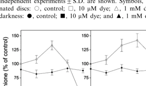
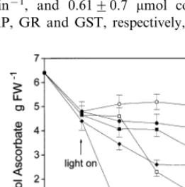
The application of xanthene dyes to pea leaf discs resulted in phytotoxic symptoms (bleaching) in the light, but not in darkness. The exposure of illuminated pea leaf tissue to 10 mM eosin led to only a very weak bleaching effect after 24 h, but 1 mM eosin was markedly phytotoxic. Rose bengal was much less toxic than eosin: only as high as 1 mM rose bengal concentration caused visible symptoms (moderate bleaching) in pea leaf tissue. Eosin, and to a lesser extent also rose bengal decreased the ascorbate content in a concentra-tion-dependent manner. Xanthene dye treatments led to ascorbate degradation in both illuminated and dark-incubated leaf discs (Fig. 1). Since singlet oxygen can not be produced by xanthene dyes without the excitation energy of light, these results proved that the ascorbate degradation was not related to singlet oxygen quenching. The dehy-droascorbate (DHA) levels also markedly de-creased in the leaf discs exposed to xanthene dyes (data not shown). In untreated pea leaves DHA accounted for 2696% (n=5) of the total ascorbic acid content. The light-independent reactions lead-ing to the depletion of ascorbate and dehy-droascorbate pools in the presence of xanthene dyes are not known. In darkness, ascorbate levels markedly declined also in untreated pea leaf discs as it had been observed earlier [19] showing that light was necessary for its synthesis or recycling. In contrast to ascorbate, both the non-protein thiol and the total glutathione contents increased in pea leaf discs exposed to subtoxic or slightly toxic concentrations of the xanthene dyes under illumination. The maximal increases were found after 24 h (Fig. 2), later the thiol and GSH levels slowly returned to control levels. However, at the very toxic 1 mM eosin concentration a dramatic GSH loss was found. Similar results were obtained when GSH levels were calculated on dry weight basis (data not shown). In darkness, the GSH content of untreated leaf discs slightly decreased as observed earlier in other plants [20]. The xanthene dyes did not alter the total glutathione levels in darkness as compared to dark controls.
Increases in GSH levels were often observed in plants in response to oxidative stimuli [14,21,22]. The rate-limiting step of GSH biosynthesis is
cata-Fig. 1. Changes in the ascorbate content of pea leaf discs exposed to xanthene dyes (eosin or rose bengal) at various concentrations. All discs were preincubated for 24 h in dark-ness then illuminated (400 mmol m−2 s−1). Parallel
experi-ments were carried out in darkness. Means of three independent experiments9S.D. are shown. Symbols, illumi-nated discs: , control; , 10 mM dye; , 1 mM dye. In darkness: , control;, 10 mM dye; and , 1 mM dye.
Fig. 2. Changes in the total glutathione level of pea leaf discs incubated with eosin or rose bengal at various concentrations for 24 h under illumination (400mmol m−2s−1) preceeded by
24 h preincubation in darkness. The values presented are percentages of the glutathione level in illuminated control leaf discs (0.4690.04mmol glutathione g FW−1). Means of three
Table 1
Activities of ascorbate peroxidase (AP), glutathione reductase (GR), and glutathione S-transferase (GST) in pea leaf discs exposed to various concentrations of eosina
Enzyme activities (% of illuminated Eosin concentration
aLeaf discs were preincubated in darkness for 24 h then
illuminated (400mmol m−2s−1) for 24 h. Parallel incubations
were carried out in darkness. Means of three different experi-ments9S.D. are shown.
bEnzyme activities in illuminated control leaves were 2.69
0.3mmol ascorbate g FW−1min−1, 1.590.1mmol NADPH
g FW−1 min−1, and 0.6190.7 mmol conjugate g FW−1
min−1for AP, GR and GST, respectively, (n=5).
fate reduction is also activated in plants exposed to stress effects [21]. The effects of singlet oxygen generators on these biosynthetic pathways are not known.
In our experiments, the total glutathione con-tent accounted for 9295% (n=4) of the non-protein thiol level in the untreated leaves. Alterations of the non-protein thiol level after dye treatments were nearly identical to those of the total glutathione (GSH+GSSG) level (data not shown). These results showed that no substantial increase in the ratio of GSSG to GSH was brought about by the xanthene dyes.
Eosin significantly induced also the activities of AP, GR and GST after 24 h in the light at 10 – 100
mM concentrations, but at 1 mM it strongly inhib-ited each enzyme activity (Table 1). After 48 and 72 h incubations the activities shifted toward con-trol values (data not shown). The induction of the enzymes was light-dependent (Table 1). The induc-tion of AP, GR and GST was already observed earlier in various plants exposed to oxidative stress [1,13,23]. It is interesting to note that rose bengal treatments led to the accumulation of salicylic acid and the development of systemic induced resis-tance in tobacco leaves [7].
3.2. Monuron
In contrast to xanthene dyes, monuron treat-ments substantially decreased the ascorbate level in pea leaf tissue in a light-dependent manner. After 24 h of illumination, 100 mM monuron, which caused weak visible bleaching, reduced the ascorbate level by 88% (Fig. 3). In the darkness, the ascorbate level decreased only by 23%. These results show that the ascorbate pool was depleted by a photodynamic reaction, but further studies are necessary to confirm the role of singlet oxygen in this process. It is conceivable that the decrease of ascorbate level by monuron was mediated not only by degradation but also by changes in the ascorbate biosynthesis. A substantial amount of new information has been published recently on the biosynthetic pathways of ascorbate [24,25], but no information is available on the effects of sin-glet-oxygen generating compounds on these path-ways. No significant increase of DHA level was found neither in dark nor in light experiments (data not shown).
Fig. 3. Ascorbate levels in pea leaf discs exposed to various concentrations of monuron. All discs were preincubated for 24 h in darkness then illuminated (400mmol m−2s−1), except
in the parallel dark incubations. Means of three independent experiments9S.D. are shown. Symbols, illuminated discs:, control; , 10mM monuron; and2, 100 mM monuron. In darkness: , control; , 10mM monuron; and", 100 mM monuron.
sul-In contrast to the xanthene dyes, monuron treatments led to a slow decrease of GSH content in pea leaf tissues incubated in the light. At 100
mM concentration monuron caused a significant decrease of GSH only after 48 h of illumination (to 62% of control), but after 72 h the GSH levels shifted back toward control values. At 10 mM monuron concentration no effect was found. In dark incubations 100mM monuron did not change significantly the GSH levels as compared to dark controls. Changes of non-protein thiol levels were similar to those of total glutathione levels after monuron treatments (data not shown).
3.3. Tetrapyrrole accumulation
Applications of both acifluorfen (at 10 mM or higher concentrations) and ALA (at 1 mM and higher concentrations) led to visible bleaching symptoms on pea leaf discs after 24 h of illumina-tion. The photodynamic herbicides did not cause any visible symptoms in dark incubations during a 96 h experimental period.
In the light, the exposure of leaf discs to 100mM acifluorfen strongly decreased the ascorbate level (to 31% of control after 48 h of illumination) in accordance with earlier results obtained with cu-cumber [26]. ALA (1 mM) decreased the ascorbate level only by 18% after 72 h of incubation. The herbicides did not modify the ascorbate level of pea leaf tissues in dark incubations. Acifluorfen treatments (10 – 100 mM) decreased slightly the DHA levels in the light (data not shown). In contrast, DHA levels were not influenced signifi-cantly by ALA treatments.
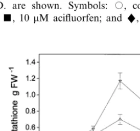
Acifluorfen (10 – 100 mM) markedly increased the GSH level in illuminated pea leaf discs. In darkness only 100 mM acifluorfen elevated the GSH level (Fig. 4). Changes of non-protein thiol levels were similar to those of total glutathione levels after acifluorfen treatments (data not shown). GSH accumulation was already observed in acifluorfen-treated plant tissues [23,27]. Our re-sults showed that the elevation of GSH level by acifluorfen in pea leaf discs was only partially light-dependent. It is known that during the degra-dation of acifluorfen in plants, the first cleavage product forms conjugates with GSH or homoglu-tathione [28]. It is possible that the plant tissue synthesize more GSH also in the darkness in order to meet greater GSH demand as a result of detox-ification reactions. Our results confirm those of Kno¨rzer et al. [13] who also found different re-sponses of ascorbate and GSH in soybean cells exposed to the peroxidizing herbicide oxyfluorfen. GSH levels were considerably elevated only by high ALA concentrations (1 – 3 mM). After 48 h of illumination the GSH content rose to 265% of the control in the presence of 3 mM ALA (Fig. 5). In darkness, ALA (0.1 – 3 mM) exerted no effect on GSH levels. Changes of non-protein thiol levels were similar to those of total glutathione levels after ALA treatments (data not shown). The ef-fects of ALA on the enzymes of GSH biosynthesis are not known yet.
Fig. 4. Effect of acifluorfen on the total glutathione level in pea leaf discs. All discs were preincubated for 24 h in dark-ness then illuminated (400 mmol m−2 s−1), except in the
parallel dark incubations. Means of three independent experi-ments9S.D. are shown. Symbols: , control; , 1 mM acifluorfen; , 10mM acifluorfen; and", 100 mM acifluor-fen.
Fig. 5. Effect of 5-aminolevulinic acid (ALA) on the total glutathione level in pea leaf discs. All discs were preincubated for 24 h in darkness then illuminated (400 mmol m−2 s−1),
In summary, both ascorbate and GSH levels were strongly altered in pea leaf discs by very different singlet oxygen generating reactions. Ascorbate levels markedly decreased in all treat-ments independently of the site (or mode of ac-tion) of singlet oxygen generation. The xanthene dyes are known to act predominantly in the cyto-plasm of plant cells [6]. Monuron exerts its oxida-tive effect in the chloroplasts [6]. Tetrapyrrole derivatives initially accumulate in the plastids (pre-dominantly in the chloroplasts) after acifluorfen and ALA treatments [4,9,29], but the accumulated tetrapyrroles may subsequently leak out of the plastids and photosensitize other cellular compart-ments [29]. In contrast to ascorbate, the foliar GSH levels were considerably increased by singlet oxygen generators (except by monuron). Singlet oxygen production in both the cytoplasm (by xan-thene dyes) and in the plastids (by acifluorfen and ALA) led to marked GSH accumulations. Further studies are necessary to elucidate the effects of singlet oxygen generators on the biosynthetic pathways of both ascorbate and GSH.
Acknowledgements
The financial support of the Royal Society Hun-garian Postdoctoral Fellowship Programme to G. Gullner is gratefully acknowledged.
References
[1] G. Noctor, C.H. Foyer, Ascorbate and glutathione: keeping active oxygen under control, Annu. Rev. Plant Physiol. Plant Mol. Biol. 49 (1998) 249 – 279.
[2] J.P. Knox, A.D. Dodge, Singlet oxygen and plants, Phytochemistry 24 (1985) 889 – 896.
[3] C.H. Foyer, M. Lelandais, K.J. Kunert, Photooxidative stress in plants, Physiol. Plant. 92 (1994) 696 – 717. [4] N. Chakraborty, B.C. Tripathy, Involvement of singlet
oxygen in 5-aminolevulinic acid-induced photodynamic damage of cucumber (Cucumis sati6us L.) chloroplasts, Plant Physiol. 98 (1992) 7 – 11.
[5] K. Briviba, L.O. Klotz, H. Sies, Toxic and signaling effects of photochemically or chemically generated sin-glet oxygen in biological systems, Biol. Chem. 378 (1997) 1259 – 1265.
[6] A.D. Dodge, Herbicide action and effects on detoxifica-tion processes, in: C.H. Foyer, P.M. Mullineaux (Eds.), Causes of Photooxidative Stress and Amelioration of Defense Systems in Plants, CRC Press, Boca Raton, 1994, pp. 219 – 236.
[7] A.J. Enyedi, Induction of salicylic acid biosynthesis and systemic acquired resistance using the active oxygen spe-cies generator rose bengal, J. Plant Physiol. 154 (1999) 106 – 112.
[8] S.O. Duke, C.A. Rebeiz, Porphyrin biosynthesis as a tool in pest management, in: S.O. Duke, C.A. Rebeiz (Eds.), Porphyric Pesticides: Chemistry, Toxicology, and Phar-maceutical Applications, Symposium Series 559, Ameri-can Chemical Society, Washington, DC, 1994, pp. 1 – 16. [9] M. Matringe, R. Scalla, Studies on the mode of action of acifluorfen-methyl in nonchlorophyllous soybean cells, Plant Physiol. 86 (1988) 619 – 622.
[10] T. Ko¨mives, G. Gullner, Mechanisms of plant tolerance to photodynamic herbicides, in: S.O. Duke, C.A. Rebeiz (Eds.), Porphyric Pesticides: Chemistry, Toxicology, and Pharmaceutical Applications, Symposium Series 559, American Chemical Society, Washington, DC, 1994, pp. 177 – 190.
[11] H. Matsumoto, Y. Tanida, K. Ishizuka, Porphyrin inter-mediate involved in herbicidal action ofd-aminolevulinic acid on duckweed (Lemna paucicostataHegelm.), Pestic. Biochem. Physiol. 48 (1994) 214 – 221.
[12] R.S. Bodannes, P.C. Chan, Ascorbic acid as a scavenger of singlet oxygen, FEBS Lett. 105 (1979) 195 – 196. [13] O.C. Kno¨rzer, J. Durner, P. Bo¨ger, Alterations in the
antioxidative system of suspension-cultured soybean cells (Glycine max) induced by oxidative stress, Physiol. Plant. 97 (1996) 388 – 396.
[14] T. Ko¨mives, G. Gullner, Z. Kira´ly, Role of glutathione and glutathione-related enzymes in response of plants to environmental stress, in: P. Csermely (Ed.), Stress of Life. From Molecules to Man, Annals of the New York Academy of Sciences, vol. 851, The New York Academy of Sciences, New York, 1998, pp. 251 – 258.
[15] L.J. De Kok, M. Graham, Levels of pigments, soluble proteins, amino acids and sulfhydryl compounds in foliar tissue of Arabidopsis thaliana during dark-induced and natural senescence, Plant Physiol. Biochem. 27 (1989) 203 – 209.
[16] I.K. Smith, Stimulation of glutathione synthesis in pho-torespiring plants by catalase inhibitors, Plant Physiol. 79 (1985) 1044 – 1047.
[17] R.R. Wise, A.W. Naylor, Chilling-enhanced photooxida-tion. Evidence for the role of singlet oxygen and superox-ide in the breakdown of pigments and endogenous antioxidants, Plant Physiol. 83 (1987) 278 – 282.
[18] C.H. Foyer, J. Rowell, D.A. Walker, Measurement of the ascorbate content of spinach leaf protoplasts and chloroplasts during illumination, Planta 157 (1983) 239 – 244.
[19] D.J. Gillham, A.D. Dodge, Chloroplast superoxide and hydrogen peroxide scavenging systems from pea leaves: seasonal variations, Plant Sci. 50 (1987) 105 – 109. [20] R. Schupp, H. Rennenberg, Diurnal changes in the
glu-tathione content of spruce needles (Picea abiesL.), Plant Sci. 57 (1988) 113 – 117.
[22] M.J. May, T. Vernoux, R. Sa´nchez-Ferna´ndez, M. Van Montagu, D. Inze´, Evidence for posttranscriptional acti-vation of g-glutamylcysteine synthetase during plant stress responses, Proc. Natl. Acad. Sci. USA 95 (1998) 12049 – 12054.
[23] G. Gullner, T. Ko¨mives, L. Kira´ly, Enhanced inducibil-ity of antioxidant systems in a Nicotiana tabacum L. biotype results in acifluorfen resistance, Z. Naturforsch. [C] 46 (1991) 875 – 881.
[24] O. Arrigoni, L. De Gara, C. Paciolla, A. Evidente, M.C. de Pinto, R. Liso, Lycorine: a powerful inhibitor of
L-galactono-g-lactone dehydrogenase activity, J. Plant Physiol. 150 (1997) 362 – 364.
[25] G.L. Wheeler, M.A. Jones, N. Smirnoff, The biosyn-thetic pathways of vitamin C in higher plants, Nature 393 (1998) 365 – 369.
26] W.H. Kenyon, S.O. Duke, Effects of acifluorfen on en-dogenous antioxidants and protective enzymes in cu-cumber (Cucumis sati6us L.), Plant Physiol. 79 (1985)
862 – 866.
[27] A. Schmidt, K.J. Kunert, Lipid peroxidation in higher plants, Plant Physiol. 82 (1986) 700 – 702.
[28] D.S. Frear, H.R. Swanson, E.R. Mansager, Acifluorfen metabolism in soybean: diphenylether bond cleavage and the formation of homoglutathione, cysteine and glucose conjugates, Pestic. Biochem. Physiol. 20 (1983) 299 – 310.
[29] H.-P. Mock, U. Keetman, E. Kruse, B. Rank, B. Grimm, Defense responses to tetrapyrrole-induced oxi-dative stress in transgenic plants with reduced uropor-phyrinogen decarboxylase or coproporphyrinogen oxidase activity, Plant Physiol. 116 (1998) 107 – 116.