Monitoring drug resistance
in influenza viruses
Report of the Regional Workshop
Nonthaburi, Thailand 23-28 August 2010
© World Health Organization 2010
All rights reserved.
Requests for publications, or for permission to reproduce or translate WHO publications – whether for sale or for noncommercial distribution – can be obtained from Publishing and Sales, World Health Organization, Regional Office for South-East Asia, Indraprastha Estate, Mahatma Gandhi Marg, New Delhi 110 002, India (fax: +91 11 23370197; e-mail: publications@searo. who.int).
The designations employed and the presentation of the material in this publication do not imply the expression of any opinion whatsoever on the part of the World Health Organization concerning the legal status of any country, territory, city or area or of its authorities, or concerning the delimitation of its frontiers or boundaries. Dotted lines on maps represent approximate border lines for which there may not yet be full agreement.
The mention of specific companies or of certain manufacturers’ products does not imply that they are endorsed or recommended by the World Health Organization in preference to others of a similar nature that are not mentioned. Errors and omissions excepted, the names of proprietary products are distinguished by initial capital letters.
All reasonable precautions have been taken by the World Health Organization to verify the information contained in this publication. However, the published material is being distributed without warranty of any kind, either expressed or implied. The responsibility for the interpretation and use of the material lies with the reader. In no event shall the World Health Organization be liable for damages arising from its use.
This publication does not necessarily represent the decisions or policies of the World Health Organization.
Contents
Background
1. ...1
Objectives
2. ...3
Inaugural session
3. ...4
Proceedings of the workshop
4. ...5
Conclusions and recommendations
5. ...24
Annexes
List of participants
1. ...27
Programme of work
1
Background
Influenza has been one of the most enigmatic of all diseases. It is caused by a variety of strains of influenza viruses. In any given year, some strains of influenza virus can cause seasonal disease while others may cause epidemics, or even a pandemic. During the past decade the world has witnessed global occurrence of seasonal influenza, the appearance of avian influenza due to the influenza A (H5N1) virus with a high mortality rate in many countries, and the eruption of a pandemic due to a novel strain of influenza called as pandemic (H1N1) 2009. While seasonal influenza occurs every year, epidemics may occur after several years and pandemics strike once in several decades. Influenza viruses are known for their high evolutionary rate and tendency to acquire point mutations at different positions in their genomes. Some mutations can result in amino acid substitutions at key locations in proteins, such as antigenic sites or the receptor binding site of the haemagglutination antigen (HA), and can alter properties such as those associated with the virus antigenicity or pathogenicity.
Development of resistance to antiviral drugs is often seen when these drugs are used in substantial quantities. The speed with which such resistance develops, the percentage of viruses developing resistance and the ability of the new virus to spread are influenced by several factors, including genetic stability of the virus, how the antiviral drug is used, and whether the new (mutated) drug-resistant virus can compete well enough to spread.
phase, there is a need to build capacity of the National Influenza Centres (NICs). This will also help in understanding the genetic changes in the influenza viruses (with or without manifestation of resistance), since the same technique is applied to characterize the virus in totality.
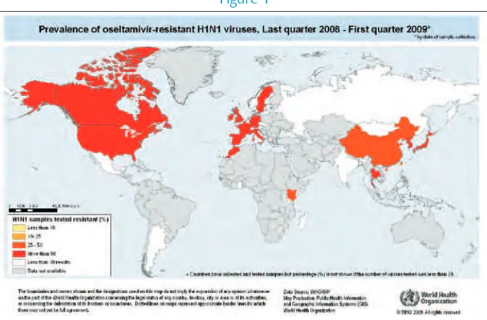
Figure 1 (REPLACE WITH LA
To fulfill this need of the Region, a workshop was organized at the Regional Influenza Reference Laboratory, National Institute of Health, Department of Medical Sciences, Ministry of Public Health, Thailand, Nonthaburi. The workshop was attended by 20 nominees from all 11 Member States of the South-East Asia (SEA) Region. It was facilitated by experts from the Regional Influenza Reference Laboratory, Global H5 Reference laboratory at National Institute of Virology, Pune, India, and the WHO Collaborating Centre for Influenza at National Institute of Infectious Diseases, Tokyo, Japan. The list of participants and the programme of work are attached as Annex 1 and 2, respectively.
[image:7.499.74.427.147.379.2]2
Objectives
The objectives of the workshop were:
To review the national response to influenza pandemics and capacity (1)
for genetic characterization;
to impart training in laboratory techniques in genotypic characterization (2)
of influenza viruses;
to agree on a mechanism of regional networking for monitoring of (3)
drug resistance in influenza viruses; and
3
Inaugural session
Dr Pathom welcomed the participants on behalf of the Director General of the Department of Medical Sciences. He highlighted the importance of building laboratory capacity to respond to outbreaks of influenza.
The message from Dr Samlee Plianbangchang, WHO Regional Director for South-East Asia, was read out by Dr Maureen Birmingham, WHO Representative to Thailand. Dr Samlee elaborated upon the diverse manifestations of influenza virus due to the unstable and segmented genetic apparatus of this virus and the need to keep these genetic changes under surveillance, as far as possible in real time, so that public health actions can be initiated or modified for an efficient response. To meet this objective, the WHO Global Influenza Surveillance Network was established in 1952. The network comprises 5 WHO Collaborating Centres, 4 Essential Regulatory Laboratories and 132 institutions in 103 WHO Member States, of which 10 are from 8 Member States in the South-East Asia (SEA) Region.
4
Proceedings of the workshop
Antiviral drugs in influenza and
resistance against them
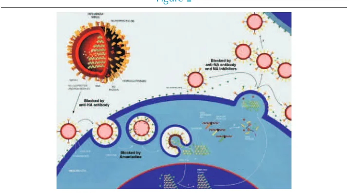
[image:10.499.71.429.425.623.2]The most widely available and licensed antiviral drugs for use in influenza are the two M2 inhibitors, amantadine and rimantadine, and the two NA inhibitors, oseltamivir and zanamivir. Amantadine was approved for prophylaxis of H2N2 influenza A infection in the United States of America in 1966, and for prophylaxis and treatment of all influenza A infections in 1976. Rimantadine was approved in the United States of America in 1994. Oseltamivir and zanamivir were introduced into clinical practice in 1999. Amantadine, rimantadine and oseltamivir are available for oral administrations, and zanamivir is administered as an inhaled powder.
Influenza viruses bind to sialic acid residues present on cell surface glycoproteins, through the receptor-binding site in the HA molecules (Figure 2). Then the viruses enter the cell by receptor-mediated endocytosis. The entry of the viral genome into the cytoplasm is dependent on the acidic pH of endosomes. The M2 protein functions as an acidic pH-activated ion channel. M2 inhibitors inhibit viral replication by blocking the ion channel activity of the M2 protein. Furthermore, NA protein enzymatically cleaves the sialic acid residues from host glycoproteins. NA inhibitors inhibit NA enzyme activity and prevent virus particles from being released from the surface of infected cells. The M2 inhibitors are active only against influenza A and not influenza B. The NA inhibitors act against both influenza A and influenza B.
Nearly all pandemic (H1N1) 2009 virus isolates and all seasonal H3N2 viruses tested have been resistant to amantadine and rimantadine. In January 2008, a high rate of resistance to oseltamivir in seasonal H1N1 viruses was officially notified to WHO by the Norwegian authorities. Ever since, oseltamivir-resistant seasonal H1N1 viruses have been circulating globally. The majority of pandemic (H1N1) 2009 viruses are susceptible to oseltamivir; however, rare sporadic cases of oseltamivir-resistant pandemic viruses have been detected worldwide. An antiviral should not be used for treatment where the virus is known or highly likely to be resistant to that antiviral.
Resistance to M2 inhibitors is very well characterized and is known to be caused by mutation in one or more of five residues in the M2 protein. Genotyping of viruses is a widely accepted monitoring method for M2 inhibitor resistance. This genotyping can be done in two ways: Sanger sequencing or pyrosequencing.
To monitor the emergence of resistant viruses, influenza virus NA inhibitor resistance is defined as a significantly raised IC50 value; that is, drug concentration inhibiting NA activity by 50%, coupled with a characterized mutation in the NA gene.
While there are a number of different laboratory methods currently available to measure NA inhibitor susceptibility, the enzyme inhibition assay is the simplest and clearest method to determine the IC50 value. There are two commercially available substrates suitable for this type of assay. The most widely used is a fluorescent substrate 2’-(4-methylumbelliferyl)-
∝
-D-N-acetylneuraminic acid ( MUNANA). The amount of released fluorescence directly relates to the amount of enzyme activity. The second assay uses a chemiluminescent substrate, abbreviated as NA-Star, which is sold as part of a dedicated kit. IC50 values generated from the different assay methods should not be directly compared because values generated by chemiluminescence are typically lower than those for the same virus and drug in the fluorescence test.Country reports
Bangladesh
Pandemic (H1N1) 2009 virus was first detected in Bangladesh on 18 June 2009. Since then a total of 1310 cases were identified upto 16 August 2010. The epidemic spread at low or moderate intensity and accounted for seven deaths among the laboratory-confirmed cases. The cases occurred throughout the year although the majority number of cases occurred in March to August 2010, the peak being observed in July.
The ages mainly affected were 15 to 40 years. The common presenting symptoms were fever, shortness of breath, rhinorrhoea, cough, sore throat, nasal congestion, fatigue, myalgia/arthralgia, vomiting and headache.
The Government of Bangladesh, in collaboration with WHO, has taken prompt action to control the pandemic. In 2009, a BSL 3 laboratory was established at the Institute of Epidemiology Disease Control and Research (IEDCR). Later, this was declared the National Influenza Centre (NIC). The NIC is performing diagnosis of H1N1 using real-time polymerase chain reaction (RT PCR) and reports results within 24 hours of receipt of specimens in the laboratory. However, virus isolation and drug resistance monitoring has not yet been started because of lack of a virus isolation facility.
Dhaka. NIC has started national influenza surveillance from April 2010 in 14 district hospitals in the first phase and it will be extended to all district hospitals in the next phase. The surveillance network is operating in collaboration with NIC and the International Centre for Diarrhoeal Disease and Research, Bangladesh (ICDDR-B).
Bangladesh is adequately prepared to deal with the H1N1 influenza pandemic—, adequate hospital facilities, patient management capabilities and community preparedness. However, apart from the NIC at IEDCR, there are no other laboratories to diagnose Pandemic (H1N1) 2009. It is recommended to establish a few other laboratories to deal with the laboratory confirmation in the event of a pandemic of greater intensity.
Bhutan
Bhutan has initiated the surveillance of influenza-like illness (ILI) in May 2009 and among them influenza A and B have received major emphasis for the surveillance. Since the declaration of an influenza pandemic in 2009, the ILI surveillance has almost assumed the role of influenza surveillance. This is coupled with the fact that in Bhutan, there is limited capacity to detect or diagnose other agents causing ILIs. About 10 sentinel sites were selected for the ILI surveillance, in addition to collection of samples from sites reporting outbreaks. The main objective of ILI surveillance is to monitor the trend of agents causing ILI in the country and to determine the shift in the prevalent species or agents causing ILI. Since May 2009, Bhutan had 98 laboratory-confirmed cases of influenza due to pandemic (H1N1) 2009 virus. The number of cases was expected to rise as sporadic outbreaks of ILI are reported around the country with the onset of the influenza season in August-October.
The ILI surveillance showed that influenza A H3 and influenza B is generally circulating in the population, causing seasonal flu. Moreover, influenza A seasonal H1N1 has also been found to be circulating in the population.
Bhutan needs to develop its capacity for establishing the monitoring of drug resistance in influenza viruses in order to contribute towards global influenza surveillance, as well as for the patient treatment purposes. To achieve this, training of microbiologists/lab technologists, and procurement of necessary reagents and equipment needs to be supported.
Guidelines on monitoring and management of drug resistance need to be developed in collaboration with WHO-recognized institutes. Linkages with regional reference laboratories need to be established to refer samples for drug resistance testing and sharing of other resources, including proficiency testing. Advocacy should be undertaken with policy-makers regarding the need for monitoring antimicrobial resistance in emerging infectious diseases, to gain their support in combating antimicrobial resistance.
DPR Korea
Surveillance for influenza in DPR Korea is coordinated by the National Centre for Disease Control under the Ministry of Public Health. At the national level, the National Institute of Epidemiology and Hygiene provides institutional support. Similar institutes are operational at local level, and in peripheral areas this support is provided through hospitals.
Laboratory techniques for diagnosis of influenza are available in the National Institute. The rest of the centres collect clinical material and send it to the National Institute for analysis. In the meantime, the diagnosis is established on the basis of clinical features.
At national level in DPR Korea, facilities are available for virus isolation, detection of viral nucleic acids through conventional and real-time PCRs; DNA sequencing based on Sanger’s method; and serological diagnosis of influenza.
DPR Korea plans to introduce pyrosequencing and advanced serological tests for influenza, for which technical support is expected from WHO.
India
extended the surveillance with four regional laboratories in different parts of the country. After the first outbreak of avian influenza, the network was strengthened by five more centres. Currently two government agencies, ICMR and the National Centre for Disease Control (NCDC), are independently conducting influenza surveillance in India. The data generation is on a weekly basis and shared with the global influenza surveillance programme through WHO’s FluNet. NIV Pune acts as referral centre for the network and is responsible for reconfirmation and molecular characterization of influenza viruses. Till December 2008, 15000 ILI specimens were collected and 749 isolates had been obtained out of which 237 were H1N1, 201 were H2N2, and 311 were type B. 535 of these isolates were shared with the WHO Collaborating Centre (CC) located at CDC.
Genetic analysis data of Indian isolates for 2004-2009 showed that each year the circulating strains were clustered, with the respective northern hemisphere vaccine component with 98 to 99 percent amino acid identity. Drug susceptibility for the seasonal influenza was assessed by genotypic assay, and since 2004 30% resistance to amantidine was observed for H3N2 viruses, and it became 100% resistant after 2008. The majority of H1N1 isolates were amatadine-sensitive, and in 2009 amantadine-resistant H1N1 viruses were found to be in circulation. Neuraminidase drug resistance susceptibility was detected for seasonal influenza using molecular techniques to detect known markers for NAI, and till date type B and H3N2 isolates were found to be sensitive to oseltamivir. For seasonal H1N1 the resistance to NAI was detected from the end of December 2008.
Beginning in May 2009, India experienced a pandemic caused by pandemic (H1N1) 2009 virus. The network with its pandemic preparedness took a frontline role in providing diagnosis, as well as carrying out genetic analysis and pandemic mitigation activities, undertaken together with the local and central health authorities. With information on the novel influenza in Mexico and the United States, the influenza department stepped up surveillance activities in Pune. Diagnosis was provided for patients from various parts of the country. Clinical samples were received from the Andaman and Nicobar Islands, Tamil Nadu, Delhi, Karnataka, West Bengal, Maharashtra, Goa and Kerala. India got its first sample from an incoming traveler on 2 May 2009. The first positive was detected from Hyderabad on 14 May to date, NIV and the National Centre for Disease Control, New Delhi tested and other state laboratories as well as few private laboratories tested
154 259 samples and got 23.5% (36 240) positive cases, with 1833 deaths.
samples by real-time PCR. 281 isolation had been done and full genomes were done for 16 isolates. All the 281 isolates and more than 600 clinical samples were tested for NAI drug susceptibly and found to be sensitive. Sero-surveillance study showed that approximately 30% herd immunity is developed in different social groups. Vaccination for the pandemic influenza is effectively administrated in India.
Indonesia
Since 1975, The Center for Biomedical and Pharmaceutical Research and Development (CBPRD) under the National Institute of Health Research and Development (NIHRD), Ministry of Health, Republic of Indonesia acted as a national influenza centre (NIC) for Indonesia. At the beginning of this activity, influenza-like illness (ILI) surveillance had not been intensively conducted and influenza was not considered a priority disease. In 1999, influenza virology surveillance in Indonesia was re-initiated in three major cities on Java Island (Jakarta, Bandung, Jogjakarta). Since the detection of highly pathogenic avian influenza in poultry (2003) and the identification of H5N1 infection in humans (2005) in Indonesia, the Indonesia Ministry of Health has been concerned about influenza in Indonesia. Therefore, to monitor circulating strains, the Indonesia Ministry of Health monitors patients with ILI in a network of primary health centres (PHCs) in diverse regions of the country.
For surveillance of influenza, the country uses the WHO case definition of ILI. Medical staff of PHCs have been trained to identify and collect nasal and throat swabs from patients with ILI symptoms. Specimens are then sent to the CBPRD-NIHRD, which are equipped with PCR conventional and real-time PCR. The CBPRD-NIHRD has facilities available for virus isolation (BSL2 and BSL3) and virus sequencing. All ILI specimens are tested for all influenza strains with real-time PCR using primers and probes which are provided by US-CDC. For specimens giving negative result for avian influenza, these are cultured for virus isolation in MDCK cells for further characterization.
Sulawesi or Celebes island; and the CBPRD-NIHRD is as a referral laboratory for ILI surveillance.
Surveillance data from 2007 to 2009 show that the proportion of influenza A cases increased (5%, 17%, 20% respectively), while influenza B cases increased from 2007 (4%) to 2008 (11%), but decreased in 2009 (3%). The distribution subtype variation from 2007 to 2009 shows seasonal H1N1 increase (22, 68, and 96 cases, respectively). However, significantly decrease was seen in 2010 (4 cases up to July 2010). In contrast, influenza A/H3N1 increased from 2009 (41 cases) to July 2010 (213 cases). The flu season based on influenza-like iIlness and influenza case data, cannot be predicted in Indonesia since the dry and rainy seasons are unpredictable due to the global warming. The data show that the proportion of positive samples fluctuated every year. The highest numbers of influenza cases are in the age group 5 to 15 years old. However, there is no significant difference in the number of cases between male and female. In June 2009, the WHO announced the beginning of the H1N1 pandemic. Indonesia is one of the countries hit by the influenza pandemic. The annual report to WHO show that Indonesia reported 188 cases due to pandemic (H1N1) 2009 virus during 2009 by the ILI surveillance programme and 20 cases from January until July 2010.
Maldives
PCR diagnostic facilities using real-time PCR were established in Maldives in 2009. BSL2 facilities have also been established. Influenza surveillance has been recently initiated. Testing is done at Indira Gandhi Memorial Hospital. Specimens are collected at various peripheral sites and shipped to Male. The surveillance data are analysed at the Centre for Community Health and Disease Control (CCHDC). Every fortnight data are shared with WHO.
In the recent pandemic the first case was detected in July 2009. After this, 65 568 suspected cases were detected. The age group predominantly affected was 15-29 years. Of these, almost 20 ,000 were from Male alone. Only one death was reported. The national laboratory could confirm diagnosis in 41 cases by PCR.
Maldives proposes to strengthen the capacity for influenza testing by PCR, to build a network for influenza virus monitoring, and to strengthen the regional laboratories.
Myanmar
The National Influenza Centre was established in Myanmar in February 2008. Facilities available include conventional PCR, real-time PCR, virus isolation, and resistance studies (in collaboration with Niigata University, Niigata, Japan). At present virus sequencing and electron microscopic studies are not undertaken. The National Health Laboratory has BSL2 facilities, and, for avian influenza and pandemic influenza BSL 2 facilities plus BSL3 practices were followed.
A national surveillance mechanism exists only for avian influenza and pandemic influenza, but not for seasonal influenza. A national network of laboratories on seasonal influenza is operational with six laboratories as members.
The number of confirmed cases of pandemic influenza was 177 without any deaths. The first case of pandemic influenza was detected in June 2009. The major age group affected was 11-25 years.
While influenza viruses are being shared with the WHO Collaborating Centre, Tokyo, Japan, no studies are being done to determine the drug resistance in influenza viruses.
Nepal
The National Public Health Laboratory (NPHL) was established in 1968 and it has been identified as the referral laboratory for influenza with direct linkages to 291 government and more than 1024 private laboratories within the country. NPHL has been providing routine/special diagnostic facilities, including laboratory-based surveillance such as influenza surveillance which started in 2007.
A novel influenza A /H1N1 virus was detected in June, 2009 in a patient who had returned from the United States, and evidence of local transmission was established within the country in October 2009. A total of 609 samples were submitted to NPHL virology laboratory for pandemic (H1N1) 2009 virus testing of which 172(28.3%) were positive for pandemic influenza. Pandemic (H1N1) 2009 virus was detected most frequently among patients in the 11–20 year age group (30%), followed by 21–30 years (27%) and 0-10 years (24%). The lowest number of cases was detected in the 51–60 year age group (2%) and over 60 years (2%).
All confirmed positive and negative cases were reported to WHO/IHR through the Avian Influenza Control Project (AICP) and Epidemiology and Disease Control Division (EDCD), Department of Health Services. After the completion of refurbishment and operationalization of surveillance network, NPHL will share the information and virus isolates with WHO and disseminate the results to WHO’s Global Influenza Surveillance Network (GISN) regularly.
Major challenges in pandemic and response include proper transportation of specimens, shortage of trained technical staff, maintenance of machines, problems in procurement and supplies, as well as availability of limited budget.
Sri Lanka
The National Influenza Center (NIC) of Sri Lanka was established in 1968. It was in an inactive state until 1998 when laboratory investigations of out-breaks were started. It was strengthened recently under the National Pandemic Preparedness Plan. There are facilities for virus isolations in eggs and tissue culture and molecular techniques of conventional and real-time PCR, but sequencing and antiviral susceptibility facilities are not yet available. The laboratory activities are done under the BSL-2 level.
The NIC received 6543 specimens from suspected pandemic influenza cases, of which 647 were confirmed by real-time PCR. Of these two died. Most of these patients presented with fever, cough, body aches and difficulty in breathing. Some had vomiting and diarrhoea. The predominant age group affected was 11-20 years. Males and females were equally affected.
The first case of influenza due to pandemic ( H1N1) 2009 virus was diagnosed on 16 June 2009 among arrivals from abroad. Community transmission started in October. The peak activity was seen in December. The cases started disappearing from February 2010. The most number of cases were presented from the western and central parts of the country.
Virus isolations were not attempted during the pandemic period. In 2009, there were 41 isolates of Influenza A H3N2 and 11 isolates of seasonal H1N1 and 10 Influenza B isolates. There were no avian influenza (H5N1 ) viral isolates or PCR-positive human cases in Sri Lanka.
Samples were shared with WHO reference laboratories at the University of Hong Kong and Melbourne, Australia. Drug resistance studies were done at the WHO CC in Australia by the fluorescence-based technique and found 5 of the isolates of H3N2, 3 of the pandemic (H1N1) 2009 virus and 1 of the B isolate were sensitive to oseltamivir. Seasonal H1N1 ( 2 isolates) were highly resistant to oseltamivir.
Thailand
The National Influenza Centre was designated by WHO in 1972 at the National Institute of Health, Thailand. On 22 July 2010, Thai NIC was designated as the Regional Influenza Reference Laboratory (RIRL) for the SEA Region. The laboratory has undertaken various tests on influenza viruses for their detection, isolation and characterization. The unusual isolates are immediately shipped to the WHO CC and information is shared at weekly intervals with FluNet.
To respond effectively to the pandemic, it was decided to implement standard methods for all members of the laboratory network; set up guidelines for laboratory preparedness during the pandemic; and provide a refresher training course on diagnosis, handling and transportation of specimens under WHO guidelines. For rapid diagnosis, mobile laboratories were dispatched to the site of the outbreak.
The challenges in the first wave of influenza pandemic in Thailand were the large number of specimens and insufficient of laboratory service and hence efforts were soon initiated to upgrade laboratory facilities, increase the number of automated extractors and real-time PCR machines for Regional Medical Science Centers. At the same time, it was decided to initiate a national network for strengthening public health laboratories, provide an additional training course on PCR for 25 regional hospital labs, prepare and provide minimum requirement to set up a PCR laboratory, assure quality through proficiency testing, strengthen sentinel pandemic influenza surveillanceand integrate sentinel sites of Department of Medical Sciences (DMSc) with those of the Bureau of Epidemiology (BOE) surveillance to have a total of 24 sentinel sites in the country.
A rapid reporting system to monitor the trend of influenza activity and novel influenza strains was established. Information was disseminated through weekly reports which monitored the trend of the outbreak by calculation of the baseline and epidemic threshold.
Five of 470 isolates of pandemic influenza virus were found to be resistant to oseltamivir. The enhanced system was able to effectively inform about the second wave of the pandemic which began in November 2009 and peaked in mid-February 2010, and the discovery of five oseltamivir- resistant pandemic (H1N1) 2009 strains with H275Y mutation. During April-May 2010, overall pandemic and seasonal activity declined to a very low level, but still there was sporadic pandemic (H1N1) 2009 in some areas. In July 2010, there was an upward trend in the percent ILI visit and an increase in the proportion of patients with ILI testing positive for pandemic flu (12.71%), seasonal influenza B (10.65%) and H3N2 (2.4%). This may be a sign of the seasonal Influenza peak in Thailand when the rainy season is coming Enhancement of the influenza surveillance system assists the surveillance teams in identifying clusters of influenza activity and outbreak trends of providing timely surveillance data for health policy-makers to determine effective control measures.
Timor-Leste
The National Laboratory was established in Timor Leste in 2000. The existing laboratories in Timor-Leste are one National Laboratory, one National Hospital Laboratory, four Referral Hospital laboratories and 72 Comumunity Health Center Laboratories (small laboratories with facilities available for only basic tests.
Timor-Leste initiated the surveillance of influenza virus in June 2009. Facilities for PCR are not yet available. Virus isolation, resistance studies for drugs and a virus sequencing have not been started yet there.
Since the declaration of an influenza pandemic in June 2009 the number of cases reported of H1N1 are 31 suspected cases, 7 of them confirmed positive, but no deaths.
Specimens were sent for testing and confirmation to WHO CC in Melbourne, Australia with assistance from WHO. Major clinical features for seasonal flu were fever, sore throat, nasal discharge, body pain and headache. Seasonal variations occur between the rainy season in October-February. Timor-Leste did not have many samples, staff were adequate, and reagents were available for screening using rapid test.
in influenza virus, Timor-Leste needs to establish and strengthen a capacity to test influenza viruses for antimicrobial resistance.
Timor-Leste also needs support to establish PCR testing including training to staff, expert assistance to do the setup, and help in getting other equipment and reagents required to do the set-up. The country also needs to develop capacity for establishing the monitoring of drug resistance in influenza viruses .
Drug resistance assays to monitor resistance in
influenza viruses
There are two major categories of assays to determine resistance in influenza viruses. These are:
Genotypic-based assays
•
Nucleotide sequencing
–
Real-time PCR (RT-PCR)
–
Pyrosequencing
–
Phenotypic-based assay
•
Plaque reduction assay
–
Neuraminidase inhibition assay
–
Viral protein reduction assay
–
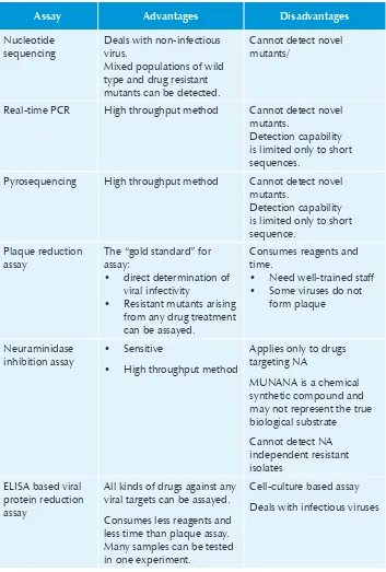
Table 1: Drug resistance assays for influenza viruses
Assay Advantages Disadvantages
Nucleotide sequencing
Deals with non-infectious virus.
Mixed populations of wild type and drug resistant mutants can be detected.
Cannot detect novel mutants/
Real-time PCR High throughput method Cannot detect novel mutants.
Detection capability is limited only to short sequences.
Pyrosequencing High throughput method Cannot detect novel mutants.
Detection capability is limited only to short sequence.
Plaque reduction assay
The “gold standard” for assay:
direct determination of •
viral infectivity
Resistant mutants arising •
from any drug treatment can be assayed.
Consumes reagents and time.
Need well-trained staff •
Some viruses do not • form plaque Neuraminidase inhibition assay Sensitive •
High throughput method •
Applies only to drugs targeting NA
MUNANA is a chemical synthetic compound and may not represent the true biological substrate
Cannot detect NA independent resistant isolates
ELISA based viral protein reduction assay
All kinds of drugs against any viral targets can be assayed.
Consumes less reagents and less time than plaque assay. Many samples can be tested in one experiment.
Cell-culture based assay
Techniques for monitoring resistance in influenza
viruses
DNA sequencing
DNA sequencing has been a molecular biology method that has developed from a technically difficult procedure to a method that is indispensable for all molecular biology labs, as well as many other kinds of labs and classes. Automated DNA sequencing has become an increasingly popular choice for many labs.
The method of sequencing DNA (Sanger 1977) uses a polymerization reaction (using DNA polymerase) in conjunction with a mix of deoxynucleotide triphosphates (dNTPs) and dideoxynucleotide triphosphates (ddNTPs). Since dideoxynucleotides terminate the growth of the DNA polymer once they are incorporated (because the hydroxyl at the 3’ position is absent), a series of fragments is produced depending on the dideoxynucleotide used and the DNA sequence of the template. This method has also been adapted for use with PCR. In this case, a small amount of DNA is used in conjunction with dNTPs, and a fluorescent labeled ddNTP. Each of the ddNTPs is labeled with a different fluorescent dye (one color for each ddNTP). Only one reaction mix is needed. The single lane is run on a polyacrylamide gel which is connected to a laser light source and a light sensor and computer. The sensor and computer determine which color of light is being emitted, and record the corresponding nucleotide at that position. Recorded data were analysed and interpreted into DNA sequences.
The sequencing method enables identification of point mutations in other residue that may confer resistance to either oseltamivir or zanamivir. Furthermore, it may be possible to classify novel point mutation in the residue that may confer neuraminidase resistance by using sequencing combined with neuraminidase inhibition assay.
Detection of the H275Y Mutation in Pandemic (H1N1) influenza
virus by allelic discrimination real-time PCR
mutation (N1 numbering) in the NA gene of pandemic (H1N1) 2009 influenza viruses.
Allelic discrimination assay or allelic discrimination RT-PCR assay is a multiplex one-step RT-PCR which uses a pair of primers with two fluorogenic probes (TaqMan MGB probe). This assay can be successfully used for discriminate alleles that differ by a single base substitution or single nucleotide polymorphism (SNP).
The primers and probes used in this assay are specific for pandemic (H1N1) influenza viruses (A/California/04/2009) and were designed in the Regional Influenza Reference Laboratory, Thailand, National Institute of Health, Department of Medical Sciences, Ministry of Public Health, Thailand. The assay uses a pair of primers and two TaqMan MGB probes, one specific for the H275 wildtype (labeled with FAM), and the other specific for the Y275 mutant (labelled with VIC). An end-point analysis (known as “allelic discrimination”) is then performed to determine the genotype of the virus with respect to amino acid 275 of the viral NA gene. A substantial increase in FAM-labeled probe fluorescence indicates the presence of the wild-type H275, whereas a substantial increase in VIC-labeled probe fluorescence indicates the presence of the Y275 mutation.
It is important to remember that this assay detects the presence or absence of the H275 →Y275 mutation in Pandemic (H1N1) 2009 influenza virus, and does not detect any other mutations in other residues that may confer resistance to either oseltamivir or zanamivir.
Fluorometric neuraminidase inhibition assay
incubation with a range of NI drug concentrations, it is possible to determine an IC50 value which indicates the drug concentration required to reduce NA activity by 50% as the IC50 value.
Pyrosequencing for the detection of the H275Y mutation in
pandemic (H1N1) 2009 virus
Pyrosequencing is a method to determine the order of nucleotides in DNA based on the “sequencing by synthesis" principle. It differs from Sanger sequencing, relying on the detection of pyrophosphate release on nucleotide incorporation rather than chain termination with dideoxynucleotide. The technique was developed by Mostafa Ronaghi at the Royal Institute of Technology in Stockholm in 1996.
The pyrosequencing method is based on detecting the activity of DNA polymerase with another chemiluminascent enzyme. Essentially, the method allows sequencing of a single strand of DNA by synthesizing the complementary strand along it, one base pair at a time, and detecting which base was actually added at each step. The template DNA is immobile, and solutions of A, C, G, and T nucleotides are added and removed after the reaction, sequentially. Light is produced only when the nucleotide solution complements the first unpaired base of the template. The sequence of solutions that produce chemiluminescent signals allows the determination of the sequence of the template.
Currently, a limitation of the method is that the lengths of individual reads of DNA sequence are in the neighborhood of 300-500 nucleotides, shorter than the 800-1000 obtainable with Sanger methods.
It is important to remember that this assay detects the presence or absence of the H275 →Y275 mutation in pandemic (H1N1) 2009 influenza virus and does not detect any other mutations in other residues that may confer resistance to either oseltamivir or zanamivir.
Hands on Practice
The participants were shown all the techniques through demonstrations. Subsequently they themselves performed the techniques under the supervision of facilitators. A plenary session was organized to discuss the issues raised by the participants and difficulties encountered by them.
The participants expressed gratitude to WHO for organizing this workshop. They felt this activity will act as a catalyst in initiating or expanding facilities for monitoring drug resistance in influenza viruses in their respective countries. They made following recommendations:
Participants should:
Advocate with senior management for the establishment/expansion of
•
facilities for monitoring of resistance in influenza viruses.
Assess the infrastructure that is available within the laboratory that can
•
be utilized for initiating monitoring resistance in influenza viruses.
Share information with other laboratories in the country on this subject
•
and explore collaboration/networking with them that can benefit the country.
Share data on resistance in influenza viruses with designated national
•
influenza/EID/CD focal points.
Member States should:
Provide appropriate resources for the establishment/expansion of facilities
•
for monitoring of resistance in influenza viruses.
Share information on detection and prevalence of drug-resistant influenza
•
viruses with WHO and with other countries.
5
WHO should:
Provide technical support to Member States in establishment/expansion
•
of facilities for monitoring of drug resistance in influenza viruses.
Share global data on the emergence and spread of resistance in influenza
•
viruses.
Forge a network of laboratories undertaking resistance studies in influenza
•
viruses for promoting scientific knowledge and regional capacity in this area.
Organize regular regional workshops on resistance in influenza viruses.
1
List of participants
Annex
Bangladesh
Prof. Dr Shahina Tabassum 1.
Professor and Chairman Department of Virology BSMMU
Dhaka
Email: shahina.tabassum@yahoo. com
Dr Monira Pervin 2.
Assistant Professor of Virology (Incharge)
Dhaka Medical College Dhaka Email: [email protected]
Bhutan
Mr. Dorji 3. Microbiologist Head of DepartmentDepartment of Laboratory, 4.
Jigme Dorji Wangchuk National Referral Hospital
Thimphu, Bhutan Email: [email protected]
DPR Korea
Dr Pak Yong Su 5.
Researcher,
Generic Medicine Research Institute Pyongyang Medical University
Dr Kim Yong Min 6.
Researcher,
Generic Medicine Research Institute Pyongyang Medical University Email: [email protected]
Dr Yun Song Jin 7.
Researcher,
Generic Medicine Research Institute Pyongyang Medical University Email: [email protected]
Indonesia
Dr Arie Bratasena 8.
Head
Sub Directorate of Upper Respiratory Tract Infection Ministry of Health
Indonesia
Email: [email protected]
Dr Pretty Multihartina D. Sasono 9.
Head
Dr Hana Apsari Pawestri 10.
National Laboratory Staff Designated for Drug Resistance Determination
Ministry of Health Indonesia
Email: [email protected]
India
Dr Varsha Potdar 11.
Research Scientist
National Institute of Virology 20-A, Ambedkar Road Pune
Maldives
Ms. Fathimath Ibrahim Manik 12.
Senior Lab Technologist
Indira Gandhi Memorial Hospital Male
Republic of Maldives
Email: [email protected], [email protected]
Myanmar
Dr Ommar Swe Tin (Mrs) 13.
Medical Officer
National Health Laboratory Yangon, Myanmar
Email: [email protected]
Dr Win Win yee (Mrs) 14.
Medical Officer
National Health Laboratory Yangon, Myanmar
Email: [email protected]
Nepal
Mr. Bishnu Prasad Upadhyay 15.
Senior Medical Technologist National Public Health Laboratory DHS
Nepal
Email: [email protected] Mr Ram Krishna Bhandari 16.
Medical Technologist
National Public Health Laboratory DHS
Email: [email protected], [email protected]
Sri Lanka
Dr (Mrs) G.A. Wickramasinghe 17.
Consultant Virologist Medical Research Institute Colombo 8
Email: geethaniwvirology@yahoo. com
Mrs. G.D.W.S. Gunathilake 18.
Medical Laboratory Technologist Medical Research Institute, Colombo 8
Email: [email protected]
Thailand
Dr Darika Kingnate 19.
Director
Bureau of Emerging Infectious Diseases
Department of Disease Control Ministry of Public Health Tivanond Road
Timor-Leste
Dr Maria Santina de Jesus Gomes 20.
General Director of Laboratory National
Ministry of Health Dili
Email: drasantinagomez@yahoo. co.id
I. Observer
Ms. Hatairat Lerdsamran 1.
Department of Microbiology Faculty of Medicine Siriraj Hospital Mahidol University
Bangkok
Tel : 662-4197059 Fax : 662-4182663
II. Temporary Advisor
s
Dr Pathom Sawanpanyalert 1.
Director of National Institute of Health, Department of Medical Sciences,
Ministry of Public Health, Nonthaburi
Thailand
Tel. 662-5890022 ext. 99354-5, Fax. 662-5911912
E-mail: [email protected] (local organizer)
Ms. Malinee Chittaganpitch 2.
Chief of RIRL for SEA Region National Institute of Health, Department of Medical Sciences Ministry of Public Health, Nonthaburi
Thailand
Tel. 662-5890022 ext. 98408, Fax. 662-5915449
E-mail: [email protected]
Ms.
3. Sirima Pattamadilok
Head, WHO Measles RRL in SEAR Deputy Director
National Institute of Health, Department of Medical Sciences Ministry of Public
Health,Nonthaburi Thailand
Tel. 662-5890022 ext. 99312, Fax. 662-5915449
E-mail: [email protected], [email protected]
Prof. Dr Pilaipan Puthavathana 4.
Department of Microbiology Faculty of Medicine Siriraj Hospital Mahidol University
Bangkok
Tel : 662-4197059 Fax : 662-4182663
Email : [email protected]
Dr AC Mishra 5.
Director
H5 Global Reference Laboratory National Institute of Virology, Pune India
Email : [email protected] Dr Emi Takashiya
6.
Principal Research Staff Influenza Virus Surveillance Laboratory (Lab No.1) WHO Collaborating Centre for Reference and Research on Influenza
Influenza Virus Research Center National Institute of Infectious Diseases, Japan
III. Guest lecturer / facilitator
Prof. Dr Ruengpung Sutthent 1.
Department of Microbiology Faculty of Medicine Siriraj Hospital Mahidol University
Bangkok ,Thailand Tel. : 662-4198409 Fax : 662-4113921
E-mail : [email protected] Dr Navin horthongkham 2.
Department of Microbiology Faculty of Medicine Siriraj Hospital Mahidol University
Bangkok , Thailand Tel. : 662-4198409 Fax : 662-4113921
Email : [email protected]
Ms. Niracha Athipanyasilp 3.
Department of Microbiology Faculty of Medicine Siriraj Hospital Mahidol University
Bangkok , Thailand Tel. : 662-4198409 Fax : 662-4113921
Dr Jiranan Warachit De silva 4.
National Institute of Health, Department of Medical Sciences Ministry of Public
Health,Nonthaburi Thailand
Tel. 662-5890022 ext. 98408, Fax. 662-5915449
E-mail: [email protected], [email protected]
Dr Siriphan Saeng-Aroon 5.
National Institute of Health, Department of Medical Sciences Ministry of Public
Health,Nonthaburi Thailand
Tel. 662-5890022 ext.98384 , Fax. 662-9659729
E-mail: [email protected]
Ms. Sunthareeya Waicharoen 6.
National Institute of Health, Department of Medical Sciences Ministry of Public
Health,Nonthaburi Thailand
Tel. 662-5890022 ext. 98408, Fax. 662-5915449
E-mail: [email protected]. go.th
Mrs. Atchariya Lukebua 7.
National Institute of Health, Department of Medical Sciences Ministry of Public
Health,Nonthaburi Thailand
Tel. 662-5890022 ext. 99312, Fax. 662-5915449
E-mail: [email protected]. go.th
Dr Rajesh Bhatia Regional Adviser
Blood Safety & Laboratory Technology
2
Programme of work
Annex
Monday, 23 August 2010 Seminar Room, Bld 10, NIH
9.00-9.30 Opening Welcome and opening remarks
Dr Maureen Birmingham Dr Pathom Sawanpanyalert
10.00-10.30 Lecture Country reports Every participants 10.45-12.00 Lecture Country reports continue Every participants
13.00-13.45 Lecture Global influenza drug resistant monitoring
Dr Emi Tsuchiya
13.45-14.30 Lecture The Pandemic H1N1 Experience from National Institute of Health, Thailand
Dr Pathom Sawanpanyalert
14.45-15.30 Lecture Situation update: Seasonal/ pandemic influenza and drug resistance monitoring in India
Dr AC Mishra
15.30-16.15 Lecture Situation update: Seasonal/ pandemic influenza and drug resistance monitoring in Indonesia
Representative from, Indonesia
Tuesday, 24 August 2010 Seminar Room & Training Room, Bld 1, NIH
9.00-9.45 Lecture Overview of antiviral drug resistance assay
Prof. Dr Pilaipan Puthavathana
9.45-10.00 Lecture Overview of today’s lab. activities
10.00-10.30 Lecture Real-time PCR detection of resistance mutation
Dr Jiranan Warachit de Silva/ facilitators
10.45-11.15 Lab intro-duction
Real-time PCR detection of resistance mutation
Dr Jiranan Warachit de Silva
10.45-12.00 Lab practical
Testing of unknown samples using Real-time PCR
Dr Jiranan Warachit de Silva/ facilitators
13.00-14.30 Lab practical
Continue testing of unknown samples using Real-time PCR
Dr Jiranan Warachit de Silva/ facilitators
14.45-15.30 Lecture Detection of resistance mutation by partial NA gene sequencing
Dr Jiranan Warachit de Silva/ facilitators
15.30-16.30 Lab dem-onstration
Testing of influenza isolate by partial NA gene sequencing
Dr Jiranan Warachit de Silva/ facilitators
Wednesday, 25 August 2010 Seminar Room & Training Room, Bld 1, NIH
9.00-9.45 Lecture NA Inhibition Fluorescent based assay
Ms. Sunthareeya Waicharoen/ facilitators
9.45-10.30 Lab intro-duction/ Lab practice
NA Inhibition Fluorescent based assay
Ms. Sunthareeya Waicharoen/ facilitators
10.45-12.00 Lab practice
Continue NA Inhibition Fluorescent based assay
Ms. Sunthareeya Waicharoen/ facilitators
13.00-14.00 Lab practice/ Lab discussion
IC50 determination using software
Ms. Sunthareeya Waicharoen/ facilitators
14.00-15.30 Lab discussion
Analysis of Real-time PCR data from earlier practical
Dr Jiranan Warachit de Silva/ facilitators
15.45-16.00 Lab discussion
Analysis of NA gene sequence data from earlier practical
Dr Jiranan Warachit de Silva/ facilitators
Thursday, 26 August 2010 Seminar Room & Training Room, Bld 1, NIH
9.00-9.45 Lecture Overview influenza drug resistant monitoring at Siriraj Hospital and Implementation of Pyrosequencing technique
9.45-10.30 Lab intro-duction
Pyrosequencer : Detection of influenza drug resistant mutation
Dr Navin horthongkham / Dr Jiranan Warachit de Silva
10.45-12.00 Lab practical
Pyrosequencer : Detection of influenza drug resistant mutation
Dr Navin horthongkham / Dr Jiranan Warachit de Silva
13.00-14.00 Lab practical
Analysis of pyrosequencing result
Dr Navin horthongkham / Dr Jiranan Warachit de Silva
14.00-15.00 Lab practical
Analysis of pyrosequencing result
15.15-16.30 Discussion Evaluation of the workshop and discussion
facilitators
Friday, 27 August 2010 Seminar Room & Training Room, Bld 10, NIH
9.00-9.45 Lecture Update on pandemic preparedness in SEARO
Dr Rajesh Bhatia
9.45-10.30 Lecture Enhancement of National Influenza drug resistant Network
Ms. Malinee Chittaganpitch
10.45-12.00 Lecture Experience from enhancement of national influenza surveillance system in Thailand
Ms. Krongkaew Supawat Ms. Sirima Patthamadilok Dr Thitipong Yingyoung
13.00-14.30 Discussion Identification of needs/gaps, proposal of solutions for efficient networking
facilitators
14.45-16.00 Discussion Conclusions and Recommendation
16.00-16.30 Closing session Dr Pathom Sawanpanyalert/ Dr Rajesh Bhatia
Saturday, 28 August 2010 Department of Microbiology, Siriraj Hospital, Mahidol University
9.00-12.00 Field visit Facility of Molecular biology lab. and
Specimens receiving at OPD ward