CEREBELLAR STROKE WITHOUT MOTOR DEFICIT: CLINICAL
EVIDENCE FOR MOTOR AND NON-MOTOR DOMAINS WITHIN THE
HUMAN CEREBELLUM
J. D. SCHMAHMANN,a* J. M
ACMOREaAND M. VANGELb aAtaxia Unit, Cognitive/Behavioral Neurology Unit, Department of Neu-rology, Massachusetts General Hospital and Harvard Medical School, Suite 340, Charles River Plaza South, 175 Cambridge Street, Boston, MA 02114, USA
bMartinos Center for Biomedical Imaging, Massachusetts General Hospital and Harvard Medical School, Boston, MA 02114, USA
Abstract—Objective. To determine whether there are non-motor regions of cerebellum in which sizeable infarcts have little or no impact on motor control. Experimental proce-dures. We evaluated motor deficits in patients following cer-ebellar stroke using a modified version of the International Cooperative Ataxia Rating Scale (MICARS). Lesion location was determined using magnetic resonance imaging (MRI) and computerized axial tomography (CT). Patients were grouped by stroke location—Group I, stroke within the ante-rior lobe (lobules I–V); Group 2, anteante-rior lobe and lobule VI; Group 3, posterior lobe (lobules VI–IX; including floccu-lonodular lobe, lobule X); Group 4, posterior lobe but ex-cluding lobule VI (i.e. lobules VII–X); Group 5, stroke within anterior lobe plus posterior lobe. Results. Thirty-nine pa-tients were examined 8.0ⴞ6.0 days following stroke. There were no Group 1 patients. As mean MICARS scores for Groups 2 through 5 differed significantly (one-way analysis of variance,F(3,35)ⴝ10.9, Pⴝ0.000 03), post hoc Tukey’s least significant difference tests were used to compare individual groups. Group 2 MICARS scores (nⴝ6; meanⴞ SD, 20.2ⴞ6.9) differed from Group 3 (nⴝ6; 7.2ⴞ3.8;Pⴝ0.01) and Group 4 (nⴝ13; 2.5ⴞ2.0;Pⴝ0.000 02); Group 5 (nⴝ14; 18.6ⴞ12.8) also differed from Group 3 (Pⴝ0.009) and Group 4 (Pⴝ0.000 02). There were no differences between Groups 2 and 5 (Pⴝ0.71), or between Group 3 and Group 4 (Pⴝ0.273). However, Group 3 differed from Group 4 when analyzed with a two-sample t-test unadjusted for multiple comparisons (Pⴝ0.03). Thus, the cerebellar motor syndrome resulted from stroke in the anterior lobe, but not from stroke in lobules VIIⴚX (Groups 2 plus 5, nⴝ20, MICARS 19.1ⴞ11.2, vs. Group 4; Pⴝ0.000 002). Strokes involving lobule VI produced minimal motor impairment. Conclusion. These findings demonstrate that cerebellar stroke does not always result in motor impair-ment, and they provide clinical evidence for topographic orga-nization of motor versus nonmotor functions in the human cerebellum. © 2009 IBRO. Published by Elsevier Ltd. All rights reserved.
Key words: cerebellum, ataxia, motor control, functional to-pography.
The notion that the cerebellum is devoted purely to the
coordination of gait, extremity and oculomotor
move-ment, and articulation has been deeply entrenched in
medical and neurological texts. Evidence pointing to
non-motor functions of the cerebellum (see
Schmah-mann, 1991
) is beginning to alter this conventional
wis-dom. Recent findings include the description of the
cer-ebellar cognitive affective syndrome in adults (CCAS;
Schmahmann and Sherman, 1998
) and children (
Levi-sohn et al., 2000
), the demonstration of reciprocal
con-nections between cerebellum and cerebral association
and paralimbic cortices (
Schmahmann and Pandya,
1997; Kelly and Strick, 2003
), and functional imaging
studies (see
Desmond and Fiez, 1998; Stoodley and
Schmahmann, 2009
) showing cerebellar activation in
cognitive and emotional paradigms. These observations
notwithstanding, some clinical neurologists and
neuro-scientists remain skeptical of a cerebellar contribution to
functions beyond motor control.
We have proposed that there is topographic
organiza-tion of funcorganiza-tion in the cerebellum such that sensorimotor
function is represented predominantly in the anterior lobe
(lobules I–V) with a second representation in lobule VIII;
cognitive processing is subserved by the posterior lobe
(lobules VI and VII in particular); and the cerebellar vermis
and fastigial nuclei constitute the limbic cerebellum
(
Schmahmann, 1991, 1996, 2004
).
There is a time-honored tradition in clinical neurology
of lesion-deficit correlation in order to derive new insights
into the functions of cerebral cortical and white matter
structures (e.g.
Wernicke, 1874; Broca, 1878; Dejerine,
1892; Geschwind, 1965a,b
) as well as of cerebellum in
humans and animals (e.g.
Luciani, 1891; Ferrier and
Turner, 1893; Russell, 1894; Bolk, 1906; Holmes, 1930
).
We draw on this method here, using the tools of the
neurological examination in patients with focal strokes to
test our anatomical–functional hypothesis. If the traditional
view that the role of the cerebellum is confined to motor
control is correct, then acute stroke anywhere in
cerebel-lum should, by definition, impair motor function. In contrast,
if the topography hypothesis is correct, then there should
be non-motor regions of cerebellum in which a sizeable
infarct would have no impact on motor control. In this study
we examined patients with cerebellar stroke, documented
their motor impairments using an ataxia rating scale, and
*Corresponding author. Tel:⫹1-617-726-3216; fax:⫹1-617-724-7836.E-mail address:[email protected](J. D. Schmahmann). Abbreviations:AICA, anterior inferior cerebellar artery; ANOVA, an-alysis of variance; CCAS, cerebellar cognitive affective syndrome; fcMRI, functional connectivity magnetic resonance imaging; fMRI, functional magnetic resonance imaging; MICARS, International Coop-erative Ataxia Rating Scale; PICA, posterior inferior cerebellar artery; SCA, superior cerebellar artery.
0306-4522/09 $ - see front matter © 2009 IBRO. Published by Elsevier Ltd. All rights reserved. doi:10.1016/j.neuroscience.2009.06.023
analyzed the relationships between motor scores and
lo-cations of the infarcts.
EXPERIMENTAL PROCEDURES
Patient recruitment
We prospectively reviewed the clinical records of adults admitted to inpatient neurology in Partners Health Care hospitals over a 4-year period, with the clinical and/or radiographic diagnosis of stroke in-volving cerebellum. Computer-generated lists of admissions to the Massachusetts General Hospital neurology wards were monitored daily, and those with cerebellar stroke as part of the admitting diag-nosis were screened for entry in the study. Patients with cerebellar stroke were also referred by residents and faculty of the Brigham and Women’s Hospital and Newton Wellesley Hospital, and screened for possible inclusion in the study. Neuroimaging was performed as part of routine clinical service. Magnetic resonance imaging (MRI) scans included diffusion weighted imaging (DWI), T1-weighted and T2-weighted sequences, and fluid attenuated inversion recovery se-quencing (FLAIR). Computerized axial tomography (CT) scans only were performed in some patients unable to undergo MRI. Screening of records and imaging was performed by J.M., and appropriateness for the study was confirmed by J.D.S. Detailed evaluation of the neuroimaging, including identification of lobules and assignment to groups, was performed by J.D.S. after completion of the clinical study and without regard to the clinical findings.
Inclusion criteria
Patientsⱖ18 years of age were evaluated for inclusion into the study if there was neuroimaging evidence of recent stroke in the cerebellum, regardless of their clinical presentation.
Exclusion criteria
Patients were excluded if neuroimaging revealed (1) evidence of previous cerebral, brainstem or cerebellar infarcts, and (2) acute stroke outside the cerebellum involving the brainstem and/or ce-rebral hemispheres.
Assessment of cerebellar motor impairment
Patients eligible for the study were examined (J.D.S.) on the ward at the time of their hospitalization. A number of patients had minimal motor impairments and were discharged from the hospital by the clinical house staff prior to being evaluated for this study; these subjects returned for examination in the outpatient clinic. This accounted for the time difference from stroke onset to exam-ination between groups. We set a cutoff time from stroke onset to examination at 1 month post-stroke.
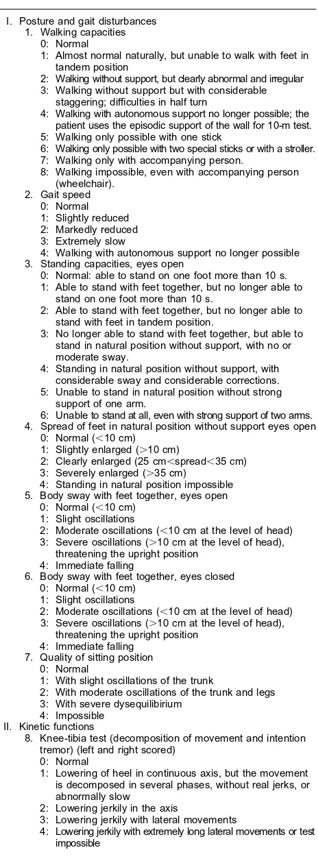
Patients were evaluated using the Modified International Coop-erative Ataxia Rating Scale (MICARS;Schmahmann et al., in press; Table 1). This validated semi-quantitative 120-point rating scale for the assessment of ataxia is based upon the International Coopera-tive Ataxia Rating Scale (ICARS;Trouillas et al., 1997; Storey et al., 2004). MICARS measures posture and gait, kinetic function of the arms and legs (appendicular dysmetria), speech disorders, and oc-ulomotor impairment. Normal subjects scoreⱕ4 (Schmahmann et al., in press), and therefore patients with a MICARS scoreⱕ4 were regarded as motorically normal.
This study was approved by the Partners Human Research Committee at the Massachusetts General Hospital, and all partic-ipants provided written, informed consent.
Lesion localization
After the clinical examinations were performed and the collec-tion of data was completed, the neuroimaging studies (MRI in
Table 1.The Modified International Cooperative Ataxia Rating Scale (MICARS)
I. Posture and gait disturbances 1. Walking capacities
0: Normal
1: Almost normal naturally, but unable to walk with feet in tandem position
2: Walking without support, but clearly abnormal and irregular 3: Walking without support but with considerable
staggering; difficulties in half turn
4: Walking with autonomous support no longer possible; the patient uses the episodic support of the wall for 10-m test. 5: Walking only possible with one stick
6: Walking only possible with two special sticks or with a stroller. 7: Walking only with accompanying person.
8: Walking impossible, even with accompanying person (wheelchair).
4: Walking with autonomous support no longer possible 3. Standing capacities, eyes open
0: Normal: able to stand on one foot more than 10 s. 1: Able to stand with feet together, but no longer able to
stand on one foot more than 10 s.
2: Able to stand with feet together, but no longer able to stand with feet in tandem position.
3: No longer able to stand with feet together, but able to stand in natural position without support, with no or moderate sway.
4: Standing in natural position without support, with considerable sway and considerable corrections. 5: Unable to stand in natural position without strong
support of one arm.
6: Unable to stand at all, even with strong support of two arms. 4. Spread of feet in natural position without support eyes open
0: Normal (⬍10 cm) 1: Slightly enlarged (⬎10 cm)
2: Clearly enlarged (25 cm⬍spread⬍35 cm) 3: Severely enlarged (⬎35 cm)
4: Standing in natural position impossible 5. Body sway with feet together, eyes open
0: Normal (⬍10 cm) 1: Slight oscillations
2: Moderate oscillations (⬍10 cm at the level of head) 3: Severe oscillations (⬎10 cm at the level of head),
threatening the upright position 4: Immediate falling
6. Body sway with feet together, eyes closed 0: Normal (⬍10 cm)
1: Slight oscillations
2: Moderate oscillations (⬍10 cm at the level of head) 3: Severe oscillations (⬎10 cm at the level of head),
threatening the upright position 4: Immediate falling
7. Quality of sitting position 0: Normal
1: With slight oscillations of the trunk
2: With moderate oscillations of the trunk and legs 3: With severe dysequilibirium
4: Impossible II. Kinetic functions
8. Knee-tibia test (decomposition of movement and intention tremor) (left and right scored)
0: Normal
1: Lowering of heel in continuous axis, but the movement is decomposed in several phases, without real jerks, or abnormally slow
2: Lowering jerkily in the axis
3: Lowering jerkily with lateral movements
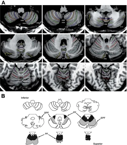
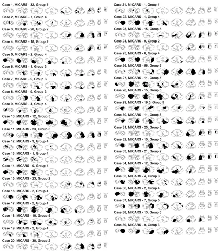
32 cases; CT scan in seven) were reviewed in detail and the location of the infarct was determined (J.D.S.) with reference to the MRI Atlas of the Human Cerebellum (Schmahmann et al., 2000). The anatomical localization was performed at the con-clusion of this 4-year study without reference to the MICARS score obtained in each subject. In most cases, nine to 10 images of the cerebellum in the axial plane from superior to inferior were available for comparison with equivalent sections in the atlas. For each case, we used the atlas to identify the primary fissure that demarcates the anterior lobe from lobule VI, and the superior posterior fissure that separates lobule VI from lobule VII (Fig. 1). Lobule VII comprises lobule VIIA, including the vermis regions VIIAf and VIIAt, and hemispheric regions crus I and II; and lobule VIIB at the vermis and hemi-spheres (Schmahmann et al., 2000). Lesioned areas were recorded on a standard template of axial sections of cerebellum derived from the atlas (Fig. 2). Blood vessel territory (superior cerebellar artery [SCA]), anterior inferior cerebellar artery [AICA] and posterior inferior cerebellar artery [PICA]) was not used for lesion localization, as the territories are not lobule-specific, relative sizes and territories irrigated are not constant, and anastomoses may occur between terminal branches (Tatu et al., 1996).
The locations of the strokes in all 39 cases were also ana-lyzed with respect to the degree of involvement of the deep cerebellar nuclei. This analysis was performed blinded to MICARS score or group membership. Cerebellar nuclei are poorly visible on MRI, and not identifiable on CT. As determined with reference to the cryosection data in the MRI atlas, however, the axial sec-Table 1.continued
9. Action tremor in the heel-to-knee test (left and right scored) 0: Normal
1: Tremor stopping immediately when the heel reaches the knee
2: Tremor stopping⬍10 s after reaching the knee 3: Tremor continuing⬎10 s after reaching knee 4: Uninterrupted tremor or test impossible
10. Decomposition of leg movement (left and right scored) 0: Normal
1: Corners or edges on the circle
2: Markedly decomposed attempts at circle 11. Decomposition of leg tapping (left and right scored)
0: Normal
1: Slightly slow and irregular 2: Clearly slow and irregular
12. Finger-to-nose test: decomposition and dysmetria (left and right scored)
0: Normal
1: Oscillating movement without decomposition of the movement
2: Segmented movement in two phases and/or moderate dysmetria in reaching nose
3: Segmented movement in more than two phases and/or considerable dysmetria in reaching nose
4: Dysmetria preventing the patient from reaching nose. 13. Finger-to-nose test: Intention tremor of the finger (left and
right scored) 0: Normal
1: Simple swerve of the movement
2: Moderate tremor with estimated amplitude⬍10 cm 3: Tremor with estimated amplitude between 10 cm and 40 cm. 4: Severe tremor with estimated amplitude⬎40 cm 14. Finger-finger test (action, tremor and/or instability) (left and
right scored) 0: Normal 1: Mild instability
2: Moderate oscillations of finger with estimated amplitude
⬍10 cm
3: Considerable oscillations of finger with estimated amplitude between 10 and 40 cm
4: Jerky movements⬎40 cm of amplitude
15. Pronation-supination alternating movements (left and right scored)
0: Normal
1: Slightly irregular and slowed
2: Clearly irregular, and slowed movement, but without elbow sway
3: Extremely irregular, and slowed, but with sway of the elbow 4: Movement completely disorganized or impossible 16: Rebound of the arms (left and right scored)
0: None
1: Less than 10 cm 2: Greater than 10 cm
17. Overshoot of the arms (left and right scored) 0: None
1: Less than 10 cm 2: Greater than 10 cm
18. Drawing of Archimedes’ spiral on a predrawn pattern 0: Normal
1: Impairment and decomposition, the line quitting the pattern slightly, but without hypermetric swerve 2: Line completely out of the pattern with recrossings and/
or hypermetric swerves
3: Major disturbances due to hypermetria and decomposition 4: Drawing completely disorganized or impossible
Table 1.continued
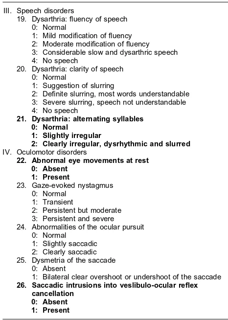
III. Speech disorders
19. Dysarthria: fluency of speech 0: Normal
1: Mild modification of fluency 2: Moderate modification of fluency 3: Considerable slow and dysarthric speech 4: No speech
20. Dysarthria: clarity of speech 0: Normal
1: Suggestion of slurring
2: Definite slurring, most words understandable 3: Severe slurring, speech not understandable 4: No speech
21. Dysarthria: alternating syllables 0: Normal
1: Slightly irregular
2: Clearly irregular, dysrhythmic and slurred IV. Oculomotor disorders
22. Abnormal eye movements at rest 0: Absent 24. Abnormalities of the ocular pursuit
0: Normal 1: Slightly saccadic 2: Clearly saccadic 25. Dysmetria of the saccade
0: Absent
1: Bilateral clear overshoot or undershoot of the saccade 26. Saccadic intrusions into veslibulo-ocular reflex
cancellation 0: Absent 1: Present
tions in this series that contain the cerebellar nuclei are sections 5 and 6 (from superior to inferior), equivalent toz⫽⫺29 andz⫽⫺37 in the atlas. The fastigial and dentate nuclei are identified in these horizontal sections in the atlas with confidence, but the globose
(fastigial, interpositus, dentate) was then examined by compar-ison of levels 5 and 6 on the MRI with the horizontal sections z⫽⫺29 andz⫽⫺37 in the MRI atlas to determine whether the infarct included the expected location of the nuclei as defined in the atlas. A measure of extent of involvement of the nuclei was determined: grade 1, minimal encroachment on any nucleus in either of the two levels; grade 2, clear involvement of any nucleus in one of the two levels; grade 3, clear involvement of any nucleus in both levels; grade 4, apparent complete involve-ment of the nuclei in both levels.
We also tested whether medial versus lateral location of the cerebellar stroke influenced the MICARS score. We used the
coor-dinate system in the atlas to identify the midline for each of the nine horizontal sections, and then measured 10 mm laterally in each direction from the midline. The cerebellum was thus divided into a medial versus a lateral zone for each hemisphere. Location of the stroke in the medial sector, the lateral sector, or both was then determined.
Data analysis
ters were (i) the cerebellar anterior lobe (lobules I–V); (ii) lobule VI; and (iii) the cerebellar posterior lobe and flocculonodular lobe without lobule VI (i.e. lobules VII⫺X).
This determination of the groups was driven by the hypothesis of the study. Specifically, we wished to determine whether lesions in lobules I–V (Group 1) would result in characteristic cerebellar motor impairments as opposed to lesions in lobules VII⫺X (Group 4) that we predicted would not. We were unable to distinguish lobule X (flocculonodular lobe) from adjacent lobules in this study, and thus included lobule X with our evaluation of the posterior lobe. The literature is mixed regarding lobule VI. Connectional and physiological studies indicate that lobule VI is part of the motor system (Schmahmann and Pandya, 1997; Kelly and Strick, 2003), whereas functional magnetic resonance imaging (fMRI) (see Stoodley and Schmahmann, 2009) and functional connectivity magnetic resonance imaging (fcMRI) studies (Buckner and Krienen, in press) suggest that it plays a role in cognition. We therefore defined Group 2 as having a lesion in the anterior lobe (lobules I–V) plus lobule VI; and Group 3 with lesion involving the remainder of the posterior lobe (lobules VII⫺X) plus lobule VI, to determine whether the addition of lobule VI adds to the motor deficit in either group. Finally, to determine whether a lesion in lobules VI–X would compound the motor deficit from lesions of the anterior lobe, we included a group, Group 5, in which stroke was present in some part of all three clusters—namely, anterior lobe, lobule VI, and lobules VII⫺X.
Statistical methods
We used one-way analysis of variance (ANOVA) to determine whether the mean MICARS scores for the different groups were statistically significantly different. We then performed pairwise post hoccomparisons of all groups using Tukey’s least signifi-cance difference test to determine which groups were different from each other. The utility of group, age of patient, and time from stroke onset to examination as predictors of MICARS scores was assessed using multiple linear regression. Pairwise comparisons of mean MICARS scores between groups were then made using two-tailedt-tests. Comparisons in 2⫻2 tables were made using Fisher’s exact test. The null hypothesis of the same proportion of males as females in the population of eligible cases was evaluated using an exact binomial test. An ANOVA model was used with MICARS score as response and group membership, nuclear in-volvement, and their interaction as factors, to test whether nuclear involvement is related to MICARS score. ANOVA was also used to test whether there was a relationship between degree of involve-ment of the nuclei and MICARS score.
RESULTS
Participants
The clinical records and neuroimaging findings of 110
patients with stroke that involved the cerebellum were
evaluated. Of these, 39 patients (ages 50.8
⫾
16.6, range
20 – 83) met the inclusion criteria of stroke isolated to
cer-ebellum with no prior neurological events clinically or on
imaging, no ischemic deficit outside of cerebellum, and
examination within 1 month of stroke onset (
Fig. 2
).
Patients were examined (mean
⫾
SD) 8.0
⫾
6.0 days
following the stroke (range 1–30 days; mode 5 days).
Craniotomy for prevention of herniation was necessary in
Case 5, who was examined 11 days following surgery, and
in Case 25, examined 12 days post-operatively.
There were more males than females in the cohort of
39 patients (M
⫽
27 [69%],
P
⫽
0.01). All six patients in
Group 3 were male. Gender division was not significantly
different however, in the patients in Groups 2 and 5 (65%
male) and those in Group 4 (62% male).
Data analysis by lesion location
There were no patients in this cohort in whom the stroke
was present exclusively rostral to the primary fissure, i.e.
restricted to any part of lobules I–V of the anterior lobe.
The number of patients in Group 1 was therefore 0.
Table 2
shows the number of participants and MICARS scores for
Groups 2–5.
The MICARS score means for the four Groups (2
through 5) were significantly different (one-way ANOVA,
F(3,35)
⫽
10.9,
P
⫽
3.4
⫻
10
⫺5). Pairwise post hoc
compari-son of all Groups using Tukey’s least significant difference
test was then performed. Four of the mean MICARS score
pairings were significantly different: Group 2 differed from
Group 3 (P
⫽
0.01) and from Group 4 (P
⫽
0.00002); and
Group 5 differed from Group 3 (P
⫽
0.009) and from Group
4 (P
⫽
0.00002). The other two group comparisons were
not significant: Group 2 was not different from Group 5
(P
⫽
0.71); and Group 3 was not different from Group 4
(P
⫽
0.273). Note however, that a two-sample
t-test for
Groups 3 and 4 alone, unadjusted for multiple
compari-sons, did reach statistical significance (t
⫽
2.8,
P
⫽
0.03).
The discrepancy between this test and the corresponding
post hoc
result is due to the fact that the SDs for the two
groups (3.8 for Group 3 and 2.0 for Group 4) were
sub-stantially less than the residual ANOVA SD of 8.4 used in
the Tukey LSD test.
The MICARS scores for patients with anterior lobe
involvement (Groups 2 plus 5,
n
⫽
20; 19.1
⫾
11.2) differed
from the mean MICARS scores in Group 3 (n
⫽
6;
P
⫽
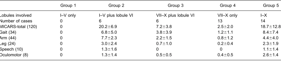
Table 2.Data grouped by anatomical location of lesion and MICARS scores for patients in these groups
Group 1 Group 2 Group 3 Group 4 Group 5
Lobules involved I–V only I–V plus lobule VI VII–X plus lobule VI VII–X only I–X
Number of cases 0 6 6 13 14
MICARS-total (120) 0 20.2⫾6.9 7.2⫾3.8 2.5⫾2.0 18.7⫾12.8
Gait (34) 0 6.8⫾5.0 3.8⫾3.9 1.2⫾1.1 8.4⫾7.4
Arm (44) 0 7.7⫾2.3 2.2⫾1.5 0.8⫾1.2 4.4⫾4.0
Leg (24) 0 3.0⫾2.4 0.7⫾1.0 0.2⫾0.4 2.3⫾1.9
Speech (10) 0 1.3⫾1.6 0 0 1.1⫾1.4
Oculomotor (8) 0 1.3⫾1.4 0.5⫾0.5 0.4⫾0.5 2.6⫾1.4