The journal homepage www.jpacr.ub.ac.id ISSN : 2302 ‐ 4690
108
Characterization of Alkaloids from The Leaves of Psychotria
malayana Jack of Lombok Island on The Basis of Gas
Chromatography-Mass Spectroscopy
Surya Hadi1*, Dina Asnawati1, Vera Fitriya Ersalena1, Ahmadun Azwari1 and Khairul Pahmi Rahmawati1
1
Study Program of Chemistry, Faculty of Mathematics and Natural Sciences, University of Mataram, Jl. Majapahit No. 62 Lombok, Indonesia.
*
Corresponding author: [email protected]
Received as reviewed paper 18 February 2014; Re-submitted 22 February 2014; Accepted 30 April 2014; Revised form accepted 1 September 2014; Published online for edition September-December 2014
ABSTRACT
The genus Psychotria is well known as a source of alkaloids and has contributed several
novel indole-type alkaloids with quite a broad spectrum of bioactivities. This study was
focused on identifying the alkaloids from the leaves of P. malayana Jack from Lombok
island on the basis of Gas Chromatography-Mass Spectroscopy (GC-MS) data and the sample preparation followed acid-base extraction. The study showed that the major alkaloid contained by the leaves was hodgkinsine and other minor compounds, namely calycanthine, chimonantine, 2-ethyl-6-methylpyrazine, and 3-methyl-1,2,3,4-tetrahydro-gamma-carboline. Bioactivities of the compounds were also discussed.
Key word: alkaloids, Psychotria malayana, Lombok
INTRODUCTION
Psychotria malayana Jack is the plant of family Rubiaceae known as “lolon jarum” by Lombok people. This plant grows to a height of 1-4 m, and is largely distributed in the west Indonesian archipelago. The water extract of this plant have been used in Lombok for protecting the skin from infection, treating open wounds, and for other skin diseases [1]. Antiproliferatif activities of the genus Psychotria were reported by Frescura et al. in 2013, when they used the methanol extract of the P. bacypoda and P. birotula leaves collected from Brazil [2].
In 2001, Hadi and Bremner [3] reported that the alkaloid compounds from Psychotria malayana Jack showed antimicrobial activities. There is therefore a good reason for an intensive investigation of the alkaloids of the plant. This paper discloses an alkaloids characterization from leaves of Psychotria malayana Jack on the basis of GC-MS.
EXPERIMENT
Chemicals and instrumentation
The leaves of Psychotria malayana Jack was collected from Suranadi, West Lombok. The sample was prepared and analyzed at the Laboratory of Chemical Analysis, Faculty of Mathematics and Natural Sciences, the University of Mataram, Lombok.
The journal homepage www.jpacr.ub.ac.id ISSN : 2302 ‐ 4690
109
(2)
(1) (3)
°C/min to 265oC and heated until 270 oC for 2 min, (2) injection temperature 260 oC. Initial column temperature is started from 150 oC for 5 min and programmed to increase 5 °C/min to 265 oC and heated until 270 oC for 2 min. Meanwhile MS is conditioned at temperatures of transfer line at 260 oC and ion source at 200 oC. Ion was obtained by electron ionization mode. The RTX-5MS capillary column was applied (length 30 m, diameter 0.25 mm and film thickness 0.25 µm). Helium is used as gas carrier with flow rate 3 mL/min. Molecular mass range of ions was identified at 35 - 600 m/z.
Procedure of Analysis
Finely-powdered, air-dried young leaves (2.7 kg) of P. malayana Jackwere extracted with a cold MeOH (3 x 15 L) with occasional swirling. After filtration, the solvent was removed under reduced pressure at 40 oC yielding a dark-green MeOH extract. The crude alkaloid mixture was then separated from neutral and acidic materials, and water soluble fraction, by initial extraction with aqueous acetic acid (400 ml; 5% v/v) followed by dichloromethane (DCM) extraction (3 x 800 mL) of the aqueous acid extract, which was then basified with aqueous sodium carbonate solution (10%) to pH 10, and then extracted with dichloromethane (5 x 50 mL). The solvent was then evaporated under reduced pressure to get a crude alkaloid. A crude alkaloid was identified by GCMS.
RESULT AND DISCUSSION
Chromatogram results of gas chromatography analysis showed that there were 7 components of alkaloid on the base fraction of the leaves P. malayana Jack at two different programs of temperature. There were five alkaloid compounds indicated from the leaves of P. malayana Jack at injection temperature 100 oC (compound 1-5) and the other two (6-7) were obtained at the injection temperature 260 oC. The following is to discuss the ionization characters of the compounds and reported bioactivities.
1. Chimonantine
From the analysis of GC, appeared two peaks that have the same ion fragmentation pattern. The first compound’s retention time is 32.575 with area percentage at 12.36, whereas the second compound’s retention time is 32.962 with area percentage at 21.29. The estimated compounds are (-)-chimonanthine (1), (+)-chimonanthine (2), and meso-chimonanthine (3). Those three species of chimonanthine exist naturally.
Based on the structure, it was possible that meso-chimonantine was identified earlier because it had less steric inhibition in the GC colomn compared to (+/-)- chimonanthine, thus the peak earlier was meso-chimonanthine and the following peak was (+/-)- chimonanthine.
The journal homepage www.jpacr.ub.ac.id ISSN : 2302 ‐ 4690
110
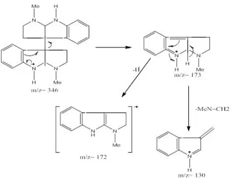
157, 143, and 130. Cleavage of the 3a, 3’a bond in chimonanthine to produce the fragment ion at m/z 173, which then may lose a hydrogen atom to give a radical ion fragment at m/z 172 (100%) which is the base peak. Fragment m/z 130 is the loss of -CH2NMe (M+●-43) from
m/z 173 [4].
Through x-ray analysis of the dihydrobromide salt, Hamor and Robertson subsequently elucidated the structure of (-)-chimonanthine [6] which was isolated from Chimonanthus fragrans [5]. Chimonanthine has two indoline units that 3,3’ connected. Chimonanthine, meso-chimonanthine and psycotridine have been isolated by Verrota et al in 1999 [7]. These compounds showed an analgesic activity [8-9].
2. Calycanthine
This compound, peaked at number 19, appeared at 210 oC with retention time 33.775 min and area percentage at 6.68. Calycanthine (4) is pyrrolidinoindoline type alkaloid and has a complex structure. Ionization data of GC-MS showed three major fragment peaks at m/z 346, 288, and 231. Parent peak of this compound is M+● 346. Fragment m/z 288 is the loss of –[MeNHCH2CH2]+• (M+●-58), and fragment m/z 231 is the loss of –[MeNCH2CH2]]+• (M+●
-57) [10].
Figure 1. Ion fragmentation of Calychantine
Chimonanthine and calycanthine gave the same molecular mass for the parent ion at m/z 346 in the GCMS spectra. However, GC-MS produced a different fragmentation pattern
The journal homepage www.jpacr.ub.ac.id ISSN : 2302 ‐ 4690
111
for each. The GC-MS spectrum of chimonanthine suggested that the molecule is fragmented symmetrically with the major peak at m/z 172 [11]. Calycanthine, on the other hand, yielded a fragment ion at m/z 231 corresponding to a singly protonated calycanine molecule.
Calycanthaceous alkaloids, which have been recognized as a central convulsant, were first isolated from the plant genus Calycanthus. The (+)-calycanthine was established chemically by Robinson and Woodward, and crystallographically using the dihydrobromide dehydrate salt by Hamor and Robertson [12].
3. Hodgkinsine (5)
Hodgkinsine (5) was identified as major alkaloid component (52.245 %). The signals from three protonated fragment ions were observed at m/z 519, 345, and 173. Ionization by MS showed five major fragment peaks at m/z 518, 345, 344, 173, 172, and 130. Cleavage of the 3a, 3’a-bond in hodgkinsine may occur more readily than that of the 7’, 3”a-bond to produce the fragment ion at m/z 173, which then may lose a hydrogen atom to give a radical ion fragment at m/z 172. The remainder of the molecule gave firstly a peak at m/z 345, which then loses a hydrogen atom to give a fragment at m/z 344 by a similar process, as shown in Scheme 7 [4].
Anet et al [13] initially isolated hodgkinsine following an observation in 1949 by Webb, who obtained a positive test for alkaloids in the leaves of the plant Hodgkinsonia frutescens. The X-ray of crystal structure was later reported by Fridrichsons et al [14]. The presence of two N-methyl groups and a significantly different specific rotation from that of chimonanthine (a dimeric compound which was also isolated from this plant), suggested hodgkinsine was a stereoisomer of chimonanthine [15]. Hodgkinsine is a potent analgesic, with results comparable to morphine in murine models using the hot-plate and tail-flick tests [16-17].
4. 2-Ethyl-6-methylpyrazine (6)
This compound appeared at column temperature 130oC, and its retention time is 3.208 with area percentage at 0.47. The signals fragmentation ions of this compound showed two major fragment peaks at m/z 121 and 94. Parent peak of this compound, M+●121 (100%), is the base peak. Fragment m/z 94 is the loss of -HCN●+ (M+●-27).
There are various applications of pyrazine (6) and their derivatives, such as in synthesis of perfumery, pharmaceutical, and agricultural chemical industries, which make them
The journal homepage www.jpacr.ub.ac.id ISSN : 2302 ‐ 4690
112
become very valuable compounds [18-20]. For example, 2-methylpyrazine which is an important lower alkyl-substituted pyrazine, is widely used as a key intermediate for pyrazineamide which is an effective anti-tubercular drug [21-22]. Da-Hong and Wen Yi Tao have identified 2-ethyl-6-methylpyrazine which is a new pyrazine compound in mycobacteria Stigmatella WXNXJ-B [21].
5. 3-Methyl-1,2,3,4-tetrahydro-gamma-carboline (7)
This compound appeared at 155 oC with retention time 15.373 min and area percentage at 3.28. The signals ionization of this compound showed two major fragment peaks at m/z 186 and 143. Parent peak of this compound is M+● 186. Fragment m/z 143 is the loss of -[MeNCH2]●+ (M+●-43) (100%), which is the base peak.
Rihui cao et al in 2007 repoted β-carboline alkaloids have various biological ativities. These compounds particularly have been shown to intercalate into DNA, and to inhibit CDK, isomerase, and monoamin oxidase. It also demonstrated many pharmacological properties including sedative, anxiolytic, hypnotic, antitumor, antiviral, antiparacitic, and antimicrobial activities [22].
CONCLUSION
There were 7 components of alkaloid that have been characterized from the leaves of P. malayana Jack of Lombok on the basis of GCMS at two different programed temperatures. Hodgkinsine was characterized as major and (+/-)-chimonanthine, meso-chimonanthine and calycanthine were indicated as minor from the leaves of P. malayana Jack at injection temperature 100 oC and 2-ethyl-6-methylpyrazine, 3-methyl-1,2,3,4-tetrahydro-γ-carboline were indicated at injection temperature 260 oC.
ACKNOWLEDGEMENT
We would like to give appreciation and thanks for the Ministry of Research and Technology of Republic Indonesia for funding the study.
(6)
The journal homepage www.jpacr.ub.ac.id ISSN : 2302 ‐ 4690
113 REFERENCES
[1] Yohannes Shin Kasahara, Medicinal Herb Index in Indonesia, 1985, P.T. Eisai Indonesia.
[2] Frescura, Viviane Dal-Souto, Andrielle Wouters Kuhn, H. D. Laughing House IV; Jucara Terezinha Paranhos; Solange Bosio Tedesco, J. Biocell., 2013, 37, 23-28.
[3] Hadi, S. and Bremner J.B., Molecules. 2001, 6, 117-129.
[4] Hart, N. K.; Johns, S. R.; Lamberton, J. A.; Summons, R. E., Aust. J. Chem. 1974, 27, 639-646.
[5] Hodson, H. F.; Robinson, B.; Smith, G. F. Proc. Chem. Soc. London, 1961, 465. 7. [6] (a) Grant, I. J.; Hamor, T. A.; Robertson, J.M.; Sim, G. A. Proc. Chem. Soc., London,
1962, 148; (b) Grant, I. J.; Hamor, T. A.; Robertson, J. M.; Sim, G. A. J. Chem. Soc.
1965 , 5678.
[7] Verotta, L.; Peterlongo, F.; Elisabetsky, E.; Amador, T.A; Nunes, D.S., J. Chromatogr. A, 1999, 841, 165-176.
[8] Verotta, L.; Orsini, F.; Sbacchi, M.; Scheildler, M. A.; Amador, T.A.; Elisabetsky, E.
Bioorg. Med. Chem., 2002, 10, 2133-2142.
[9] Amador, T.A.; Verotta, L.; Nunes, D.S.; Elisabetsky, E. Phytomedicine, 2001, 8 (3), 202-206.
[10] Manske, R.H.F.;Editor The Alkaloids Vol. 8: The Indole Alkaloids,1965, 861
[11] Hadi, S., Bioactive Alkaloids From Medicinal Plants Of Lombok, 2002; Department of Chemmistry, The University of Wollongong, Australia.
[12] Chebib, Mary; K. Duke, Rujee; Duke, Colin C. ; Connor, Mark; N. Mewett, Kenneth and A.R. Johnston, Graham. Toxicol. App. Pharmacol., 2003, 190, 58–64.
[13] Anet, E. F. L. J.; Hughes, G. K.; Ritchie, E. Aust. J. Chem., 1961, 14, 173-174. [14] Fridrichsons, J.; Mackay, M. F.; Mathieson, A. M., Tetrahedron, 1974, 30, 85-92. [15] Hendrickson, J. B.; Goeschke, R.; Rees, R. Tetrahedron, 1964, 20, 565-579.
[16] Kodanko, J. J.; Hiebert, S.; Peterson, E. A.; Sung, L.; Overman, L. E.; Linck, V. M.; Goerck, G. C.; Amador, T. A.; Leal, M. B.; Elisabetsky, E. J. Org. Chem., 2007, 72 (21), 7909-7914.
[17] Amador, T. A.; Verotta, L.; Nunes, D. S.; Elisabetsky, E. Planta Med., 2000, 66, 770- 772.
[18] Seetharamanjaneya S P, Raghavan K V, Rao P. K. and Kulkarni S. J., 2002 US 143180 A1
[19] Forni L, Stern G and Gatti M., Appl. Catal., 1987, 29, 16.
[20] Murray KE, and Whitfield FB., J. Sci. Food Agric., 1975, 26, 973-986. [21] Wang, Da-Hong and Wen-Yi Tao. A., J. Microbiol. Res., 2009, 3, 755-760.