HIF-1α Concentration and Heart Muscle Histopathology of Wistar Rats Induced by Aerobic and Anaerobic Activities
DR.Rostika Flora, S.Kep.M.Kes.AIF
Lecturer. Department of Physiology, Program Study of Nursing, Universitas Sriwijaya, Palembang, South Sumatera, Indonesia
Correspondence address: rostikaflora@gmail
ABSTRACT
Background Physical activity besides improving cardiac function, can also lead to sudden death, which is often occurred in the sport event itself. Some studies suggested that most are caused by myocardial infarct. During physical activity increased demand of oxygen required for energy metabolism. If the increasing demand is not fulfilled by the oxygen supply, then the organism will undergo hypoxia state. It is not yet known, whether hypoxia occurs in heart muscle, considering the heart must works hard in this condition. During physical activity molecular adaptation and systemic adaptation will occur. Response of heart muscle to systemic adaptation is well known. However the molecular adaption of this organ still need to be studied in details.
Method In this study, certain number of rats were divided into 3 major groups namely control group, aerobic activity group and anaerobic group . As physical activity each animal must run on a treadmill (20 m / min for 30 minutes in aerobic and 35 m / min for 20 minutes in anaerobic) in 1 day, 3 days, 7 days and 10 days. We observed hypoxia parameters (HIF-1α content) and parameter of cardiac muscle cell structure changes (histopathological). Result Rats heart muscle underwent aerobic and anaerobic physical activity increased HIF-1 α content (p <0.05), with the highest value in day-1 and in the anaerobic group is higher than aerobic group (156.8 ± 33.1 vs. 116.03 ± 5.56). Histopathology observation showed areas of infarct in both groups in day-10.
Conclusion: In rat though aerobic and anaerobic physical activity will result in heart hypoxia. The effects more severe in anaerobic than in aerobic activity. In most parameters observed, HIF-1α play important roles.
Background
Physical exercise is regular physical activity performed in certain period of time and intensity which aims to maintain the body always in well condition of health and fitness. In addition to keep body fitness, physical exercise is extremely recommended for preventive, curative, and rehabilitative program in order to maintain and enhance the health, especially the health of cardiovascular system.1 There are two types of physical
activities, i.e. aerobic and anaerobic physical activities. Aerobic physical activities are physical activities that utilize ATP energy generated by oxidation process of glycogen phosphorylase and free fatty acid. Metabolism process depends on oxygen availability. Anaerobic physical activities are physical activities that do not utilize oxygen in the metabolism process of energy production. The energy is generated by ATP production consists of 25-30% aerobic physical activities and 70-75% anaerobic physical activities. Sprint run chasing a ball and hitting ball with full power are anaerobic physical activities. During the sprint, there is no oxygen uptake; oxygen uptake occurs after the run. If there is impaired heart function and the oxygen supply during the sprint could not meet cardiac oxygen demand; therefore, acute heart attack may occur. Ignoring acute heart attack may cause extensive damage to the cardiac muscles and resulting in sudden death. 1,2
Despite enhancing the heart function, physical activity may also cause sudden death. Sudden death in athletes frequently occurs during the competition. It is assumed that performing high intensity exercise without taking rest days for the upcoming competition
has great impact in cardiac muscles damage and sudden death during the competition. 1,2
Sudden death may also occur not only in athletes but also in non-athlete or layman when performing physical exercises. Lack of knowledge in selecting the appropriate type of physical exercises conforming to their health condition may cause increased incidence of sudden death. Sudden death will not occur if the heart successfully adapts with such condition so that it may compensate the load taken during physical exercise. Therefore, the cardio-protection effect exerted by the physical exercise can be achieved.
In order to get cardio-protection effect of the physical exercise and to avoid the adaptation. The systemic adaptation reaction of the cardiac muscles on physical exercises has been extensively known; however, the molecular adaptation should be studied further.
In cardiac muscles, oxygen is the main determination factor in expressing myocardial genes. If hypoxia also occurs in cardiac muscle during physical exercise, it is assumed that HIF-1α has a role in molecular adaptation response of cardiac muscles. During hypoxia state, HIF-1α will be translocated to the nucleus of the cells in order to have dimerization with HIF-1β, producing HIF-1.3
Through the activity of HIF-1, there would be increased expression of some genes to reduce cell dependency on oxygen as well as to increase the oxygen supply in the tissues. HIF-1 arrange expressions of various genes with a very wide range. It has role in maintaining energy metabolism and oxygen balance including the genes involved in erythropoiesis, glucose metabolism and angiogenesis.4
Moreover, in order to recognize whether the hypoxia that occurs during physical activity may cause protection effect on cardiac muscles or on the contrary, it may result in cardiac muscle cell damage which may lead to sudden death; therefore, we should conduct further studies.
Material and Methods
In this study, experiments were performed using male Wistar rats (aged 6-8 weeks with weight of 60-100 gram), which were divided into 9 groups (P1-P9). P1 group was the control group receiving no treatment of aerobic and anaerobic physical activity; while the P2-P9 were groups that were treated with aerobic and anaerobic physical activity for 1, 3, 7 and 10 day(s). The treatment for physical activity was performed by putting the rats on animal treadmill and setting the treadmill on the speed rate of 20 m/minute about 30 minutes for the aerobic physical activity and 35 m/ minute about 20 minutes for anaerobic physical activity. After each experiment, tissue specimens from cardiac muscle were collected for HIF-1α assay and histological evaluation. Cardiac muscle tissue was weighed and homogenates prepared by
adding lysis buffer solution. HIF-1α concentrations were measured by sandwich ELISA method using kits Surveyor TM IC Human / Mouse Total HIF-1α immunoassay (Cat. SUV 1935, R & D Systems). Histological evaluation was accomplished in the laboratory of the Department of Pathological Anatomy, Faculty of Medicine, Universitas Padjajaran, Bandung. Cardiac muscle tissues were stained by using hematoxylin and eosin (HE). The tissue preparations were evaluated by light microscopy and categorized as normal, hypertrophic, ischemic, or infarcted:
1. Results
3.1. The concentration of HIF-1α
The result of HIF-1α concentration measurement on cardiac muscles showed that there was significant increase on HIF-1α concentration in the aerobic and anaerobic physical activity groups compared to the control group (45.7 ± 4.72 vs. 116.03 ± 5.66 vs. 156.8±33.1) on the first day of treatment. It indicates that physical activity also causes hypoxia on cardiac muscle. (Figure 1).
Figure 3.2. The concentration of VEGF
*significant difference compared with control group (p <0.05)
Δsignificant difference between
Histology of myocardium
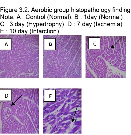
Histology of cardiac muscles on day 1 did not show any signs of hypertrophy, ischemia or infarction. On day 3, there were signs of cardiac muscle cell hypertrophy. Further histological examination revealed signs of cardiac muscle cell ischemia on day 7 and of infarction on day 10 in both groups (Figures 2 and 3).
Figure 3.2. Aerobic group histopathology findings Note: A : Control (Normal), B : 1day (Normal) C : 3 day (Hypertrophy) D : 7 day (Ischemia) E : 10 day (Infarction)
Figure 3.3. Anaerobic group histopathology findings
Note : A : Control (Normal), B : 1 day (Normal) C : 3 day (Hypertrophy) D : 7 day (Ischemia) E : 10 day (Infarction)
2. Discussion
The measurement of HIF-1α concentration in heart muscle showed a significant increase on the first day of aerobic and anaerobic physical activity compared to the control group (Figure 3.1). This indicates that physical activity also leads to hypoxia in cardiac muscle. The state of hypoxia would affect several genes that play a role in the regulation of oxygen homeostasis, such as erythropoeietin (EPO) and VEGF. Their transcription is controlled by HIF-1. Hypoxia inhibits the activity on the tissue PHD and FIH-1 (factor inhibiting HIF-1), because both of these enzymes require oxygen as co-substrate. Inhibition of the PHD results in HIF-1α stabilization to bind CBP/p300 and run a perfect transcription activity. Accumulation of HIF-1α in the cytoplasm under hypoxic condition occurs rapidly. After experiencing phosphorylation, HIF-1α is translocated into the cell nucleus for its dimerization partner HIF-1β forming the transcription factor HIF-1. Through the activation of HIF-1, expression of a number of genes that encode enzymes, growth factors and transporters such as glycolytic enzymes, vascular endothelial growth factor (VEGF), glucose transporters and erythropoietin reduce the dependence of the cells on oxygen and simultaneously increase the supply of oxygen
to the system.3,5
Increased concentrations of HIF-1α occur because of the high oxygen demand during physical activity to meet energy needs. Hypoxia is known to occur in skeletal muscle tissue under exercise, but the heart muscle plays a special role. During life, the heart has always to contract. To maintain its function in the face of various loads, continuous availablity of oxygen and substrate is necessary. Increased oxygen demand during physical activity will also result in increased oxygen delivery in heart.6,7
A B C
D E
C
D E
This study shows that concentrations of HIF-1α on the first day are higher in the anaerobic than in the aerobic group, but differences were statistically not significant. In both groups HIF-1α concentrations were significantly increased vs. controls. Anaerobic condition would trigger the accumulation of HIF-1α and HIF-1 activation. In addition, the results of blood gas analysis showed that the decrease in PaO2 was lower in the aerobic than in the through anaerobic glycolysis.4 Transition to
anaerobic metabolism is an adaptive response for energy to remain available even if through the circumstances oxygen does not sufficiently exist.7,8 Transcription factor HIF-1α is very
important in maintaining ATP levels in the cells9 e.g., its role in inducing glucose
transporter and glycolytic enzymes such as aldolase A, pyruvate kinase M, which helps to produce energy in a state of unavailability of oxygen.10 HIF-1 increases the expression of
these enzymes and activates pyruvate dehydrogenase kinase I so that the citric acid cycle inactivated.11
The results are similar to induced systemic hypoxia in mice using a mixture of 98% nitrogen and 8% oxygen. A sharp increase in HIF-1α mRNA of liver tissue on the first day was followed by a gradual decline until day 14.12 According to Shao et al. (2005), in acute
hypoxia HIF-1α mRNA expression increased, whereas in hypoxia over time it declined again.13 Research on heart and brainwith
induced systemic hypoxia in rat indicate that the occurrence of elevated levels of HIF-1α first day and a sharp increase occurred at day 7 and after that there is a decrease.16 Research
conducted by Ferdinal (2009) in mice induced systemic showed that the concentration of HIF-1α in cardiac muscle there was an increase from day 7 and peaked on day 21 (8.2 times compared to control). Allegedly, the day-to-21 is the beginning stage of cardiac dysfunction is more severe.16 From several
studies in mice induced systemic hypoxia, showed that increased HIF-1α mRNA occurs gradually. In contrast to this research model of aerobic and anaerobic physical activity increases immediately HIF-1α protein in cardiac muscle on the first day of treatment. This is because aerobic and anaerobic physical activity lead to increased oxygen demand is very high and immediate for the continuity of physical activity. This situation causes increased HIF-1α that is sensitive to decreased levels of O2. In addition, during aerobic and anaerobic physical activity the heart muscle works harder to pump blood to meet the needs of the body. Mechanical stress can cause the induction and accumulation of HIF-1α. Induction of HIF-1α occurs as a result of stretching the heart wall.17
indicates the occurrence of cardiac contraction dysfunction, although the mechanisms of contraction dysfunction is unclear. Presumably, this is caused by the presence of hypovascularisation, management changes and changes in calcium metabolism resulting in a decrease of ATP.18 According to
Seagroves et al. (2001) and Cramer et al. (2003), although HIF-1α expression should occur only in hypoxic conditions, but the reality in some tissues shows that HIF -1α is not only influential in hypoxic but also under normoxic conditions.9,19
It is necessary to maintain metabolic functions. Research conducted by Stroka et al (2001) on the heart muscle in a normoxic state found HIF-1α in the nucleus of endothelial cells and arterial myocytes. In the heart muscle, the stability of HIF-1α is required for activation of transcription of several genes that are sensitive to normoxic oxygen conditions and hypoxia.20
In normoxic circumstances, there are several mechanisms that can increase the stability and activity of HIF-1α. Activation of HIF-1α may occur through the p44/42 MAPK (Mitogen Activated Protein Kinases) by various cytokines and growth factors through tyrosine kinase receptors. The path is the MAPK signal transduction through the receptor (tyrosine kinase) which in turn can activate phosphatidyl inositol 3-kinase thus increasing transcriptional activity of HIF. In addition, according to Metzen et al (2003) NO (nitric oxide) also can lead to accumulation of HIF-1α in a normoxic state. HIF-HIF-1α accumulation is due to the inhibition of HIF-1α hydroxylation, so there is no binding of HIF-1α with pVHL and HIF-HIF-1α degradation occurs.21
Histology of cardiac muscles on day 1 did not show any signs of hypertrophy, ischemia or
infarction. On day 3, there were signs of cardiac muscle hypertrophy. Such histological finding demonstrated that there was an adaptation response to increased heart load during exercise. Continuous physical activity would cause greater muscle mass and volume, in other words, heart muscle hypertrophy called athlete’s heart, which is considered a normal adaptation response. Physical load would cause left ventricle (LV) to pump harder, which may change its size. The changes of ventricle size depend on the type of physical activity or exercise.22
Aerobic or endurance type of exercise would increase LV filling; first, it would increase plasma volume and therefore, also LV end-diastolic volume (increased preload). Next, the duration of diastolic filling would increase. The increase of plasma volume and diastolic filling duration would change the end-diastolic size of LV.22
On the contrary, during anaerobic or
resistance type of exercise, LV has to work hard to push through high pressure circulation (increased afterload). To compensate increased afterload, myocardium would increase the thickness of the muscle in order to enhancee contraction power. Increase of cardiac muscle mass is a direct adaptation to anaerobic or resistance type of exercise. In fact, increased thickness of the cardiac muscle wall (and thus, increased muscle mass) will not only be found in anaerobic or
resistance type, but also in the aerobic or
endurance type of exercise, as recent studies reported.22
Cardiac muscle cell hypertrophy was considered a mechanism of adaptation to increase muscle contractility and decrease ventricle wall pressure.23 Long-lasting
subsequently, remodeling of the ventricle would occur. Ventricle remodeling is a transition phase towards ventricle dilation and heart failure.23
Histological examination revealed signs of cardiac muscle ischemia on day 7 and infarction on day 10. Hypertrophy of cardiac muscle would cause capillary compression through increased extravascular pressure leading to ischemia. Erythrocytes are trapped in compressed capillary vessels, which along with the ischemic condition would disrupt capillaries to become more permeable to erythrocytes and increase local oxygen supply.24
On 10 day, in aerobic and anaerobic groups, cardiac muscle fibers were stretched. This lengthening of muscle fibers may occur as an adaptation response to increased preload in order to maintain the LV end-diastolic volume. Histopathology on day 10 found cardiac muscle damage both in aerobic and anaerobic groups, certainly caused by heavy exercise load without any rest-day. The damage in the anaerobic group was more severe than in the aerobic group.
Every cardiac muscle cell has Z lines that separate sarcomeres from each other. High intensity exercise would lead to over-stretched muscle fibres, Z-line disruption and protein leakage from cardiomyocytes, such as myoglobin and creatinekinase.25,26 During
heavy load or high intensity physical activity, cardiac muscle cell leakage occurred due to increased cell membrane (= sarcolemma) damage and permeability.27
Conclusion: Aerobic and anaerobic exercise performed for 10 days without any rest-day may cause hypoxia and damages of cardiomyocytes and whole cardiac muscle. These damages are more severe with anaerobic exercise in our animal model.
References:
1. Åstrand Per-Olof MD, Kaare Rodahl MD, Hans A Dahl MD, Textbook of Work Physiology : Physiological basis of exercise, fourth edition, 2003, United States.
2. Kusmana D, Olahraga untuk orang sehat dan penderita penyakit jantung. 2006. Edisi 2; FKUI; Jakarta.
3. Wang GL, Jiang BH, Semenza GL, Effect of protein kinase and phosphatase inhibitors on expression of hypoxia-inducible factor-1. Biochem Biophys Res Commun. 1995; 216:669-75
4. Semenza GL. Hidroxylation of Hif-1α : Oxygen sensing at the molecular level.
Physiology.2004;19:176-82.
5. Zagorska A and Dulak J. Hif-1 : the knowns an unknowns of hypoxia sensing. Acta Bhiochemia Polonica
2004; 51 (3):563-78
6. Foss ML, Keteyian SJ. Physiological basis for exercise and sport, Mc. Graw Hill New York.2006: 59-64
7. Mooren FC, Volker K. Human Kinetics. Molecular and Cellular Exercise Physiology. USA; 2005.
8. Guyton AC, Hall JE. Textbook of Medical Physiology 11th edition.
Elsevier Saunders, Philadelphia, Pensylvania.2006;79-82;530;1056-60
9. Seagroves TN, Ryan, HE, Lu H, Wouters, BG, Knapp M, Thibault P. Laderoute K, Johnson RS. Transcription factor HIF-1 is a necessary mediator of the pasteur effect in mammalian cells .
Mol. Cell. Biol. 2001; 21:3436-44
Ratcliffe P, Moons L, Jain RK, Collen D, and Keshet E. Role of HIF-1α in hypoxia-mediated apoptosis, cell proliferatiom and tumour angiogenesis.
Nature 394: 485-490, 1998.
11. Vaupel P. The Role of Hypoxia-Induced Factors in Tumor Progression.
Oncologist. 2004;9:10-17
12. Jusman SWA, Halim A, Wanadi SI, Sadikin M. Expression of hipoxia-inducible factor-1α (HIF-1α) related to oxidative stress in liver of rat-induced by systemic chronic normobaric hypoxia. Acta Med Indones-Indones J Intern Med. 2010;42(1): 17-23.
13. Shao G, Gao CY, Lu GW.
Neurosignal.14.2005.255.
14. Salceda S. Caro J.J.Biol Chem.272.1997.22642.
15. Prijanti AR.Peran HIF-1α dalam pengaturan ekspresi renin pada hipoksia.Disertasi.2010.FKUI.Jakarta.
16. Ferdinal F, SuyatnaFD, Wanadi SI, Sadikin M. Expression of B-type natriuretic peptide-45 (BNP-45) gene in the ventricular myocardial induced by systemic chronic hypoxia. Acta Med Indones-Indones J Intern Med.2009;4(13):136-43.
17. Metzen E, Ratcliffe PJ. HIF hydroxylation and cellular oxygen sensing.2004;385:223-30.
18. Huang LE, Gu J, Schau M, Bunn HF. Regulation of Hipoxia-inducible factor-1α is mediated by an O2 dependent degradation domain via the ubiquitin-proteosome pathway. Prog Natl Acad Sci.1998;95:7987-92.
19. Kjaer M, Farrell, PA, Christensen NJ, Galbo H. Increased epinephrine response and inaccurate glucoregulation
in exercising athletes. J Appl Physiol. 1986; 61:1693–1700.
20. Stroka D, Candinas D. Hipoxia-inducible factor-1 signaling system. In : Dufour JF, Clavien PA, Trautwein C, Graf R, eds. Signaling pathways in liver disease part II. Berlin:Heidelberg Springer . 2005; 26:311-23.
21. Metzen E, Ratcliffe PJ. HIF hydroxylation and cellular oxygen sensing.2004;385:223-30.
22. Wilmore Jack H, Costill David L, Physiology of Sport and Exercise, Third Edition, Human Kinetics, United States, 2004.
23. Selvetalla G, Lembo G.Mechanism of cardiac hypertrophy. 2005;1:263-73.
24. Flameng W, Vanhaecke.J, Borgers M. Histology of the postischaemic myocardium and its relation to left ventricular function Br. J. Anaesth.
(1988), 60, 14S-22S.
25. Wu Q, Tao L, Heng L, Guochang Z, Unsaturated fatty acid: Metabolism, synthesis and gene regulation. African J Biotech. 2009; Vol. 8 (9): 1782–85.
26. Braccacio P, Maffuli N, Limomgelli FM. Creatinr kinase monitoring in sport
medicine. British
Med.Bull.2007:81,82.209-230.