Guidelines on
Establishment of
Virology Laboratory in
Developing Countries
Guidelines on
Establishment of
Virology Laboratory in
Developing Countries
ISBN 9789290223351
World Health House, Indraprastha Estate
Guidelines on
Establishment of
W H O Library Cataloguing-in -Publication D ata
Guidelines on establishment of virology laboratory in developing countries. 1. Virology – methods. 2. Virus Diseases – diagnostic – microbiology. 3. Laboratory, Techniques and Procedures. 4. Equipment and Supplies, H ospital. 5. Specimen H andling. 6. Developing Countries. 7. Guidelines. ISBN 978-9 2-9022-335-1 (NLM classification: Q W 25)
© W orld H ealth O rganization 2008 All rights reserved.
Requests for publications, or for permission to reproduce or translate W H O publications – whether for sale or for noncommercial distribution – can be obtained from Publishing and Sales, W orld H ealth O rganization, Regional O ffice for South-East Asia, Indraprastha Estate, M ahatma Gandhi M arg, New Delhi 110 002, India (fax: + 91 11 23370197; e-mail: [email protected]).
The designations employed and the presentation of the material in this publication do not imply the expression of any opinion whatsoever on the part of the W orld H ealth O rganization concerning the legal status of any country, territory, city or area or of its authorities, or concerning the delimitation of its frontiers or boundaries. D otted lines on maps represent approximate border lines for which there may not yet be full agreement.
The mention of specific companies or of certain manufacturers’ products does not imply that they are endorsed or recommended by the W orld H ealth O rganization in preference to others of a similar nature that are not mentioned. Errors and omissions excepted, the names of proprietary products are distinguished by initial capital letters.
All reasonable precautions have been taken by the W orld H ealth O rganization to verify the information contained in this publication. H owever, the published material is being distributed without warranty of any kind, either expressed or implied. The responsibility for the interpretation and use of the material lies with the reader. In no event shall the W orld H ealth O rganization be liable for damages arising from its use. This publication does not necessarily represent the decisions or policies of the W orld H ealth O rganization.
Contents
Page
Preface...v
Abbreviations... vii
Introduction ...1
Key elements of a virology laboratory ...7
Viral diagnostic tests...19
Specimens management...27
Quality systems...39
Further reading...45
Annexes Annex 1 ...46
Preface
Emerging infectious diseases caused by viruses have assumed great public health significance in the recent past. During the last three decades, almost 20 new viral pathogens have been detected. Some of these–e.g. human immunodeficiency virus (HIV) and hepatitis viruses–have already caused substantial mortality, morbidity and economic loss all over the world. The pandemic of serious acute respiratory syndrome (SARS) unequivocally demonstrated the rapidity with which new viruses can travel across the world and inflict misery. The threat of a pandemic with one of the subtypes of influenza virus is considered the greatest public health challenge in the current millennium so far.
Laboratories play a critical role in surveillance, diagnosis and monitoring of viral disease as well as in the understanding of the genetic changes in the viral genome. Laboratory tools are also essential for the diagnosis of dengue fever and chikungunya fever–two of the important vector-borne viral diseases–and for differentiating from other fevers for institution of rational and specific therapy and control measures.
Establishment of a reli able viral laboratory is a prerequisite for a strong public health response to emerging viral diseases. Unfortunately, several developing countries lack adequate capacity to diagnose viral infections. These guidelines are therefore intended to provide the minimum requirements for establishing a national virology laboratory, keeping in view the emergence of new viral pathogens. It is not meant to serve as a comprehensive laboratory manual for diagnosis of viral infections.
These guidelines cover key aspects of establishing a virology laboratory in a developing country and address issues pertaining to policy and programme, infrastructure, human resources, technologies available, and creating high-quality systems.
Abbreviations
ACD Anticoagulant citrate dextrose
BSC Biological safety cabinet
BSL Biosafety level
CM V Cytomegalovirus
CPE Cytopathic effect
DNA Deoxyribonucleic acid
EBV Epstein-Barr virus
ECG Electrocardiogram
ECHO Enteric cytopathic human orphan
EDTA Ethylenediaminetetraacetic acid
EIA Enzyme immunoassay
ELISA Enzyme linked immunosorbent assay
FA Fluorescent antibody
FBC Full blood count
GM T Good microbiological techniques
HEPA High efficiency particulate antigen
HHV6 Human herpes virus
HIV Human immunodeficiency virus
HSV Herpes simplex virus
IHR International Health Regulations
JE Japanese encephalitis
PCR Polymerase chain reaction
QC Quality control
RNA Ribonucleaic acid
SARS Severe acute respiratory syndrome
SOP Standard operating procedures
VTM Viral transport medium
Chapter 1
Introduction
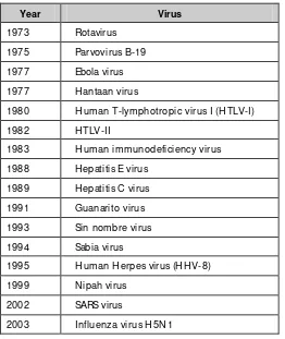
Diseases caused by viruses have assumed great public health significance in the recent past and an increase in the frequency and spread of such diseases observed globally. Several new viruses have been isolated. During the past three decades, of the 30 new pathogens discovered, 16 are viruses (Table 1.1). These emerging viruses have added a new paradigm to public health concepts. Not only the health but the economic and social fabric of global communities have been affected by viruses such as HIV, hepatitis B and C, severe acute respiratory syndrome (SARS) and avian influenza.
Table 1.1: Newly discovered viruses of public health importance
Year Virus
1973 Rotavirus 1975 Parvovirus B-19 1977 Ebola virus 1977 H antaan virus
1980 H uman T-lymphotropic virus I (H TLV-I)
1982 HTLV-II
1983 H uman immunodeficiency virus 1988 Hepatitis E virus
1989 Hepatitis C virus 1991 Guanarito virus 1993 Sin nombre virus 1994 Sabia virus
1995 H uman H erpes virus (H H V-8) 1999 Nipah virus
2002 SARS virus
Guidelines on establishment of virology laboratory in developing countries
Page 2
Ever since its discovery, the human immunodeficiency virus (HIV) has emerged as a global disaster. Around the world AIDS has led to more than 20 million deaths. Over 33 million people are living with HIV today and by 2010 it is estimated that more than 40 million children will have one or both parents dead from AIDS. People in productive age groups are predominantly affected by AIDS and hence in some countries the impact of AIDS has led to a major decrease in their respective gross national products. Till date the only tool available to ascertain the presence of HIV in an otherwise healthy looking individual is a laboratory assay.
A global view of HIV infection
33 million people
[
30–36 million
]
living with HIV, 2007
Source: 2008 Report on the global AIDS epidemic
The beginning of the millennium saw the appearance of Nipah virus in Malaysia which caused the deaths of 100 people in the country as well as significant economic losses due to the slaughter of pigs. It is estimated that the outbreak cost was nearly USD 500 million. Subsequently, Nipah virus outbreaks were identified in India and Bangladesh.
Guidelines on establishment of virology laboratory in developing countries
diagnostic services. The isolation of the virus and understanding its genetic characteristics were major contributors in curtailing the spread of this disease.
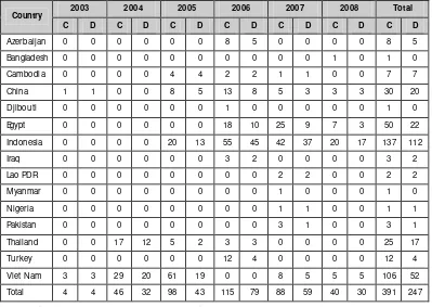
Outbreaks of highly pathogenic influenza A (H5N1) occurred in Asia beginning in early 2004. As on October 2008, 387 cases with 245 deaths have been reported from 15 countries (Table 1.2). In Indonesia, the case fatality ratio is as high as 85%. More than 250 million birds have either died from the disease or were culled in efforts to contain the outbreaks. The world continues to live under the threat of a pandemic of influenza. Laboratory support is critical not only to detect the presence of the virus in clinical material and birds but, more importantly, to understand the genetic changes in the viral genome which shall be the harbinger of the pandemic.
Table 1.2: Cumulative number of confirmed human cases of avian influenza A/(H5N1) reported to WHO
2003 2004 2005 2006 2007 2008 Total
Country
C D C D C D C D C D C D C D
Azerbaijan 0 0 0 0 0 0 8 5 0 0 0 0 8 5 Bangladesh 0 0 0 0 0 0 0 0 0 0 1 0 1 0 Cambodia 0 0 0 0 4 4 2 2 1 1 0 0 7 7 China 1 1 0 0 8 5 13 8 5 3 3 3 30 20 Djibouti 0 0 0 0 0 0 1 0 0 0 0 0 1 0 Egypt 0 0 0 0 0 0 18 10 25 9 7 3 50 22 Indonesia 0 0 0 0 20 13 55 45 42 37 20 17 137 112 Iraq 0 0 0 0 0 0 3 2 0 0 0 0 3 2 Lao PDR 0 0 0 0 0 0 0 0 2 2 0 0 2 2 Myanmar 0 0 0 0 0 0 0 0 1 0 0 0 1 0 Nigeria 0 0 0 0 0 0 0 0 1 1 0 0 1 1 Pakistan 0 0 0 0 0 0 0 0 3 1 0 0 3 1 Thailand 0 0 17 12 5 2 3 3 0 0 0 0 25 17 Turkey 0 0 0 0 0 0 12 4 0 0 0 0 12 4 Viet Nam 3 3 29 20 61 19 0 0 8 5 5 5 106 52 Total 4 4 46 32 98 43 115 79 88 59 40 30 391 247
Guidelines on establishment of virology laboratory in developing countries
Page 4
Apart from new viruses, several other viruses–especially those that cause dengue fever and chikungunya fever–have emerged as major public health issues. Both these diseases are vector-borne and have a complex epidemiology. Laboratory tools are essential for their diagnosis and for differentiating from other fevers for institution of rational and specific therapy.
It is in this context that the establishment and strengthening of existing national virology laboratories gains paramount importance. A national viral surveillance system needs to be established. The epidemiology of virus diseases needs to be studied in depth. The need to be able to easily, cheaply and quickly diagnose these and other potential outbreaks of viral infections as public health problems in specific geographic regions, in additi on to travel and bio-terrorism concerns, is crucial. Apart from development of diagnostic reagents and their supply to investigating centres, a central serum bank, and a virus repository are important factors. Research on viruses, as regards the epidemiology, diagnosis, pathogenesis and vaccinology of virus infections, needs to be strengthened. An international network of databases of virus infections, needs to be instituted. A global network for the diagnosis and containment of emerging viral diseases is the need of the hour. This document has therefore been conceived and developed to provide to policy makers, administrators and public health professionals in developing countries, an overview of the requirements for establishing a national virology laboratory.
Guidelines on establishment of virology laboratory in developing countries
Scope
These guidelines cover key aspects of establishing a virology laboratory in a developing country and address issues pertaining to policy and programme, infrastructure, human resources, technologies available, and high-quality systems. The guidelines do not describe the technique for processing the specimens because every laboratory has to develop its own standard operating procedures (SOP). Also, various standard textbooks are available for these techniques.
These guidelines shall principally assist the national laboratory programmes in expanding their diagnostic profile and should be used in conjunction with any other national directives on International Health Regulations (IHR 2005) or disease surveillance programmes.
Guideline development process
The W HO Regional Office for South-East Asia commissioned the Department of Virology, National Institute of Mental Health and Neurosciences, Bangalore, India (NIMHANS) to develop the first draft of the guidelines. The objectives were to assist developing countries in establishing practical diagnostic facilities for viral diseases based upon a pragmatic approach and available scientific evidence.
The draft guidelines were first reviewed by WHO and subsequently peer reviewed by two eminent virologists from Sri Lanka and Thailand.
The guidelines were revised by NIMHANS as per the observations made by the peer reviewers.
The guidelines also provide information for the procurement of products that can be used in laboratories. However, the information is only suggestive and W HO does not specifically endorse these products.
Guidelines development team
Chapter 2
Key elements of a virology laboratory
The key elements for the establishment of a virology laboratory and diagnostic services are:
(1) Physical infrastructure
(2) Human resources
(3) Equipment and supplies
Physical infrastructure
Virusisolation and a numberof methods for detection of viral antigens, nucleic acids, and antibodies (serology)are the core repertoire of techniques used in a diagnostic virology laboratory. Virusisolation using cell culture is always performed in designatedvirology laboratories although the othermethods may be performedin diverse laboratory settings such asclinical microbiology, serology, blood bank, clinical chemistry, pathologyor molecular biology. In future, thelikelihood of viral diagnostictesting being conducted outside the traditional virologylaboratory setting is likely to increase as rapid diagnostic techniques based on immunologic and nucleic acid detection methodsgain greater acceptance.
A diagnosti c virology laboratory should ideally be located in a separate, multi -storied building. If this is not possible, it must be separated from other areas and facilities that are open to unrestricted staff movement within the building. It is therefore ideal to have the virology laboratory situated at the end of a corridor in a building where other laboratories are located. This would restrict entry of visitors, prevent contamination and facilitate maintaining biosafety standards.
Biosafety requirements
Guidelines on establishment of virology laboratory in developing countries
Page 8
Box 1: Classification of infective micro organisms by risk group
Risk Group 1 (no or low individual and community risk)
• A microorganism that is unlikely to cause human or animal disease.
• Example - Adeno -associated virus (AAV) types 1-4 Risk Group 2 (moderate individual risk, low community risk)
• A pathogen that can cause human or animal disease but is unlikely to be a serious hazard to laboratory workers, the community, livestock or the environment.
• Laboratory exposures may cause serious infection, but effective treatment and preventive measures are available and the risk of spread of infection is limited.
• Examples include herpes viruses, foot-and- mouth disease viruses, adenoviruses and unconventional slow viruses.
Risk Group 3 (high individual risk, low community risk)
• A pathogen that usually causes serious human or animal disease but does not ordinarily spread from one infected individual to another. Effective treatment and preventive measures are available.
• Examples include human immunodeficiency virus (H IV), hepatitis B virus (H BV), hantaviruses, Japanese encephalitis virus (JEV), rabies, rift valley fever and yellow fever virus
Risk Group 4 (high individual and community risk)
• A pathogen that usually causes serious human or animal disease and that can be readily transmitted from one individual to another, directly or indirectly.
Effective treatment and preventive measures are not usually available.
• Examples include Lassa fever, filoviruses, smallpox, Crimean -Congo
haemorrhagic fever, avian influenza viruses, Nipah virus, Russian spring-summer encephalitis and Kyasanur forest disease viruses.
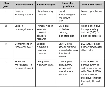
The biosafety infrastructure must be designed on the basis of risk assessment for handling specific pathogens. An agent that is assigned to Risk Group 2 may generally require Biosafety Level 2 facilities, equipment, practices and procedures for safe conduct of work. The desired biosafety levels are established on the basis of professional judgement based on risk assessment.
Guidelines on establishment of virology laboratory in developing countries
Table 2.1: Relation of risk groups to biosafety levels, practices and equipment (WHO Manual on Biosafety, 2004)
Risk
group Biosafety level Laboratory type
Laboratory
practices Safety equipment
1. Basic–in with Class II BSCs, double-ended autoclave (through the wall), filtered air
Diagnostic virology laboratories must be designed for Biosafety level 2 (BSL 2) or above and special attention should be paid to conditions that are known to pose biosafety problems. It is desirable that every country have at least one national laboratory that is equipped with Biosafety level 3 (BSL 3) facility especially because most of the viral pathogens that have emerged in the recent past have been agents that require BSL 3 facilities for handling specimens and cultures. However, if resources are limite d, a BSL 2 laboratory with negative pressure (BSL 2) is essential to handle most emerging pathogens.
Guidelines on establishment of virology laboratory in developing countries
Page 10
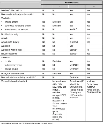
Table 2.2: Summary of biosafety level requirements
Biosafety level
1 2 3 4
Isolationa of laboratory No No Yes Yes
Room sealable for decontamination No No Yes Yes Ventilation:
• inward airflow
• controlled ventilating system
• H EPA-filtered air exhaust
No
• in laboratory room
• double-ended Biological safety cabinets No D esirable Yes Yes Personal safety monitoring capabilityd No No D esirable Yes
Viruses that can be handled herpes viruses (H SV, VZV,
a Environmental and functional isolation from general traffic b Dependent on location of exhaust
c Dependent on agent(s) used in the laboratory.
Guidelines on establishment of virology laboratory in developing countries
A typical biosafety level 3 laboratory
The laboratory is separated from general traffic flow and accessed through an anteroom (double door entry or basic laboratory – Biosafety Level 2) or an airlock. An autoclave is available within the facility for decontamination of wastes prior to disposal. A sink with hands-free operation is available. Inward directional airflow is established and all work with infectious materials is conducted within a biological safety cabinet.
In principle, unless the recommended biosafety level facilities are available the laboratory should not handle a particular virus. In situations where there is an outbreak of a viral illness and the required biosafety levels are not available, it is advisable to collect and refer the specimens to the nearest laboratory in the Region that has the required BSL laboratory. If specimens are collected from patients who are suspected to be suffering from viral infections caused by agents which need BSL 3 facilities (e.g. avian influenza) it is advisable that they be directly forwarded to the reference laboratory which has facilities for handling such agents.
Apart from the biosafety requirements mentioned above, the following are some of the essential features that need to be incorporated in the design of a virology laboratory.
Ø Adequate space must be provided for the safe conduct of laboratory work and for cleaning and maintenance. Designated cubicles, rooms or areas should be available to carry out different activities, e.g. office rooms, specimen collection cubicle, specimen reception and processing room, serology laboratory, cell culture cubicle, molecular diagnostic laboratory comprising three cubicles, cold room, dark room for fluorescent microscopy, common equipment room, media preparation room, washing and sterilization section, store room, toilets and lunch room.
Ø Each laboratory room should have space for housing a BSL 2 cabinet, one workbench, a sink, discard bins, wall cabinets to store consumables, a centrifuge, an incubator and a refrigerator.
Ø Ideally the entire laboratory should be air conditioned to maintain a dust-free environment and an ambient temperature of 22–25 °C. If this
Guidelines on establishment of virology laboratory in developing countries
Page 12
Ø Walls, ceilings and floors should be smooth, easy to clean, impermeable to liquids and resistant to chemicals and disinfectants normally used in the laboratory. Floors should be slip-resistant.
Ø Bench-tops should be impervious to water and resistant to disinfectants, acids, alkalis, organic solvents and moderate heat.
Ø Illumination should be adequate for all activities. Undesirable reflections and glare should be avoided.
Ø Laboratory furniture should be sturdy. Open spaces between and under benches, cabinets and equipment should be accessible to cleaning.
Ø Storage space must be adequate to hold supplies for immediate use and thus prevent clutter on bench tops and in aisles. Additional long-term storage space conveniently located within or outside the lab area should also be provided.
Ø All doors should have vision panels, appropriate fire ratings and preferably be self-closing.
Ø Facilities for storing outer garments, personal items as well as having tea and lunch and restrooms should be provided outside the laboratory working area.
Ø Hand-washing basins with running tap water should be provided in each laboratory room, preferably near the exit door. A dependable supply of good quality water is essential. There should be no cross-connections between sources of laboratory water supply and drinking water supplies.
Ø There should be reliable and adequate electricity supply and emergency lighting for safe exits. A stand-by generator is essential at least for some equipment such as incubators, biosafety cabinets, freezers etc.
Ø Safety systems should cover fire, electrical emergencies, emergency shower and eyewash facilities. These include:
Guidelines on establishment of virology laboratory in developing countries
H uman resources
A diagnostic virology laboratory should be adequately staffed. The minimum staff requirements for a diagnostic virology laboratory should include:
Ø A qualified virologi st possessing a postgraduate qualification in virology with three to five years experience in diagnostic virology who will be the chief of the laboratory. This officer will have overall responsibility for activities of the laboratory and will supervise all the staff working at the centre. In addition, this officer will be directly responsible for reporting results of the various diagnostic assays performed in the laboratory.
Ø Two junior microbiologists possessing a M aster’s degree in Medical M icrobiology with one to two years experience in diagnostic virology. These microbiologists will be responsible for the day-to-day functioning of the diagnostic laboratory such as supervision of technical staff who carry out the various diagnostic tests, stock management and procurement of lab supplies and diagnostic kits, quality control and quality assurance.
Ø Two laboratory technologists possessing a graduate degree in science with a diploma in Medical Laboratory Technology (one to be trained in cell culture and virus isolation methods and the other to be trained in serology). The technicians will be responsible for specimen processing, testing, laboratory safety, maintenance of laboratory records and media preparation.
Guidelines on establishment of virology laboratory in developing countries
Page 14
Equipment and supplies
The virology laboratory should be provided with adequate equipment which should take into account certain general principles, i.e. it should be:
Ø Designed to prevent or limit contact between the operator and the infectious material (e.g. biosafety cabinets, electronic pipetting aids, etc.)
Ø Constructed of materials that are impermeable to liquids, resistant to corrosion and meet structural requirements.
Ø Should be free of sharp edges, burrs and unguarded moving parts.
Ø Designed, constructed and installed to facilitate simple operation and provide for ease of maintenance, cleaning, decontamination and certification testing. Glassware and other breakable materials should be avoided wherever possible.
A list of essential and desirable equipment required for a virology laboratory is listed below. Details of equipment specifications and sources of laboratory equipment required for a virology laboratory can be obtained from a variety of sources and some of these are appended to this report as Annex 1.
Essential equipment
Ø Biosafety cabinets Class II – Three (one for handling cell cultures, one for handling stock viruses and one for processing clinical specimens)
Ø One incubator and two CO2 incubators (one for uninfected cell cultures and the other for infected cell cultures)
Ø 20 °C and –70 °C freezers
Ø Inverted light microscope
Ø Fluorescent microscope with photography attachments
Ø Filtration apparatus for preparation of tissue/cell culture media
Ø Refrigerate centrifuge
Ø water bath
Guidelines on establishment of virology laboratory in developing countries
Ø M agnetic stirrer
Ø Vortex mixer
Ø Electronic balance for weighing chemicals
Ø Elisa Reader and washer
Ø Micropipttes ( 100ul, 200 ul, 20 ul)
Ø Multi-channel pipettes – 8 and 12 channel pipettes (20-200 ul and 50-300 ul)
Ø Autoclave – Two ( one for decontamination and one for sterilization)
Ø Hot air oven for sterilizing glassware
Ø One personal computer with printer, photocopier, fax machine and telephone lines
Ø PCR machine ( conventional and real-time)
Ø Gel electrophoresis apparatus
Ø UV transilluminator
Ø Ice-making machine
Ø Liquid nitrogen containers
Ø W ater purification/distillation system, which provides high-grade water suitable for ti ssue culture work
Ø Glassware such as volumetric flasks, measuring cyli nders, pipettes (1 ml, 2 ml, 5 ml and 10 ml), conical flasks, reagent storage bottles (50 ml, 100 ml, 250 ml, 500 ml and 1000 ml)
Ø Electric brushing machine and automatic pipette washer
D esirable equipment
Ø Shaker water bath
Ø Rocking platform
Guidelines on establishment of virology laboratory in developing countries
Page 16
Reagents and supplies
A diagnostic virology laboratory requires a whole range of reagents and supplies. Most of the reagents and supplies listed below are generic and would suffice for the diagnosis of a variety of viral infections.
Ø Diagnostic kits as per requirements of the laboratory
Ø Tissue culture media
Ø Foetal bovine serum
Ø Fluorescent conjugates
Ø ELISA plates, antibodies and conjugates
Ø Analytical-grade fine chemicals for preparation of buffers
Ø Sterile tissue culture plastic ware (25 cm2 and 75cm2 flasks, 24 and 96 well plates, petridishes, tissue culture tubes centrifuge tubes and pipettes)
Ø V-bottom polystyrene microtitre plates for haemagglutination
Ø Serum storage cryovials and boxes
Ø Micropipette tips
Ø PCR tubes
Ø PCR reagents (Taq polymerase, reverse transcriptase, primers, probes and agarose)
Ø DNA and RNA extraction kit
It is critical to procure high-quality reagents and also confirm the same before use in routine diagnostic work.
Functions
Guidelines on establishment of virology laboratory in developing countries
(1) To provide technical advice to national authorities on the progress, needs and aspirations of the virology laboratory network in the country.
(2) To assist national authorities in planning, organization and supervision of the virology laboratory network.
(3) To undertake referral services for providing efficient virology laboratory services.
(4) To develop technical/training material for use in laboratories in the network to enhance their quality.
(5) To assess needs and impart training to staff of laboratories in provincial and country laboratories in quality testing for viral pathogens.
(6) To validate reagents and kits that may be used nationally.
(7) To validate new technologies that may become available and recommend their implementation in the country.
(8) To develop minimum standards for virology laboratories and assist laboratories in their implementation.
(9) To develop national database on the laboratory results.
(10) To organize external quality assessment schemes to periodically assess the quality of testing in networks and suggest remedial measures to those laboratories that show poor performance.
(11) To undertake research to improve the quality and cost-effectiveness of virology laboratory services in the country.
Chapter 3
Viral diagnostic tests
Virtually all diagnostic techniques used in virology are based on the following principles:
(1) Isolation and identification of viruses
(2) Detection of virus antigens
(3) Detection of viral nucleic acid
(4) Detection of virus-specific antibodies
The choice of the system used in the laboratory for confirmation or ruling out of a viral infection would primarily depend on the facilities and resources available and linkages with national and international laboratories. A broad outline of the suggested methods available for common viral infections is presented in Tables 3.1– 3.6. However, the choice of the methods to be used should be determined by the laboratory depending on the available infrastructure, resources and trained manpower.
Isolation and identification of viruses
The relative importance of viral isolation as a diagnostic method is rapidly diminishing: however, itstill remains necessary because it is the onlytechnique capable of providinga viable isolate that can be used for antiviral susceptibility testing. An additional advantageis that in contrastto most antigen andnucleic acid detection methods,viral culture allows detectionof multiple viruses, notall of which mayhave been suspected atthe time the culture was requested.
Guidelines on establishment of virology laboratory in developing countries
Page 20
of cell lines that are required for a specific specimen is determined by the information communicated from the ordering physician tothe laboratory and by knowledge of the range of viruses that can usually be isolated from a given specimen type.
Growth of viruses in cell culture is usually detected by visualizing morphological changes in the cells, known as cytopathiceffect (CPE). Cellcultures are typically viewedmicroscopically to detect CPEevery one to two days forthe first week ofincubation. W hennecessary, confirmation can beachieved by scraping the infected cells from the walls of the tubeor vessel in whichthey are growing and preparing a fluorescent antibodystain with use ofmonoclonal antibodies specific for the virus, or a neutralization test using specific antisera.
The shell vial culture method, first developed for cytomegalovirus (CMV), dramatically decreases thetime required for detectionof viruses in cellculture. The method involvescentrifugation of the specimenonto the cell culture monolayer grown on a cover slip, which is incubated for one or two days, followed by fluorescent antibody staining ofthe cell culture, regardlessof whether CPE isvisible or not. In addition todetection of CMV, shell vial cultures have alsobeen used to speed the detection of herpes simplex virus (HSV), varicella zoster virus(VZV), respiratory viruses, andthe enteroviruses.
A recent modification of the traditional cell culture involvesthe use of geneticallyengineered cell lines such as the L20B, which is used for the isolation of polio viruses, and Vero cells for isolation of measles and rubella viruses. Another example of genetically engineered cell line uses genes that are transfected into indicator celllines so thatthe cell line responds to a specificviral protein present in the specimen. Activation of the promoter triggers areporter enzyme, such as ß
-galactosidase, which acts ona substrate to indicatethe presence of thevirus being sought. Thisapproach has been mostwidely used for HSVand HIV.
D etection of virus antigens
Guidelines on establishment of virology laboratory in developing countries
multiple different antibodieslabeled with different fluorescent labels has enabled rapid detection of a variety of viruses.
Antigen detection methodsare particularly useful forviruses that grow slowly or are labile, makingrecovery in culture difficult.The most important targetshave been detection of viruses in respiratory specimens; cutaneous specimens; stool specimens; and cereberospinal fluid (CSF). Viruses such asthe enteroviruses and the rhinoviruses that have extensive antigenic heterogeneity and cross-reacting antigens are not suitable for antigen-detection techniques. The advantages of antigen-detection techniques are rapidity (resultscan be available withinhours of receipt ofthe specimen in thelaboratory) and lack ofrequirement for viral viability in the specimen, allowing greater flexibility in the handling and transport of specimens.
D etection of viral nucleic acid
The development of PCR analysis in 1985 made possible the diagnosisof viral infection through sensitive detection of specific viral nucleic acids. Anyvirus can potentially bedetected using this technology. By inclusionof a step employingthe enzyme reverse transcriptase(RT), PCR analysis canbe adapted to detectviral RNA, especially for rapid diagnosis during outbreaks of diseases such as avian influenza and Nipah virus infections.
Viral load assays for HIV and hepatitis C virus (HCV) are examples of quantitative nucleic acid detection techniques. Multiplex techniques pe rmit the simultaneous detection of morethan one virus oreven of a virusand a different classof pathogen. For example,a multiplex PCR analysisthat detects the DNAof Epstein-Barr virus (EBV)and Toxoplasma gondii has beenused for the diagnosisof mass lesions inthe brains of patientswith AIDS.
Guidelines on establishment of virology laboratory in developing countries
Page 22
Many laboratorieshave developed their own "home-brew" PCR assays. The quality and performance of these assays vary widely. Inter-laboratory certification programmes to helpstandardize these assays areonly beginning to becomeavailable.
D etection of virus specific antibodies
The diagnosis of viralinfections by detection ofmore than a four-fold increase in specific antiviral antibodies isa traditional method whoseclinical utility is limitedby the need for comparison of acute and convalescent antibody titres. However, detection of virus-specific IgMantibodies allows a diagnosisto be made froma single specimen. Virusesfor which detection of virus-specific IgM antibodies are useful include flaviviruses (JEV, dengue, West Nile), hepatitisA virus; hepatitis Bvirus (IgM antibodies tothe hepatitis B coreantigen), measles,rubella and mumps viruses.
Serological methods are also useful for diagnosis of certain chronic infections such as with HIVor HCV, in whichthe presence of anyantiviral antibodies is always (HIV) or usually (HCV)indicative of current infection.Finally, serology is uniquely useful for defining specific antiviral immunity. Viruses for which definition of immune status by serology isuseful include arboviruses (Japanese encephalitis, dengue, W est Nile and chikungunya viruses) VZV, CMV,EBV, HSV, measles and rubella viruses, parvovirus B19, hepatitis A (total antibodies), and hepatitis B (antibodiesto the hepatitis Bsurface antigen) and rabies (presence of neutralizing antibodies after vaccination).
The successful exploitation of diagnostic virology laboratory in clinical practice depends on the adherence to the following principles; which are important to both the treating clinician as well as the clinical virologist.
Ø The clinician should be aware that three components that can contribute to any diagnostic result are the information provided by the physici an, the type of assay used and the competence of laboratory personnel. It is vital that these three must communicate and perform well to ensure that proper results are obtained.
Ø False positive and false negative results are a possibility with most assays that are currently in use , and therefore it should be noted that no assay is perfect; hence it may be dangerous to base all patient management procedures on a single result.
Guidelines on establishment of virology laboratory in developing countries
laboratory. A sample of a virology request form is appended as Annex 2. Collection of specimens, storage and transport require constant dialogue between clinicians and diagnostic laboratory personnel in order to obtain rapid and accurate results.
Diagnostic assays that are currently used for several syndromes are summarized in tables 3.1 to 3.5.
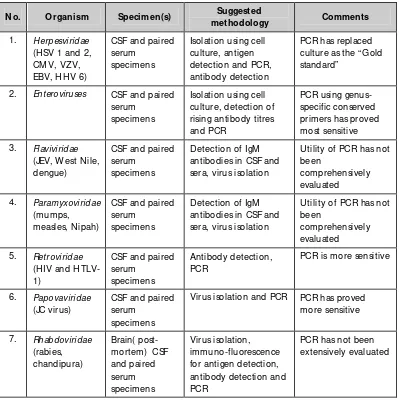
Table 3.1: Diagnostic assays for central nervous system viral infections
N o. O rganism Specimen(s) Suggested antibodies in CSF and sera, virus isolation
Utility of PCR has not been antibodies in CSF and sera, virus isolation
Utility of PCR has not been
PCR is more sensitive
6. Papovaviridae (JC virus)
CSF and paired serum
specimens
Guidelines on establishment of virology laboratory in developing countries
Page 24
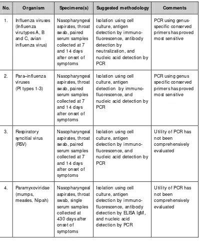
Table 3.2: Diagnostic assays for respiratory viruses
N o. O rganism Specimens(s) Suggested methodology Comments
1. Influenza viruses (Influenza nucleic acid detection by PCR nucleic acid detection by PCR nucleic acid detection by PCR
Utility of PCR has not been detection by ELISA IgM , and nucleic acid detection by PCR
Utility of PCR has not been
Guidelines on establishment of virology laboratory in developing countries
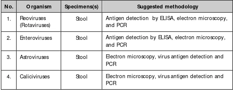
Table 3.3: Diagnostic assays for gastroenteritis viruses
N o. O rganism Specimens(s) Suggested methodology
1. Reoviruses (Rotaviruses)
Stool Antigen detection by ELISA, electron microscopy, and PCR
2. Enteroviruses Stool Antigen detection by ELISA, electron microscopy, and PCR
3. Astroviruses Stool Electron microscopy, virus antigen detection and PCR
4. Caliciviruses Stool Electron microscopy, virus antigen detection and PCR
Table 3.4: Diagnostic assays for hepatitis viruses
N o. O rganism Specimen Suggested methodology Comments
1. H epatitis A virus Serum IgM /IgG antibody detection by ELISA and nucleic acid detection by PCR
IgM antibody detection is most sensitive
2. Hepatitis B Virus Serum H BsAg antigen detection by ELISA and nucleic acid detection by PCR
H bsAg detection is most sensitive
3. Hepatitis C virus Serum Antibody detection by ELISA and nucleic acid detection by PCR
Detection antibodies most useful
4. Hepatitis D virus Serum Delta antigen by ELISA and Nucleic acid detection by PCR
PCR is the test of choice
5. Hepatitis E virus Serum IgM antibody detection by ELISA and nucleic acid detection by PCR
IgM antibody detection is very useful
6. O ther hepatitis viruse: hepatitis G virus (H GV) & transfusion
Serum N ucleic acid detection by PCR
Guidelines on establishment of virology laboratory in developing countries
Page 26
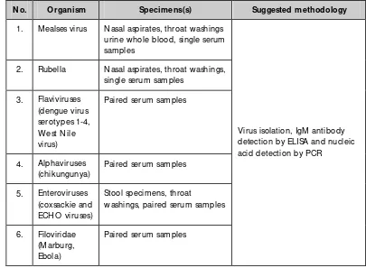
Table 3.5: Diagnostic assays for viruses that cause fever with rash or haemorrhage
N o. O rganism Specimens(s) Suggested methodology
1. M ealses virus Nasal aspirates, throat washings urine whole blood, single serum samples
2. Rubella Nasal aspirates, throat washings, single serum samples
3. Flaviviruses (dengue virus serotypes 1-4, W est Nile virus)
Paired serum samples
4. Alphaviruses (chikungunya)
Paired serum samples
5. Enteroviruses (coxsackie and ECH O viruses)
Stool specimens, throat
washings, paired serum samples
6. Filoviridae (M arburg, Ebola)
Paired serum samples
Chapter 4
Specimens management
Laboratory diagnosis of viral infections requires an understanding of the pathogenesis of the suspected agent and knowledge regarding the type and duration of illness, the stage of infection, age of the patient, immunization status, and whether outbreaks of similar illness have been recorded in the community. This information facilitates selection of the appropriate specimen, careful collection to optimize recovery of the agent, and transport of specimens in a manner that maintains viability and minimizes overgrowth with contaminating organisms.
Detailed guidelines for collection, transport and processing of specimens are available for a variety of viral diseases. However, every laboratory has to prepare its own standard operating procedures (SOPs) depending on the local conditions.
Selection of specimens
For best correlation with a particular disease, the specimen should reflect the target organ whenever possible, which is usually based on the clinical symptoms. A brief summary of the type of specimens to be collected in common viral infections has been presented in Tables 3.1 to 3.6.
M aterials required
Virus culture
Guidelines on establishment of virology laboratory in developing countries
Page 28
Ø Sterile, leak-proof, externally threaded screw-cap containers including urine cups, disposable centrifuge tubes (15 ml and 50 ml), and smaller tubes (e.g., 4-ml Nunc vials, 13x100 tubes), suitable for holding 1 ml to 2 ml of VTM.
Ø Sterile cotton swabs with plastic or alumi nium shafts; small-tip flexible (fine-shafted aluminium) swabs are used for certain samples such as urethral swabs. Calcium alginate swabs and wooden-shafted swabs are not recommended.
Ø Tuberculin syringe with 26-gauge or 27-gauge needle for aspirating vesi cular fluid.
Ø Blood collection tubes containing anticoagulant (ACD).
Specimen collection
For virus isolation, specimens should be collected as soon after onset of symptoms as possible. The chance of viral recovery is best during the first three days after onset and is greatly reduced beyond 5 days with many viruses.
For serological tests, two blood specimens need to be collected–one during the acute phase of the illness and the second sample seven to ten days after the first specimen. In some diseases only one sample is needed, which should not be collected earlier than four to seven days or later than one month after onset of disease. A minimum of 5 ml of clotted blood collected into sterile containers needs to be sent to the laboratory as early as possible; if not, serum should be separated aseptically within four to six hrs of collection and transported in screw -capped vials to the laboratory under cold conditions.
As a general rule, specimens collected for PCR should be obtained during the early part of the illness. Cotton swabs with wooden sticks should not be used for collection specimens that are processed for PCR. Similarly, heparin should not be used as anticoagulant for the PCR test on blood as it interferes with Taq polymerase enzyme. Instead, EDTA may be used as an anti coagulant.
O ptimal times for collection of specimens
Guidelines on establishment of virology laboratory in developing countries
into a tube containing a small volume of VTM, and scrapings and small pieces of tissue into a tube containing a small volume of VTM or saline. Place fluid and bulk specimens (e.g. tissue) into a sterile leak- proof container; add a volume of VTM sufficient to prevent drying of tissue. Collect up to 1 ml – 5 ml of serum or CSF.
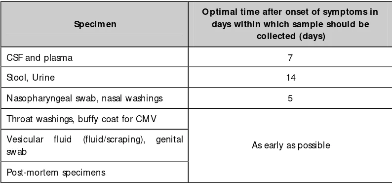
Table 4.1: O ptimal time for collection of specimens
Specimen
O ptimal time after onset of symptoms in days within which sample should be
collected (days)
CSF and plasma 7
Stool, Urine 14
Nasopharyngeal swab, nasal washings 5
Throat washings, buffy coat for CM V Vesicular fluid (fluid/scraping), genital swab
Post-mortem specimens
As early as possible
The health worker who is collecting the material should be trained to obtain a complete patient history, including the date of onset of symptoms, clinical findings, recent exposure history, animal or arthropod contacts or bites, recent travel to areas of endemi c infections, and recent vaccination.
Specimen transport
Place the tightly capped specimen container and the appropriate laboratory requisition form into separate compartments of a plastic specimen transport bag (Annex 3). All specimens should be delivered to the laboratory as soon after collection as possible, since a loss of infectivity occurs over time; samples containing labile viruses at low titres are those most likely to show loss of infectivity with delayed transport.
If immediate delivery is not possible, refrigerate specimens (2 °C to 8 °C), or
Guidelines on establishment of virology laboratory in developing countries
Page 30
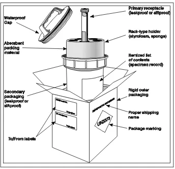
Figure 4.1: Packing of specimens for transport to laboratories
Biosafety
Safety in a virology laboratory is part and parcel of laboratory quality control to obviate laboratory-acquired infections. Each laboratory should have the W HO Laboratory Biosafety Manual/Document WHO/CDS/CSR/LYO/2004.11, available at: http://www.who.int/csr/resources/publications/biosafety/en/Biosafety7.pdf ).
Guidelines on establishment of virology laboratory in developing countries
Where pathogens that cause severe human disease are known to infect by the airborne route, primary containment using microbiological safety cabinets and the provision of secondary containment using appropriate ventilation have been recommended. For work with blood-borne organisms such as HIV and HBV, the use of "sharps" should be avoided. Where available, vaccination is recommended for people working with known organisms. Vaccination is essential for work with HBV, polio, rabies, Japanese encephalitis, and Kyasanur Forest disease virus. Vaccination is also recommended for measles, mumps and rubella.
Personal protective equipment
Over the years, three kinds of protective clothing came into general use: gowns, front-buttoned white coats, and specially designed overalls, which afforded protection up to the neck. The latter is considered the best protective clothing available since they are easy to remove, aesthetically pleasing, and afford much better protection than front-buttoned white coats. It is advisable to wear disposable plastic aprons over ordinary protective clothing when working with blood that might be infected. Gloves should be worn when handling the specimens and all hazardous agents/materials.
Amenities
Hand basins should be provided in each laboratory, lockers for staff clothing should be available near to (but not in) laboratory rooms, and rooms for eating and drinking should be provided close to laboratory rooms.
H ealth of staff
Guidelines on establishment of virology laboratory in developing countries
Page 32
M edical surveillance
A system that records and follows up all cases of illness in the laboratory staff to ascertain the source of infection and initiate any follow-up action should be developed.
Accidents
There should be a rule in all microbiological laboratories that all accidents causing personal injury with exposure to infectious agents or not should be reported to the supervisor or safety officer. Records should be kept of all staff sicknesses, injuries and accidents, and of X-rays and immunization. Accident records are particularly important for legal reasons.
It is highly desirable to have a qualified first-aid provider in the laboratory, but he/she should receive additional training in dealing with exposure to and ingestion of infectious, toxic and corrosive chemicals. All staff should know whom to contact if an accident occurs. The standard first aid kit for minor injuries should be in a prominent position and well sign-posted. Care should be taken with eye irrigation since the bottles of "sterile" water are frequently contaminated. Standard operating procedures for management of post-exposure prophylaxis should be in place.
M inimizing equipment and technique-related hazards
Pipettes
M outh pipetting is banned at all times. Pipetting devices should be inspected routinely for leakage and replaced as necessary. W hen discharging infectious material from a pipette, it is important to minimize the formation of aerosols. Contaminated pipettes should always be discarded into disinfectant fluid that is prepared daily and placed in a flat, covered metal tray. Broken and chipped pipettes should be promptly discarded into sharps containers.
H ypodermic needles and syringes
Guidelines on establishment of virology laboratory in developing countries
caused by these instruments. M any inoculations result during experiments with animals, others occur during transfer of infected material by syringes. M any needlestick accidents occur when the needle is being recapped. It is important that used needles are promptly disposed of properly into harden "sharps containers" which are then autoclaved before disposal. These sharps containers should be available wherever needed.
Centrifugation
Infectious material may be dispersed by a centrifuge either through broken tubes or other means, such as through the threads of the tubes and caps. Sealed centrifuge buckets should be used when centrifuging infectious material. Care should be taken to ensure that the centrifuge tubes are not cracked or flawed. Tubes should not be more than three-quarters full, especially if angle centrifuges are used. Tubes should be capped and they and the buckets should be balanced carefully to avoid vibration, which may lead to breakage.
Pouring infectious material
Pouring of the supernatant after centrifuging cultures or viruses should be avoided. All infectious material should be transferred from one container to another using mechanical/electrical pipetting devices.
M anagement of laboratory waste
Laboratory waste should ideally be separated into colour-coded containers. The recommended colours are listed below; however, the laboratories should follow and adhere to the national guidelines:
Ø Yellow–for incineration
Ø Light blue or transparent with blue inscription–for autoclaving (but may be incinerated subsequently)
Ø Black–normal household waste: local authority refuse collection
Guidelines on establishment of virology laboratory in developing countries
Page 34
There are three practical methods of treating contaminated laboratory waste:
Ø Sterilization by autoclaving
Ø Chemical disinfection
Ø Incineration
The first two are laboratory processes; incineration involves the transport of material off-site. Sterilization and disinfection are not synonymous: sterilization implies the killing of all microorganisms; disinfection kills most microorganisms but depends greatly on the chemical used.
Autoclaves
Autoclaving involves the timed exposure of materials to steam above atmospheric pressure and hence at temperatures above 100 oC. Autoclaves operate at high pressures and temperatures and their manufacture, installation, and use are regulated. Sterilization is achieved by exposure of material to 121 oC for 15–20 minutes. Chemical and biological indicators are also widely used, the most well-known of which is the "autoclave" tape. It was customary to place material for autoclaving in wire baskets and cylindrical buckets. Autoclavable plastic bags are now in general use but they must be permeable to steam. The mouths of plastic bags should not be closed before autoclaving since this will reduce steam penetration.
Chemical disinfection
Guidelines on establishment of virology laboratory in developing countries
D iscard jars
Discard jars should be robust and autoclavable. Glass jars should not be used because they are easily broken. Polypropylene beakers or jars are probably the most serviceable items. They are left open on the bench but are closed at the end of the working session. It is important that discard jars are filled frequently with known active dilutions of disinfectant, not overloaded with protein or floating articles. Laboratory supervisors should ensure that inappropriate articles are not placed in discard jars (e.g., a 25 ml pipette has no place in a small 1 litre jar). Funnels may be used to prevent splashing and aerosol dispersal. Discard jars should be washed with hot water before reuse. It is better still to autoclave the jar before washing. The contents of the discard jars should then be autoclaved and/or incinerated.
Reusable pipettes
After use, reusable pipettes should be completely immersed in disinfectant placed in metal/plastic trays that have lids so that no air remains within the pipette. They should remain in disinfectant for at least 18 hours before washing and/or autoclaving.
The key to effective disposal of wastes is segregation and decontamination at the point of generation. Generators of wastes are responsible for ensuring that wastes are placed in the correct receptacle. In the clinical virology laboratory this is crucial, because the generator may often be the only person who fully appreciates the risks the waste poses. The generator has a legal duty to take reasonable care for the health and safety of persons at the place of work who may be affected by acts or omissions.
W astes should be segregated on the basis of the major or primary hazard they pose. Generators need to make this assessment and decide if further segregation is necessary because of secondary hazards associated with the waste.
Infectious waste
Guidelines on establishment of virology laboratory in developing countries
Page 36
Routine contaminated wastes
Generators must also take care to segregate these wastes from general wastes because they, too, may need to be decontaminated prior to removal from the area in which they are generated. Alternative means of decontamination to autoclaving or incineration may be used, provided they are effective.
Sharps
All sharps must be segregated from other wastes and placed in dedicated and labelled single-use rigid containers immediately after use. Sharps containers must not be compacted or emptied into ordinary waste streams.
Labelling of wastes
Standard labels to identify major waste categories are available commercially. All wastes must be identified with an appropriate type of label which will remain attached to the waste. The label must state the nature of waste material, the department, date, and the name of the person responsible for the waste.
Guidelines on establishment of virology laboratory in developing countries
Flow of clinical specimens in a virology laboratory
Receive clinical specimens
Assess the quality, quantity, condition on receipt, adequacy of clinical information provided and suitability for virological testing
Accept specimen Reject specimen
Process specimen, aliquot and store at
–20 °C or –70 °C
Test specimen using appropriate laboratory algorithms
Subject results of testing to validation procedures (Internal quality control
procedures)
Release results to clinician/referral agency
Archive results in appropriate formats (registers, excel sheets etc.)
Preserve original specimen in aliquots
at –20 °C or –70 °C and send to
reference laboratory if and when required
Communicate decision to the referring clinician/agency giving precise reasons
and request for a second samp le
Document action taken for rejecting sample in appropriate laboratory
Chapter 5
Q uality systems
Quality systems in virology laboratories are of paramount importance. A system should be comprehensive and must cover all aspects from the decision to collect the specimen to interpretation of the results. Errors at any stage in the investigation can affect the outcome of the result. Further, any break in the chain can result in the generation of a faulty report. Quality does not just happen on its own.
Systematic efforts through organizational structure and efficient utilization of resources are needed to implement all the steps that will assure generation of quality reports by the laboratory. Accordingly, a quality system is a part of overall quality management that aims at consistency, reproducibility, traceability and efficaciousness of the laboratory services. A quality system therefore comprises of five essential elements:
Ø Organizational management structure
Ø Quality standards
Ø Documentation
Ø Training
Ø Monitoring and evaluation
Guidelines on establishment of virology laboratory in developing countries
Page 40
fashion while the data is still clinically relevant. A clinical virology laboratory should be designed in a manner so that biohazard risks to the laboratory personnel and the general public are minimized and that cultures are protected from environmental contamination.
D ocumentation
Routine procedures must be described in written Standard Operating Procedures (SOPs). They must be reviewed regularly and modified, if necessary. The modified versions should be signed and dated by the laboratory director. The recent most version of SOPs should be available directly at the work place. The old versions should be achieved in the laboratory and should be archived if required.
The records must be stored for long periods of time but be available for prompt retrieval, safe archiving of all records must be ensured. Archiving of the source documents and other essential documents must be such that data are kept in an integer state and can neither be lost nor altered to achieve this goal.
Records of usage, maintenance and calibration should be kept in the laboratory and should be routinely monitored. Reports of the tests should be released only after proper scrutiny and documentation of the scrutiny with the signature and date by the lab-supervisor.
Standard operating procedure (SO P
)SOP is one of the most important documents in a diagnostic laboratory. Apart form providing the complete details of how exactly a test or a procedure is carried out in a laboratory, the SOP also provides information on specimen collection, laboratory safety instructions, purpose and limitations of the procedure, turnaround times, interpretation of results, and above all, the line of authority.
Guidelines on establishment of virology laboratory in developing countries
Some of the areas where quality control checks are critical are briefly described below:
Specimen transport
Specimens for viral isolation are often held for long periods of time before they reach the laboratory. Enveloped viruses such as RSV and CMV are extremely labile at room- temperature and freeze-thaw cycles, whereas non-enveloped viruses such as enteroviruses tolerate these conditions well. As a general rule, viral specimens held for short periods should be refrigerated, while those for longer periods may be frozen at –20 oC or –70 oC.
Transport media
The composition and type of viral transport media can affect viral isolation rates. In general, the media should be a balanced isotonic solution at physiological pH. It should contain a substance that will stabilize the virus such as gelatin, fetal calf serum or bovine serum albumin, and antibiotics against bacteria and fungi. The swab should be made of a material that is non-toxic to viruses, such as dacron or rayon.
Smears
Smears are becoming increasingly popular because of the rapidity of staining techniques. The smear should contain a reasonable number of cells, be of a reasonable size, and not mixed with blood or pus, as the latter may lead to nonspecific staining.
Specimens for serology
Guidelines on establishment of virology laboratory in developing countries
Page 42
Tissue culture and media
Tissue/cell culture remains the mainstay of non-serological viral diagnosis. Therefore, adequate quality control for commercially purchased or for in-house preparation of tissue culture cells is of great importance. Within a given cell line, there may be significant variations in sensitivity to virus isolation which may depend on the particular cell line or clone and the passage number. Information on a particular cell line should be recorded accurately including the source, type, passage number, confluency and cell condition. The loss of a cell line routinely passaged for use can lead to a severe disruption of workflow, and therefore provisions must be made for back-up cells in the event of contamination or laboratory accident. These back-up systems include:
Ø Freezing and storage of low passage cells at –70 oC liquid nitrogen tanks.
Ø Use of paired stock flasks. W hen the flasks reach confluency, only one is passaged, while the second flask is held as a back-up until the new flask displays good growth.
Ø Carrying of a parallel set of stock flasks using a separate set of tissue culture reagents and glassware.
Cells purchased from a commercial company should be certified to be free from mycoplasma, fungal, and bacterial contamination as well as adventitious viral agents such as SV40. Tissue culture lines carried in-house should be subjected to:
Ø Daily checks for growth rate and contamination.
Ø Mycoplasma contamination monitored monthly by Hoechst stain or others as available.
Ø The sensitivity to viral isolation monitored by periodic TCID50 experiments with stocks of reference virus.
Guidelines on establishment of virology laboratory in developing countries
M edia
Following filter sterilization, aliquots of the media should be taken and checked for bacteriological or fungal contamination. These samples should be examined daily for five days and should be free from contamination. Aliquots of all other medium components such as fetal calf serum and L-glutamine should also be checked. New lots of medium and fetal calf serum that have passed the sterility check should be monitored for their ability to support cell growth.
Reagents and kits
Reagents and kits should be ordered from reputable manufacturers and dealers with reliable transportation systems. Upon receipt, the reagents should be checked for obvious breakage or contamination. The quantity, source, lot number and date of receipt should be entered in a logbook and the reagents stored according to the manufacturer’s storage specifications. When new lots of any reagent are opened, the date should be noted on the container. Caution must be exercised in the case of kits as different components of a kit may require different storage conditions and have different expiration dates.
Instruments
Laboratory instruments should be subjected to routine preventive maintenance and checked and calibrated on a regular basis. Some of these checks can be performed by laboratory staff and entered into a logbook. The following are some recommendations for routine laboratory maintenance and performance checks on instruments:
Ø Incubators: daily temperature, CO2 and humidity checks. Weekly decontamination of interior.
Ø Safety cabinets: daily air pressure check and cleaning of UV lamp. W ork surface should be decontaminated after each use. Annual checks for air velocity and filter integrity and paraldehyde decontamination, as applicable.
Guidelines on establishment of virology laboratory in developing countries
Page 44
Ø Refrigerators and freezers: daily temperature check; annual check of compressor and refrigerant levels.
Ø W ater baths: daily temperature checks, weekly decontamination.
Ø Refrigerate Centrifuges: weekly decontamination, annual inspection of motor, speed calibration and drive system.
Ø Autoclaves: daily temperature check and monthly spore strip testing.
Ø pH meters: single reference buffer check before each use, multiple point check monthly.
Ø Pipetting devices: gravimetric volume check monthly; annual overhaul.
Assessment of Q uality System
Guidelines on establishment of virology laboratory in developing countries
Further reading
(1) Isenberg HD, editor-in-chief. Clinical Microbiology Procedures Handbook. Second Edition. ASM Press; 2004
(2) Quality Control in a Virology Laboratory (http://virology-online.com/general/ QualityControl.htm)
(3) Safety in a Clinical Virology Laboratory (http://virology-online.com/general/ Safety.htm)
(4) Smith T, editor. Laboratory Diagnosis of Viral Infections. Third edition. Informa HealthCare; 1999
(5) Storch GA. Diagnostic Virology. Clinical Infectious Diseases 2000;31:739-51
(6) Specter SC, Hodinka RL, and Young SA. Clinical Virology Manual. Third edition. ASM Press; 2000
(7) Tibbets MW, Gomez R, Kannangai R and Sridharan G. Total quality management in clinical virology laboratories. Indian Journal of Medical Microbiology. 2006;24:258-62
(8) U.S. Department of Health and Human Services, Centers for Disease Control and Prevention, and National Institutes of Health. Biosafety in M icrobiological and Biomedi cal Laboratories (BMBL) Fifth Edition. U. S. Government Printing Office, W ashington; 2007 (http://www.cdc.gov/OD/OHS/biosfty/bmbl5/ BMBL_5th_Edition.pdf)