Eyes are among the most readily accessible organs in terms of location in the body. The physiological constraints of the eye render this organ impervious to foreign sub-stances, thus presenting a constant challenge to the formulator to circumvent the protec-tive barriers of the eye without causing permanent tissue damage (1). All ocular thera-peutics have been mostly administered to the eye as simple aqueous eye drops. About 90 % of the dose applied topically from such solutions is lost due to pre-corneal losses (lacrimation and drainage), which leads to poor availability and frequent dosing is re-quired for the instillation to achieve an adequate level and therapeutic effect (2). The standard treatment of severe bacterial keratitis requires frequent administration of fluo-roquinolone eye drops (3). However, this regimen is not only disruptive to the patient and usually necessitates hospitalization, but it has also been associated with toxicity to the corneal epithelium (4, 5). To overcome these limitations, ocular inserts seem promis-ing by prolongpromis-ing the contact time with improved efficiency of the therapy and patient compliance. Ocular inserts offer many advantages over conventional dosage forms, like
Design and evaluation of moxifloxacin
hydrochloride ocular inserts
PRAVIN K. PAWAR1*
RAJESH KATARA2
DIPAK K. MAJUMDAR2
1Chitkara College of Pharmacy, Chitkara University, Chandigarh-Patiala National Highway, Rajpura, Rajpura-140401 Patiala, Punjab, India
2Department of Pharmaceutics, Delhi Institute of Pharmaceutical Sciences and Research (formerly College of Pharmacy) University of Delhi, Pushp Vihar Sector III, New Delhi-110017, India
Accepted December 16, 2011
The objective of the present investigation was to prepare and evaluate ocular inserts of moxifloxacin. An ocular insert was made from an aqueous dispersion of moxiflo-xacin, sodium alginate, polyvinyl alcohol, and dibutyl phthalate by the film casting method. The ocular insert (5.5 mm diameter) was cross-linked by CaCl2and was coated with Eudragit S-100, RL-100, RS-100, E-100 or L-100. Thein vitrodrug drainage/permeation studies were carried out using an all-glass modified Franz diffusion cell. The drug concentration and mucoadhesion time of the ocular insert were found satisfactory. Cross-linking and coating with polymers extended the drainage from inserts. The cross-linked ocular insert coated with Eud-ragit RL-100 showed maximum drug permeation com-pared to other formulations.
Keywords: moxifloxacin, ocular insert, goat cornea
increased ocular residence, possibility of releasing drugs at a slow and constant rate, ac-curate dosing, and avoidance of toxicity due to preservative and increased shelf life (6). Ocular inserts of ciprofloxacin hydrochloride (7) and ofloxacin (8) have been designed to improve ocular availability.
Moxifloxacin,[1-cyclopropyl-6-fluoro-1,4-dihydro-8-methoxy-7-[(4aS,7aS )-octahydro-6H-pyrrolol (3,4b) pyridin-6-yl]-4-oxo-3-quinoline carboxylic acid], is a fourth generation fluoroquinolone with a methoxy group at the C-8 position and a bulky C-7 side chain (9). Moxifloxacin has increased activity againstS. aureuscompared to second and third generation fluoroquinolones. Bactericidal activity of moxifloxacin is mediated by the hibition of DNA gyrase (topoisomerase II) and topoisomerase IV, essential enzymes in-volved in bacterial DNA replication, transcription, repair and recombination (10). Effects of the formulation factor onin vitrocorneal permeation of moxifloxacin from aqueous drops have been investigated and the results showed that moxifloxacin 0.5 % (m/V) oph-thalmic solution (pH 7.2) containing benzalkonium chloride (0.01 %) (BAK) and EDTA (0.01 %) provides maximumin vitroocular availability through all excised mammalian corneas (11). Thein vitropermeation profile of moxifloxacin oil drops has been reported (12). Oil drops containing castor oil with benzyl alcohol provided maximumin vitro per-meation through all the corneas. Aqueous and oily drops of moxifloxacin showed better permeation characteristics but these formulations require frequent instillation to achieve therapeutic concentration due to shorter precorneal residence time. Taking the above in-formation in account, the purpose of the current study was to formulate ocular inserts of moxifloxacin that would be capable of prolonging the contact time, thereby potentially enhancing intra-corneal delivery of ophthalmic medicament.
EXPERIMENTAL
Materials
Moxifloxacin hydrochloride (99.97 % purity on anhydrous basis) was obtained from Ranbaxy Laboratories (India) as a gift. Sodium alginate, Eudragit S-100 (anionic crylic acid-methyl methacrylate copolymer), Eudragit RL-100 (acrylic acid and metha-crylic acid ester copolymer containing 10 % trimethylammonium methacrylate chloride), Eudragit RS-100 (acrylic acid methacrylic acid ester copolymer containing 5 % trimethyl-ammonium methacrylate chloride), Eudragit E-100 (cationic polymer based on dime-thylaminoethyl methacrylate and other neutral methacrylic acid esters), Eudragit L-100 (anionic copolymer based on methacrylic acid and methyl methacrylate) were obtained from Jubilant Organosys (India) as gift samples. Polyvinyl alcohol (PA) and dibutyl phthalate were obtained from the Central Drug House (India) and S.D. Fine chemicals Ltd (India), respectively. All other chemicals were of analytical grade. Fresh goat eyeball was obtained from a butcher’s shop. The method of cornea dissection and the apparatus used in permeation studies were the same as described previously (13).
Preparation of ocular inserts
The moxifloxacin control ocular insert (MIF1) was made from an aqueous disper-sion (40 mL) of moxifloxacin hydrochloride (400 mg), sodium alginate (0.25 g), polyvi-nyl alcohol (PVA) (1.5 g), and dibutyl phthalate (0.3 mL) by the film casting method. Oc-ular insert (5.5 mm diameter) was cross-linked (MIF2) by CaCl2(0.2 %,m/V) by dipping and drying 5 times. Similarly, the cross-linked insert was coated with different grades of Eudragit polymer 0.2 % (m/V) in isopropyl alcohol and acetone by the dip and dry me-thod (Table I). The polymers used were Eudragit S-100 (MIF3), Eudragit RL-100 (MIF4), Eudragit RS-100 (MIF5), Eudragit E-100 (MIF6) and Eudragit L-100 (MIF7). The mass gain of the ocular insert after coating with polymer was found to be in the range 23.4–29.6 %.
Physicochemical evaluation of ocular inserts
Thickness uniformity.– Insert thickness (5.5 mm diameter) was measured at five
dif-ferent points using a Micrometer screw gauge (Mitutoyo Co., Japan) and the mean insert thickness was noted (n= 3).
Drug content uniformity.–Three inserts were taken out of the film and drug
concen-tration determined. The uncoated ocular insert was dissolved in 10 mL of 0.1 mol L–1
HCl while the coated insert was dissolved in 10 mL of acetone. The volume was made up to 100 mL with distilled water and the solution was filtered. The drug in the filtrate was analyzed by measuring absorbance at 291 nm in a spectrophotometer 1601 (Shimad-zu, Japan). The experiment was done in triplicate.
Mucoadhesion time
The mucoadhesion time was determined (in triplicate) after application of coated or uncoated ocular inserts (5.5 mm diameter, 0.40–0.45 mm thickness) on a freshly cut goat eyelid. The eyelid was fixed on the bottom of a beaker with cyanoacrylate glue. Ocular insert (coated or uncoated) was attached to the mucosal surface of the eyelid by
apply-Table I. Compositions of moxifloxacin insert formulations for ocular delivery
Formulation Polymer composition (18+12+70 %,m/m) Plasticizer (dibutyl phthalate, %,V/V) Cross-linking agent CaCl2(%,m/V) Coating material (0.2 %,m/V)
MIF1 MOX + SA + PVA 0.12 – –
MIF2 MOX + SA + PVA 0.12 0.2 –
MIF3 MOX + SA + PVA 0.12 0.2 Eudragit S-100
MIF4 MOX + SA + PVA 0.12 0.2 Eudragit RL-100
MIF5 MOX + SA + PVA 0.12 0.2 Eudragit RS-100
MIF6 MOX + SA + PVA 0.12 0.2 Eudragit E-100
MIF7 MOX + SA + PVA 0.12 0.2 Eudragit L-100
ing a light force with a fingertip for 20 s. The beaker was filled with 800 mL of bicarbon-ate Ringer solution pH 7.4 and stirred at a rbicarbon-ate of 150 rpm at room temperature (14). The time needed for complete detachment of the insert from the mucosal surface was consid-ered as mucoadhesion time.
Drug permeation
Thein vitrodrug permeation studies were carried out by putting the ocular insert
(5.5 mm in diameter) on Millipore membrane filter (0.15mm); between the donor and re-ceptor compartments of an all-glass modified Franz diffusion cell. The Millipore mem-brane filter was used to simulate the corneal epithelial barrier as isolated cornea will not remain viable beyond 4 h. To simulate the tear flow, the donor compartment was infused with bicarbonate Ringer, pH 7.4, at a flow rate of 20mL min–1throughout the study. The
drained sample was monitored for moxifloxacin at 291 nm every hour. Similarly, a 1.0-mL sample was withdrawn at hourly intervals from the receptor compartment (con-taining 10 mL bicarbonate Ringer, pH 7.4, under stirring at 37 °C) and the drug permea-ted was measured. Each withdrawn sample was replaced with an equal volume of fresh bicarbonate Ringer. Drug permeation experiments were also carried out using freshly excised goat cornea for 4 h. At the end of the experiment, each cornea (freed from adher-ing sclera) was weighed, soaked in 1 mL of methanol, dried overnight at 90 °C and re-weighed. Corneal hydration was calculated from the difference in masses.
The drug permeation data was plotted according to zero-order, first-order kinetics, Higuchi equation and Korsmeyer-Peppas equation (15).
Fourier transform infrared (FTIR) spectroscopy
The FTIR spectra of the pure drug and physical mixture (moxifloxacin, polyvinyl al-cohol and sodium alginate) were taken as KBr pellets in the range of 4000–650 cm–1
(Per-kin Elmer Model 1600 FT-IR spectrophotometer, USA). The infrared analysis of all in-serts was carried out in the same range by ATR-IR spectroscopy (Perkin Elmer Model 1600 FT-IR spectrophotometer with ATR mode (Pike Miracle ATR Accessory, Perkin El-mer, USA).
In vivostudy
Selected ocular inserts (MIF4 and MIF5) sterilized usingg-irradiation were used for
in vivodrug release studies. The protocol on the use of animals (albino rabbits) was
ap-proved by the Institutional Ethics Committee. Two groups of ten healthy albino rabbits were used to study the drug releasein vivofrom formulations that showed satisfactory
in vitrodrug release (16). Each rabbit was kept under good hygienic conditions in order
to avoid vulnerability to any disease, including ophthalmic types. Selected ocular inserts were placed in thecul-de-sacof each rabbit while the other eye served as a control. At pe-riodic intervals (1–10 h) the inserts were taken out carefully from thecul-de-sacof each rabbit and analyzed for the remaining drug content. The remaining drug was subtracted from the initial drug content of the insert, which gave the amount of drug released in the rabbit eye.
Stability of ocular inserts
Stability studies were carried out on ocular insert formulations according to ICH guidelines (17).A sufficient number of ocular inserts (packaged in aluminum foils) were stored in a humidity chamber with a relative humidity of 75±5 % and temperatures of 40±2 °C or at room temperature. Samples were withdrawn at time 0, 3 weeks, 6 weeks, 3 months and 6 months. Ocular inserts were also evaluated for their physical character-istics (color) and analyzed for drug concentration. The degradation rate constant was determined from the plot of logarithm of the remaining drugvs. time.
RESULTS AND DISCUSSION
Physicochemical evaluation and mucoadhesion time of ocular inserts
The thickness of ocular inserts was found to be from 0.40±0.00 to 0.45±0.06 milli-meters. Drug concentration of the ocular insert without cross-linking and uncoated (con-trol MIF1) was maximum (i.e., 0.149 mg mm–2), followed by the cross-linked ocular
in-sert (i.e., 0.122 mg mm–2) and coated ocular insert (i.e., 0.115 mg mm–2) (Table II). Inserts
were supposed to be retained in the lower conjunctivalcul-de-sac and accordingly the mucoadhesive property of the inserts was evaluated. The mucoadhesion time of various ocular inserts was found to be 0.32–1.07 h. Control ocular inserts showed maximum mu-coadhesion, which appears to be due to the presence of hydroxyl groups in the inserts (contributed by PVA and alginate), which could form hydrogen bonding with the mu-cosa. Cross-linking by CaCl2reduced the mucoadhesion. On coating with Eudragit
poly-mers, cross-linked insert further reduced mucoadhesion as the surface hydroxyl group of the insert got coated with polymer having a carboxylic group and/or quaternary am-monium group. Thus, taking both mucoadhesion and sustained drug release into con-sideration, the cross-linked ocular insert coated with Eudragit RL-100 appears to be pro-mising (Table II).
Fourier transform infrared (FTIR) spectroscopy
FTIR spectra of moxifloxacin hydrochloride (Fig. 1) showed aromatic C=C stretch-ing at 1621, 1515 and 1454 cm–1and C–H bending for substituted benzene at 873 cm–1.
Besides, spectra also showed carboxylic acid C=O stretching at 1705 cm–1, C–N
stretch-ing at 1350 cm–1, stretching of monofluorobenzene at 1183 cm–1. Spectra of a physical
mixture of the drug, PVA and sodium alginate showed peaks at 3520 cm–1due to
stretch-ing of the hydroxyl group, 2920 cm–1 due to C–H stretching and 1023 cm–1 due to
C–O–C stretching (which appears to be contributed by PVA and alginate), along with drug peaks. The ATR spectra of the blank insert of PVA-alginate showed shifting of the hydroxyl peak from 3520 to 3306 cm–1due to intermolecular hydrogen bonding, C–H
stretching at 2913 and C–O–C stretching at 1027 cm–1. Uncross-linked and cross-linked
inserts showed hydroxyl stretching, C–H stretching along with asymmetric C–O–C stret-ching at 1247 cm–1and symmetric C–O–C stretching at 1090 and 1023 cm–1, indicating
T able II. Amount of moxifloxacin permeated and drained fr om ocular insert thr ough Millipor e membrane filter and goat cornea Formulation Thickness (mm) a Muco-adhesive time (h) a Dr ug concentration (mg mm –2) a Millipor e filter membrane Goat cornea Dr ug permeated (%) Dr ug drained (%) Dr ug permeated (%) Dr ug drained (%) Corneal hydration (%) MIF1 0.404 ± 0.003 1.07 ± 0.03 0.149 ± 0.03 34.95 ± 2.45 (5 h) 40.61 ± 0.47 (5 h) 15.57 ± 0.54 (4 h) 24.30 ± 0.34 (4 h) 76.02 ± 0.12 MIF2 0.403 ± 0.003 0.90 ± 0.03 0.122 ± 0.001 49.63 ± 1.21 (6 h) 39.81 ± 0.85 (6 h) 17.42 ± 0.20 (4 h) 21.47 ± 0.39 (4 hrs) 76.61 ± 0.47 MIF3 0.440 ± 0.003 0.35 ± 0.03 0.1 15 ± 0.002 56.77 ± 2.21 (7 h) 28.83 ± 0.81 (7 h) 17.47 ± 0.19 (4 h) 17.70 ± 0.04 (4 h) 76.72 ± 0.58 MIF4 0.444 ± 0.055 0.32 ± 0.02 0.1 15 ± 0.001 68.42 ± 1.83 (9 h) 30.38 ± 0.24 (9 h) 18.21 ± 0.1 1 (4 h) 20.66 ± 0.13 (4 h) 76.99 ± 0.85 MIF5 0.442 ± 0.003 0.33 ± 0.02 0.1 15 ± 0.001 59.98 ± 0.19 (7 h) 34.05 ± 0.70 (7 h) 17.51 ± 0.08 (4 h) 21.16 ± 0.51 (4 h) 76.58 ± 0.44 MIF6 0.441 ± 0.003 0.42 ± 0.02 0.1 15 ± 0.001 52.43 ± 2.19 (6 h) 35.61 ± 1.57 (6 h) 17.20 ± 0.27 (4 hrs) 23.76 ± 0.33 (4 h) 76.02 ± 0.12 MIF7 0.453 ± 0.003 0.35 ± 0.03 0.1 15 ± 0.001 53.29 ± 2.17 (5 h) 37.14 ± 0.55 (5 h) 17.22 ± 0.01 (4 h) 21.91 ± 0.18 (4 h) 76.39 ± 0.25 aMean ± SD, n =3 .
aromatic C=C stretching, C=O stretching and C–N stretching. All the coated inserts sho-wed aromatic C=C stretching at usual positions, indicating incorporation of moxifloxa-cin and peaks for ester at 1730 cm–1, since acrylate polymers are esters. It would be
im-portant to mention here that ATR spectra provide information on the surface functional groups. Major characteristic peaks of moxifloxacin were found in the entire coated ocu-lar insert, confirming the presence of the drug in the polymer without interaction.
In vitro/ex vivopermeation study
The results are shown in Table II, indicating that thein vitrodrug release from the plain ocular insert (control) was sustained for 5 h. The drug release from MIF2 was ex-tended up to 6 h. Formulation MIF6 did not have any effect on drug release, while MIF3 and MIF5 could sustain drug release up to 7 h. After coating with different grades of Eudragit, drug release from ocular inserts was found to be sustained due to the presence of polymeric film. Among all formulations, MIF4 showed the best sustaining effect. The MIF4 formulation gave 68.4 % drug permeated through the Millipore membrane filter after 9 hours. 4000 3000 2000 1500 1000 650 T ra n smittance (%) Wavenumber (cm )–1 a b c d e f g h i j
Fig. 1. FTIR spectra of: a) blank insert (without moxifloxacin), b) moxifloxacin, c) physical mixture of the drug, PVA and sodium alginate, d) control insert (not cross-linked and uncoated), MIF1, e) cross linked insert (CaCl2), MIF2, f) optimal formulation (Eudragit RL-100), MIF4, g) Eudragit RS-100, MIF5, h) Eudragit S-100, MIF3, i) Eudragit E-100, MIF6, j) Eudragit L-100, MIF7.
To mimic real life conditions, permeation studies were then conducted with freshly excised goat cornea and the results are shown in Table II. Considering cornea viability, the experiment was conducted for 4 h and the drug permeation from inserts ranged be-tween 15.6 and 17.5 %, which was less than the permeation observed with the Millipore membrane filter in 4 h. Millipore membrane filter acts as a mechanical barrier to drug diffusion while cornea[made of epithelium (lipophilic), stroma (hydrophilic), and endo-thelium (less lipophilic than epiendo-thelium)]acts as a lipophilic-hydrophilic barrier and the drug will have to partition through the barrier for corneal penetration. Accordingly, per-meation through the cornea would be lower compared to that across the Millipore mem-brane filter. The corneal hydration level was found to be below 80 % in each experiment, indicating the inserts would not cause any damage to the cornea.
The release profiles of ocular inserts were treated with the Korsmeyer-Peppas equa-tion and slope values were > 0.89, indicating super case II type of drug release from ocu-lar inserts through the Milipore membrane filter and excised goat cornea (15). Thus, all formulations followed the Korsmeyer-Peppas kinetics model (Table III).
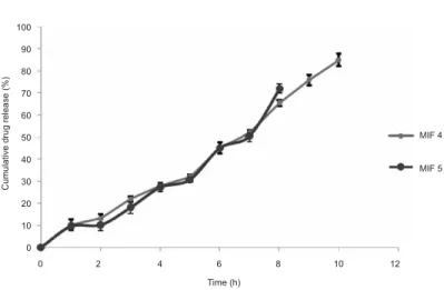
In vivodrug release study
Formulations MIF4 and MIF5 were selected forin vivodrug release studies on the basis of maximum mucoadhesion and better sustained behavior of the drug among all insert formulations. Ocular inserts were removed carefully at hourly intervals (from
Table III. Kinetics ofin vitrodrug release from ocular inserts through Millipore membrane filter and goat cornea
Formu-lation R2 Mechanism of drug releasea Zero-ordera First-ordera Higuchia Korsmeyer-Peppasa
Millipore membrane Goat cornea Millipore membrane Goat cornea Millipore membrane Goat cornea Millipore membranea Goat corneaa MIF1 0.8482 0.9938 0.9432 0.9979 0.9877 0.9511 0.9988 0.9997 Non--Fickian MIF2 0.9781 0.9919 0.9622 0.9969 0.9871 0.9536 0.9992 0.9999 Non--Fickian MIF3 0.9444 0.9930 0.9634 0.9975 0.9831 0.9514 0.9963 1.000 Non--Fickian MIF4 0.9653 0.9926 0.9625 0.9983 0.9742 0.9515 0.9982 0.9993 Non--Fickian MIF5 0.9626 0.9922 0.9748 0.9978 0.9891 0.9537 0.9991 0.9999 Non--Fickian MIF6 0.9688 0.9889 0.9654 0.9950 0.9712 0.9577 0.9964 0.9999 Non--Fickian MIF7 0.9642 0.9938 0.9678 0.9979 0.9702 0.9511 0.9964 0.9997 Non--Fickian an= 3.
1–10 hours) and analyzed for residual drug content. The drug was subtracted from the initial content of the insert and gave the amount of drug released in the rabbit eye. Fig. 3 presents the cumulative drug release (%) from formulations MIF4 and MIF5 at different time intervals. Thein vivostudy suggests the formulation MIF4 showed maximum cu-mulative drug release of 85.0 % and release was sustained up to 10 h. Formulation MIF5 showed 72.1 % cumulative release which was sustained up to 8 h. Also, formulation MIF4 had more tendency to stick to ocular mucosa due to the presence of higher concentration of quaternary ammonium groups (10 %) in Eudragit RL-100 as compared to Eudragit RS-100 (5 %). The presence of quaternary ammonium groups renders positive charge to the polymers by which it can interact with anionic mucin and thereby increase its resi-dence on corneal surface.
Stability
Finally, accelerated stability studies at elevated temperature and humidity revealed no significant changes in color in insert formulations. The moxifloxacin concentration in all formulations at accelerated and room temperature are shown in Fig. 2. The degrada-tion rate constants (kcal) and shelf life (t90) values for MIF1, MIF3, MIF5–MIF7 at room
temperature were found to range between 2.12 and 1.69 days–1, and 490–617 days. MIF2
and MIF4 showed lower degradation constants of 1.12 and 1.41 day–1and longer shelf
life of 926 and 739 days, resp. The stability study concluded that formulations MIF2 and MIF4 showed the lowest degradation and maximum shelf life as per the ICH guidelines (17) suggest there is no need to add overages to ensure 2 year shelf life.
MIF1E T MIF1R T MIF2E T MIF2R T MIF3E T MIF3R T MIF4E T MIF4R T MIF5E T MIF5R T MIF6E T MIF6R T MIF7R T MIF7E T 0.16 0.14 0.12 0.10 0.08 0.06 0.04 0.02 0 0 D 3 W 6 W 3 M 6 M Drug c onc entration (m g m m ) –2
Fig. 2. Stability of moxifloxacin ocular inserts under accelerated condition and room temperature. Mean±SD (n= 3). ET – elevated temperature (40 °C), RT – room temperature, D – days, W – weeks, M – months.
CONCLUSIONS
In conclusion, we suggest that the ocular insert formulation of moxifloxacin can be a promising vehicle for topical ocular administration of antibiotics. Its application could possibly replace the use of fortified solutions of antimicrobials and reduce the necessity for repeated drug administration at frequent intervals, thereby potentially lowering cor-neal toxicity and increasing patient compliance.
Acknowledgements.–The authors are thankful to Ranbaxy Laboratories Limited, Gurgaon, In-dia, for donating the moxifloxacin hydrochloride bulk drug. The authors thank All India Council of Technical Education (New Delhi, India) for providing a National Doctoral Fellowship to Mr. Pravin Kondiba Pawar.
REFERENCES
1. E. Barbu, I. Sarvaiya, K. L. Green, T. G. Nevell and J. Tsibouklis, Vinylpyrrolidone-co-(meth) acrylic acid inserts for ocular drug delivery: synthesis and evaluation,J. Biomed. Mater. Res.74 (2005) 598–606; DOI: 10.1002/jbm.a.30329.
2. N. Gangopadhyay, M. Daniell, L. Weih and H. Taylor, Fluoroquinolone and fortified antibiotics for treating bacterial corneal ulcers,Br. J. Ophthalmol.84(2000) 378–384; DOI: 10.1136/bjo.84.4.378. 3. A. P. Smith, M. Pennefather, S. B. Kaye and C. A. Hart, Fluoroquinolones: place in ocular
ther-apy,Drugs61(2001) 747–761.
4. P. E. Cutarelli, J. H. Lass, H. M. Lazarus, S. C. Putman and M. R. Jacobs, Topical fluoroquinolo-nes: antimicrobial activity and in vitro corneal epithelial toxicity,Curr. Eye Res.10(1991) 557–563; DOI: 10.3109/02713689109001764. 100 90 80 70 60 50 40 30 20 10 0 MIF 4 MIF 5 Cumulative dru g release (%) 0 2 4 6 8 10 12 Time (h)
5. P. L. Mallari, D. J. McCarty, M. Daniell and H. Taylor, Increased incidence of corneal perforation after topical fluoroquinolone treatment for microbial keratitis, Am. J. Ophthalmol.131 (2001) 131–133; DOI: 10.1016/S0002-9394(00)00642-5.
6. S. Barath and S. R. Hiremath, Ocular delivery system of perfloxacin mesylate,Pharmazie54(1999) 55–58.
7. A. S. Mundada and B. K. Srikhande, Formulation and evaluation of ciprofloxacin hydrochloride soluble ocular drug insert,Curr. Eye Res.33(2008) 469–475; DOI: 10.1080/02713680802023104. 8. G. Di Colo, S. Burgalassi, P. Chetoni, M. P. Fiaschi, Y. Zambito and M. F. Saettone, Gel-forming erodible inserts for ocular controlled delivery of ofloxacin,Int. J. Pharm.14(2001) 101–111; DOI: 10.1016/S0378-5173(02)00421-0.
9. J. M. Woodcock, J. M. Andrews, F. J. Boswell, N. P. Brenwald and R. Wise, In vitro activity of BAY 12-8039, a new fluoroquinolone,Antimicrob. Agents Chemother.41(1997) 101–106. 10. E. Pestova, J. J. Millichap, G. A. Noskin and L. R. Peterson, Intracellular targets of
moxaciflo-xacin: a comparison with other fluoroquinolones,J. Antimicrob. Chemother.45(2000) 583–590; DOI: 10.1093/jac/45.5.583.
11. P. K. Pawar and D. K. Majumdar, Effect of formulation factors on in vitro permeation of moxi-floxacin from aqueous drops through excised goat, sheep and buffalo corneas,AAPS PharmSci Tech.7(2006) E13.
12. P. K. Pawar and D. K. Majumdar, In vitro permeation characteristics of moxifloxacin from oil drops through excised goat, sheep, buffalo and rabbit corneas,Pharmazie 62(2007) 853–857; DOI: 10.1691/PH.2007.11.7055.
13. M. Malhotra and D. K. Majumdar, In vitro transcorneal permeation of ketorolac tromethamine from buffered and unbuffered aqueous ocular drops,Indian J. Exp. Biol.35(1997) 941–947. 14. L. Perioli, V. Ambrogi, S. Giovagnoli, M. Ricci, P. Blasi and C. Rossi, Mucoadhesive bilayered
tablets for buccal sustained release of flurbiprofen,AAPS PharmSciTech. 8 (2007) E54; DOI: 10.1208/pt0803054.
15. P. Costa and L. J. Manuel Sousa, Modeling and comparison of dissolution profiles,Eur. J. Pharm. Sci.13(2001) 123–133; DOI: 10.1016/S0928-0987(01)00095-1.
16. V. Shankar, A. K. Chandrasekaran, S. Durga, G. Geetha and V. A. Ravichandran, Design and evaluation of diclofenac sodium ophthalmic inserts,Acta Pharm. Sci.48(2006) 5–10.
17. International Conference on Harmonization,Q1A (R2): Stability Testing of New Drug Substances and Products,ICH, Geneva 2003.
S A @ E T A K
Dizajniranje i vrednovanje okularnih umetaka moksifloksacin hidroklorida
PRAVIN K. PAWAR, RAJESH KATARA i DIPAK K. MAJUMDAR
Cilj rada bio je priprava i evaluacija okularnih umetaka moksifloksacina. Okularni umetak izra|en je od vodene suspenzije moksifloksacina, natrijevog alginata, polivinil-nog alkohola i dibutil-ftalata metodom odlijevanja filma. Okularni umetak (promjera 5,5 mm) umre`en je pomo}u CaCl2i oblo`en Eudragitom S-100, RL-100, RS-100, E-100 ili
Eudragit L-100.In vitrodrena`a/permeacija lijeka prou~avana je koriste}i staklenu mo-dificiranu Franzovu difuzijsku }eliju. Koncentracija lijeka i vrijeme mukoadhezije
oku-larnih umetaka bili su zadovoljavaju}i. Umre`avanje i oblaganje polimerima produljilo je drena`u iz umetaka. Umre`eni okularni umetci oblo`eni s Eudragit RL-100 pokazali su ve}u permeaciju lijeka u odnosu na ostale pripravke.
Klju~ne rije~i:moksifloksacin, okularni umetak, kornea koze
Chitkara College of Pharmacy, Chitkara University, Chandigarh-Patiala National Highway, Rajpura, Rajpura-140401, Patiala, Punjab, India
Department of Pharmaceutics, Delhi Institute of Pharmaceutical Sciences and Research, (formerly College of Pharmacy), University of Delhi, Pushp Vihar, Sector III, New Delhi-110017, India