ISSN 1858-2419 Vol. 9 No. 1
3 Maret 2014
JURNAL TEKNOLOGI PERTANIAN
UNIVERSITAS MULAWARMAN
Mini Review
Peningkatan Produksi Padi Berkelanjutan Pada Lahan Rawa Pasang Surut (Increasing of
Sustainable Rice Production on Swampland) Nurita, Isdijanto Ar-Riza
Penelitian
Pengaruh Perbedaan Suhu Fermentasi Moromi Terhadap Sifat Kimia Dan Mikroflora Moromi Kecap Koro Pedang (Canavalia ensiformis L.) (Effect of Different Temperature
of Moromi Fermentation on Chemical and Microflora Characteristics of Jack Bean Sauce (Canavalia ensiformis L.)) Beti Cahyaning Astuti
Kajian Proses Produksi Pulp Dan Kertas Ramah Lingkungan Dari Sabut Kelapa (Study
on the Production of Environmental Friendly Pulp and Paper from Coconut Husk)
Khaswar Syamsu, Han Roliadi, Krishna Purnawan Candra, Akbar Jamaluddin Arsyad
Isolation of Cellulolytic Microbials from Several Locations were Associated with the Palm Oil Industry (Isolasi Mikroba Selulolitik dari Beberapa Lokasi yang Berkaitan
dengan Industri Minyak Sawit) Hamka Nurkaya
Keragaman dan Habitat Lebah Trigona pada Hutan Sekunder Tropis Basah di Hutan Pendidikan Lempake, Samarinda, Kalimantan Timur (Biodiversity and Habitat of
Trigona at Secondary Tropical Rain Forest of Lempake Education Forest, Samarinda, Kalimantan Timur) Syafrizal, Daniel Tarigan, Roosena Yusuf
Karakteristik Kimia Kopi Kawa Dari Berbagai Umur Helai Daun Kopi Yang Diproses Dengan Metode Berbeda (Chemical Characteristic of Coffee Kawa Produced from
Different Age of Coffee Leaf by Different Methods) Khusnul Khotimah
Bekerjasama dengan
JURNAL TEKNOLOGI PERTANIAN
PENERBIT
Jurusan Teknologi Hasil Pertanian Fakultas Pertanian Universitas Mulawarman
Jl.Tanah Grogot Kampus Gunung Kelua Samarinda 75119
KETUA EDITOR
Krishna Purnawan Candra (THP-UNMUL Samarinda)
EDITOR
Bernatal Saragih (THP-UNMUL Samarinda) Dahrulsyah (TPG-IPB Bogor)
Dodik Briawan (GMK-IPB Bogor) Khaswar Syamsu (TIN-IPB Bogor) Meika Syahbana Roesli (TIN-IPB Bogor) V. Prihananto (THP-Unsoed Purwokerto)
EDITOR PELAKSANA
Sulistyo Prabowo Hadi Suprapto Miftakhur Rohmah
ALAMAT REDAKSI
Jurusan Teknologi Hasil Pertanian Fakultas Pertanian
Universitas Mulawarman
Jalan Tanah Grogot Kampus Gunung Kelua Samarinda 75119
Telp 0541-749159 e-mail: [email protected]
JURNAL TEKNOLOGI PERTANIAN
UNIVERSITAS MULAWARMAN
Volume 9 Nomor 1
3 Maret 2014
Mini Review
HalamanPeningkatan Produksi Padi Berkelanjutan Pada Lahan Rawa Pasang Surut (Increasing of
Sustainable Rice Production on Swampland) Nurita, Isdijanto Ar-Riza ... 1-7
Penelitian
Pengaruh Perbedaan Suhu Fermentasi Moromi Terhadap Sifat Kimia Dan Mikroflora Moromi Kecap Koro Pedang (Canavalia ensiformis L.) (Effect of Different Temperature
of Moromi Fermentation on Chemical and Microflora Characteristics of Jack Bean Sauce (Canavalia ensiformis L.)) Beti Cahyaning Astuti ... 8-15
Kajian Proses Produksi Pulp Dan Kertas Ramah Lingkungan Dari Sabut Kelapa (Study
on the Production of Environmental Friendly Pulp and Paper from Coconut Husk)
Khaswar Syamsu, Han Roliadi, Krishna Purnawan Candra, Akbar Jamaluddin Arsyad ... 16-25
Isolation of Cellulolytic Microbials from Several Locations Were Associated with the Palm Oil Industry (Isolasi Mikroba Selulolitik dari Beberapa Lokasi Industri Minyak
Sawit) Hamka Nurkaya ... 26-33 Keragaman dan Habitat Lebah Trigona pada Hutan Sekunder Tropis Basah di Hutan Pendidikan Lempake, Samarinda, Kalimantan Timur (Biodiversity and Habitat of
Trigona at Secondary Tropical Rain Forest of Lempake Education Forest, Samarinda, Kalimantan Timur) Syafrizal, Daniel Tarigan, Roosena Yusuf ... 34-39
Karakteristik Kimia Kopi Kawa Dari Berbagai Umur Helai Daun Kopi Yang Diproses Dengan Metode Berbeda (Chemical Characteristic of Coffee Kawa Produced from
ISOLATION OF CELLULOLYTIC MICROBIALS FROM SEVERAL LOCATIONS WERE ASSOCIATED WITH THE PALM OIL INDUSTRY
Isolasi Mikroba Selulolitik dari Beberapa Lokasi Industri Minyak Sawit
Hamka Nurkaya
Samarinda State Polytechnic for Agriculture, Jl. Samratulangi-Kampus Poltanesa, Kotak Pos 192, Samarinda 75131, E-mail: [email protected]
Received 10 July 2013 accepted 17 November 2013 ABSTRACT
Cellulose is the main constituent component of photosynthesis in the plant biomass composed of fibrous and woody material such as straw, weeds, grass, leaves, stems and branches of plants which could be easily degraded by microbial such as bacteria, fungi and actinomycetes. With the utilization of twelve (12) samples for isolation of microbials col-lected from several locations associated with the palm oil industry which were obtained as many fifty eight (58) isolates of microbial and then classified to be the thirty nine (39) iso-lates are bacteria, ten (10) isoiso-lates of actinomycetes and nine (9) isoiso-lates of fungi. All isoiso-lates of microbial isolated were identificated by colony properties of microorganisms, Gram stain and slide culture technique.
Keywords: cellulose, bacteria, fungi, actinomycetes.
BACKGROUND
Cellulose is the main constituent com-ponent of photosynthesis in the plant biomass composed of fibrous and woody material such as straw, weeds, grass, leaves, stems and branches of plants (Sutedja et al., 1991; Jarvis, 2003; and Zhang et al., 2004). As plant biomass, cellulose often found in the biomass lignocellulosic materials such as biomass agricultural, forestry, and agro-industrial wastes that are abundant, renewable and inexpensive energy sources. Cellulose can be degraded easily and quickly just by specific organisms such as bacteria, fungi, actinomy-cetes, and the lower animals. Experts classify cellulose decomposers organisms are aerobic bacterial, myxo-bacteria, anaerobic myxo-bacteria, thermophilic group, actinomycetes, filamentous fungi, mushroom, protozoa and insects. Success in breaking down cellulose depends on the nature or circumstances of cellulose de-grading microorganisms and environ-mental factors such as humidity, aeration, tempe-rature, and nitrogen availability of
adequate and nutritional elements (Sutedja et al., 1991).
In this study, All samples used for the isolation of cellulolytic microbials (soil, leaf bamboo, rod of palm tree, etc.) were collected from several places associated with the palm oil industry. Aim of this study was to isolate and identify the cellulolytic microorganisms from samples were collected at the locations associated with the oil palm plantation and industry.
MATERIALS AND METHODS Raw materials
All of the samples for the isolation of cellulolytic microbials (soil, leaf bamboo, rod of palm tree, seed of the palm tree, fiber of the oil palm empty fruit bunch, etc.) were collec-ted from the palm oil plantation and palm mill area in Petchabury Province, Thailand.
Source of microbials
Samples for isolation of microbials used in this study collected from several places associated with the oil palm Industry. Sample properties also recorded.
Hamka Nurkaya Isolation of Cellulolytic Microbials Associated with The Palm Oil industry
Isolation of microbials
Liquid carboxymethyl cellulose medi-um (CMC medium) consist of 1 g KH2PO4, 0.5 g NaCl, 0.5 g MgSO4.7H2O, 0.01 g FeSO4.7H2O, 0.01 g MnSO4.H2O, 0.5 g (NH4)2SO4 and 10 g CMC in 1 liter of distilled water was prepared and sterilized at 121oC for 15 min, which was used to enrich microor-ganism from sample. Each sample of 1 g was added into 100 mL sterilized CMC medium and incubated at room temperature (30-32oC) under shaking condition (150 rpm) for 10 days. After incubation, microorganism; bacte-ria, fungi and actinomycetes were collected by a streak plate technique on CMC agar (1.5 % agar was added into the CMC medium). Cultured petridishes were incubated at room temperature (30-32oC) for 3 days. After incubation, colonized microorganisms were sub-sequently transferred on fresh CMC agar and incubated for 3 days to obtain pure cultures. All strains were kept in a deep freezer at -80oC and used for the future experimentation.
Pre-primary identification and characteri-zation of cellulolytic microbials
Pre-primary identification was done to separate and classify selected strains into the member of bacteria, actino-mycetes and fungi.
Colony properties of microorganism
Colony properties of microorganism were observed based on a single colony of microbes grown on a CMC medium. Cultures were incubated for 3-5 days at room temperature and then the shape, color, surface, internal structure and the structure of the edge of the colony were also observed. Based on colony proper-ties, bacteria, actinomycetes and fungi were also separated. Cell morpho-logies and spore formulation of selected isolates were also determined.
Slide culture technique for fungi
In order to accurately identify fungus, it is essential to observe the pre-cise arrangement of the conidiophores (conidial ontogeny). A simple modifi-cation of fungal slide culture technique was used to observe conidiophores of selected fungal isolates (Riddle, 1950). A method is described by Riddle (1950) and conidiophores picture was recorded and used to identify the genus of fungal isolates.
Gram staining technique
The Gram stain is the most important and universally used stained technique in the bacteriology laboratory. It is used to disti-nguish between gram-positive and gram-negative bacteria, which have distinct and consistent differences in their cell walls. Gram staining of bacteria is used Gram method and consequently, Gram-positive and Gram-negative bacteria were classified.
RESULT AND DISCUSSION
Samples for isolation of cellulolytic microbials
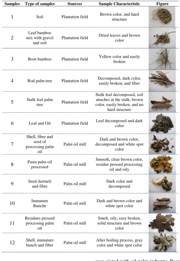
The samples that are used in this research for isolation of microbials were collected from the palm oil plantation and palm oil mill areas in Pechabury Province, Thailand. The samples were shown in Table 1.
The twelve (12) samples were selected from the palm oil plantation and palm oil mill areas such as soil, a rod palm tree, immature bunch, seed of the oil palm empty fruit bunch, shell, fiber of the oil palm empty fruit bunch which they contain normal flora that can decompose cellulosic material; cellulolytic micro-organism. According to Smruti et al. (1995) reported the cellulase producing microbes have been isolated from ligno-cellulosic materials such as decaying wood, forest residues and decomposed leaf in soil.
Table 1. Types, sources, and the characteristic of samples, which were selected from the palm oil plantation
and palm oil mill areas.
Samples Type of samples Sources Sample Characteristic Figure
1 Soil Plantation field Brown color, and hard structure
2
Leaf bamboo mix with gravel
and soil
Plantation field Dried leaves and brown color
3 Root bamboo Plantation field Yellow color and easily broken
4 Rod palm tree Plantation field Decomposed, dark color, easily broken, and fiber
5 Stalk leaf palm tree Plantation field
Stalk leaf decomposed, soil attaches at the stalk, brown color, easily broken, and no
hard structure
6 Leaf and Oil Plantation field Leaf decomposed and dark color
7
Shell, fiber and seed of processing palm
oil
Palm oil mill
Dark and brown color, decomposed and white spot
color
8 Pasta palm oil processed Palm oil mill
Smooth, clear brown color, residue pressed processing
oil and oily
9 Seed (kernel) and fiber Palm oil mill Dark color and decomposed
10 Immature Bunche Palm oil mill Dark and brown color and white spot color
11
Residues pressed processing palm
oil
Palm oil mill
Smelt, oily, easy broken, solid structure and brown
color
12 Shell, immature bunch and fiber Palm oil mill After boiling process, gray color and white spot color
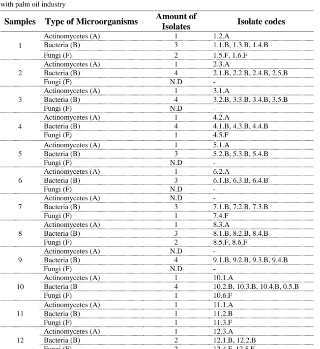
Isolation of microbials
There were fifty eight (58) isolates of microbials isolated from the samples
asso-ciated with oil palm industry. Based on colony formation and cell morphology (under microscope), those
microorga-Hamka Nurkaya Isolation of Cellulolytic Microbials Associated with The Palm Oil industry
nisms obtained from screening was separated into a member of bacteria, actinomycetes and fungi. Thirty nine isolates (39) of microorganisms belonged to member of bacteria, while ten isolates (10) was classified to be a member of actinomy-cetes. The rest isolates (nine isolates) are fungi.
According to Heck et al., (2002) and Semedo et al., (2000), reported many of cellulolytic aerobic and anaerobic bacteria could be isolated from decomposed soil and the lignocellulosic of residues (leaf, stalks, and stems of garden plants) for biotechnological applications.
Table 2. The fifty eight (58) isolates of microorganisms obtained from plant and soil samples associated
with palm oil industry
Samples Type of Microorganisms Amount of
Isolates Isolate codes
1
Actinomycetes (A) 1 1.2.A
Bacteria (B) 3 1.1.B, 1.3.B, 1.4.B
Fungi (F) 2 1.5.F, 1.6.F
2
Actinomycetes (A) 1 2.3.A
Bacteria (B) 4 2.1.B, 2.2.B, 2.4.B, 2.5.B
Fungi (F) N.D
-3
Actinomycetes (A) 1 3.1.A
Bacteria (B) 4 3.2.B, 3.3.B, 3.4.B, 3.5.B
Fungi (F) N.D
-4
Actinomycetes (A) 1 4.2.A
Bacteria (B) 4 4.1.B, 4.3.B, 4.4.B
Fungi (F) 1 4.5.F
5
Actinomycetes (A) 1 5.1.A
Bacteria (B) 3 5.2.B, 5.3.B, 5.4.B
Fungi (F) N.D
-6
Actinomycetes (A) 1 6.2.A
Bacteria (B) 3 6.1.B, 6.3.B, 6.4.B Fungi (F) N.D -7 Actinomycetes (A) N.D - Bacteria (B) 3 7.1.B, 7.2.B, 7.3.B Fungi (F) 1 7.4.F 8
Actinomycetes (A) 1 8.3.A
Bacteria (B) 3 8.1.B, 8.2.B, 8.4.B Fungi (F) 2 8.5.F, 8.6.F 9 Actinomycetes (A) N.D - Bacteria (B) 4 9.1.B, 9.2.B, 9.3.B, 9.4.B Fungi (F) N.D -10
Actinomycetes (A) 1 10.1.A
Bacteria (B 4 10.2.B, 10.3.B, 10.4.B, 0.5.B
Fungi (F) 1 10.6.F
11
Actinomycetes (A) 1 11.1.A
Bacteria (B) 1 11.2.B
Fungi (F) 1 11.3.F
12
Actinomycetes (A) 1 12.3.A
Bacteria (B) 2 12.1.B, 12.2.B
Fungi (F) 2 12.4.F, 12.5.F
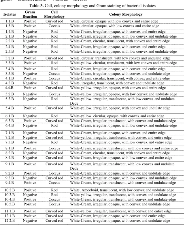
Colony properties of microbials
Table 3, showed cell and colony morphological studies of bacterial iso-lates. Under a light microscope, bacteria were clearly separated from fungi and actinomycetes. Colony morphological studies of bacteria can help at bacterial genus classification. Various factors
influence the shape and color of the colonized bacterial colony such as type of bacterial culture medium, pH, temperature and condition (aerobic, anaerobic, anoxic etc.) for incubation. In this experiment, all microorganisms were cultured on CMC agar.
Table 3. Cell, colony morphology and Gram staining of bacterial isolates Isolates Gram
Reaction
Cell
Morphology Colony Morphology
1.1.B Positive Curved rod White, circular, opaque with low convex and entire edge 1.3.B Positive Coccus White, circular, opaque, with low convex and entire edge 1.4.B Negative Rod White-Cream, irregular, opaque, with convex and entire edge 2.1.B Negative Rod White-Cream, irregular, opaque, with low convex and undulate edge 2.2.B Negative Rod White-Cream, circular, translucent, with convex and entire edge 2.4.B Negative Rod White-Cream, irregular, opaque, with convex and entire edge 2.5.B Negative Rod White-Cream, irregular, opaque, with low convex and undulate edge 3.2.B Positive Curved rod White, circular, translucent, with low convex and undulate edge 3.3.B Positive Rod White-yellow, circular, translucent, with low convex and entire edge 3.4.B Positive Rod White-Cream, irregular, opaque, with convex and entire edge 3.5.B Negative Coccus White-Cream, irregular, opaque, with convex and undulate edge 4.1.B Positive Coccus White-Cream, circular, translucent, with convex and entire edge 4.3.B Negative Rod White, irregular, translucent, with convex and undulate edge 4.4.B. Positive Curved rod White-yellow, irregular, opaque, with convex and entire edge
5.2.B Negative Coccus White-yellow, irregular, opaque, with low convex and undulate edge 5.3.B Negative Rod White-yellow, irregular, translucent, with low convex and undulate
Dede
5.4.B Positive Curved rod White-yellow, irregular, opaque, with convex and undulate edge 6.1.B Negative Rod White-yellow, circular, opaque, with convex and entire edge 6.3.B Positive Curved rod White-Cream, irregular, translucent, with convex and undulate edge 6.4.B Negative Rod White-Cream, irregular, opaque, with low convex and undulate edge 7.1.B Negative Curved rod White-Cream, irregular, opaque, with convex and entire edge 7.2.B Negative Curved rod White-yellow, irregular, translucent, with convex and entire edge 7.3.B Negative Rod White-Cream, irregular, opaque, with low convex and entire edge 8.1.B Positive Coccus White-Cream, irregular, translucent, with low convex and entire edge 8.2.B Negative Curved rod White-Cream, circular, translucent, with convex and entire edge 8.4.B Negative Curved rod White-Cream, irregular, opaque, with low convex and entire edge 9.1.B Positive Curved rod White-Cream, irregular, translucent, with low convex and undulate
Dede
9.2.B Positive Coccus White-Cream, irregular, opaque, with convex and undulate edge 9.3.B Negative Curved rod White-Cream, irregular, opaque, with low convex and undulate edge 9.4.B Positive Coccus White-Cream, irregular, translucent, with convex and undulate edge 10.2.B Positive Rod White, Amoeboid, translucent, with low convex and undulate edge 10.3.B Negative Rod White-yellow, irregular, translucent, with convex and undulate edge 10.4.B Positive Coccus White-Cream, irregular, translucent, with convex and undulate edge 10.5.B Positive Coccus White-Cream, irregular, opaque, with convex and undulate edge 11.1.B Positive Curved rod White-yellow, irregular, translucent, with convex and entire edge 12.1.B Positive Curved rod White-Cream, irregular, opaque, with convex and entire edge 12.2.B Negative Curved rod White-Cream, irregular, opaque, with convex and undulate edge
Hamka Nurkaya Isolation of Cellulolytic Microbials Associated with The Palm Oil industry
There were fifty five (58) microbials isolates obtained from plant and soil samples associated with oil palm industry. Cell morphology and colony morphology of bacterial isolates such as shape, the margins or edges of the colony, the colony’s color, colony and Gram cell wall were explained to support pre-primary identification of bacterial isolates. Under a light microscope, various shapes of bacterial cell were observed such as rod, curved rod, and coccus. There were 2 major types of bacterial colony shapes which could be seen after cultured on
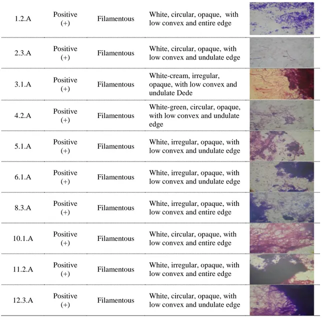
CMC agar including circular and irregular form. The color or the pigment of the colony of all bacteria were white color (opaque or translucent) and also present of cream or yellow colonies. The shape of the colony of bacteria is some circular or some irregular. The edge of bacterial colony such as low convex with entire edge and convex with undulate edge was also determined. With Gram stain technique, there were nineteen (19) isolates and twenty (20) isolates classified into a member of Gram-negative and Gram-positive bacteria, respectively. Table 4. Cell, colony morphology and Gram staining of actinomycetes isolates
Isolates Gram
Reaction
Cell
Morphology Colony Morphology Figure
1.2.A Positive
(+) Filamentous
White, circular, opaque, with low convex and entire edge
2.3.A Positive
(+) Filamentous
White, circular, opaque, with low convex and undulate edge
3.1.A Positive
(+) Filamentous
White-cream, irregular, opaque, with low convex and undulate Dede
4.2.A Positive
(+) Filamentous
White-green, circular, opaque, with low convex and undulate edge
5.1.A Positive
(+) Filamentous
White, irregular, opaque, with low convex and undulate edge
6.1.A Positive
(+) Filamentous
White, irregular, opaque, with low convex and undulate edge
8.3.A Positive
(+) Filamentous
White, irregular, opaque, with low convex and entire edge
10.1.A Positive
(+) Filamentous
White, circular, opaque, with low convex and entire edge
11.2.A Positive
(+) Filamentous
White, irregular, opaque, with low convex and entire edge
12.3.A Positive
(+) Filamentous
White, circular, opaque, with low convex and undulate edge
There were ten (10) isolates of actinomycetes which could be isolated from the samples associated with oil palm industry. Colonies of actinomycetes were white color, opaque with low convex and irregular in shape (table 4). Under microscope and Gram-stain technique, cell morphology of actinomycetes was
observed. A filamentous property and Gram stain positives were seen under light microscope (1,000×). With specific property of actinomycetes cell, all selected isolates were determined and confirmed to be actinomycetes. Charac-teristic of the colony and cell of actinomycetes represented in Table 4.
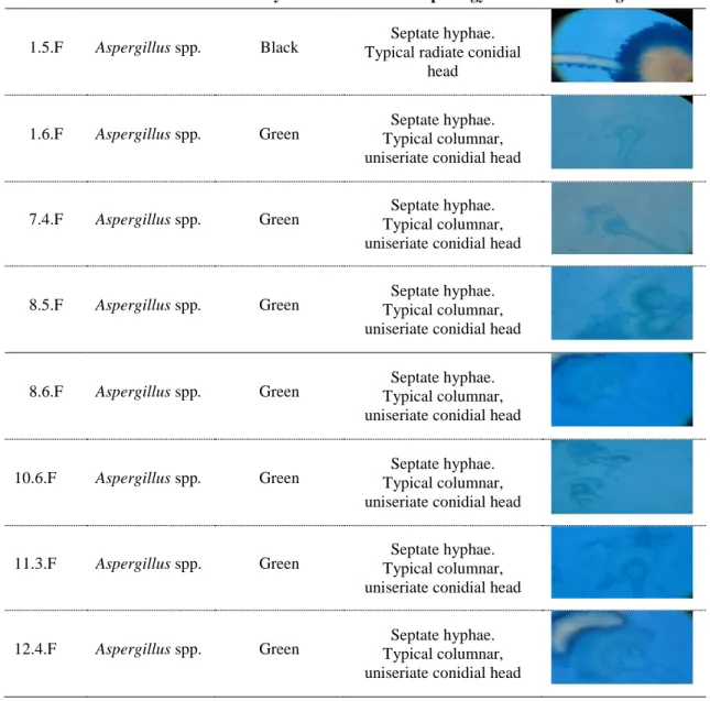
Table 5. Cell and colony morphology of fungal isolates
Isolate Genus Colony Color Morphology Figure
1.5.F Aspergillus spp. Black Typical radiate conidial Septate hyphae. head
1.6.F Aspergillus spp. Green Typical columnar, Septate hyphae. uniseriate conidial head
7.4.F Aspergillus spp. Green Typical columnar, Septate hyphae. uniseriate conidial head
8.5.F Aspergillus spp. Green Typical columnar, Septate hyphae. uniseriate conidial head
8.6.F Aspergillus spp. Green Typical columnar, Septate hyphae. uniseriate conidial head
10.6.F Aspergillus spp. Green Typical columnar, Septate hyphae. uniseriate conidial head
11.3.F Aspergillus spp. Green Typical columnar, Septate hyphae. uniseriate conidial head
12.4.F Aspergillus spp. Green Typical columnar, Septate hyphae. uniseriate conidial head
Fungi were important cellulosic microorganism associated with cellulose cycle. Fungi are known as the best source of cellulase enzyme and their applications in biological technology such as
saccharification by fungal cellulase were reported. Table 5. shows the colony and cell morphological studies of fungal isolates which were isolated from the samples associated with the oil palm
Hamka Nurkaya Isolation of Cellulolytic Microbials Associated with The Palm Oil industry
industry. Based on colony morphology, fungal isolates were selected. Pre-primary identification was done by investigation of arrangement of conidia under a light microscope (Lactophenol cotton blue as staining dye). All fungal isolates presented conidiosphore as a special characteristic of the genus Aspergillus. Therefore, nine (9) isolates of fungi were classified as member of the genus
Aspergillus. Table 5, represents cell and
colony morphology of fungal isolates.
CONCLUSION
The twelve (12) samples selected from several locations associated with the palm oil industry which were used to isolate and identificated microbials and as many fifty eight (58) isolates of microbials were obtained from the locations at the palm oil industry areas. The thirty nine (39) isolates were classified as bacteria, ten (10) isolates of actinomycetes, and nine (9) isolates of fungi. All isolates of microbial isolated were identified by colony properties of microbial, Gram stain and slide culture technique.
ACKNOWLEDGMENTS
This study was supported finan-cially by Faculty of Science, Department of Microbiology, King Mongkut’s University of Technology Thonbury, Thailand and Directorate General of Higher Education, Ministry of Education and Culture of Republic of Indonesia.
REFERENCES
Heck JX, Hertz PF, Ayub MAZ (2002) Cellulase and xylanase production by isolated Amazon Bacillus strains using soya bean industrial residue based solid-state cultivation. Brazilian Jour-nal of Microbiology 33(3): 213-218.
Jarvis M (2003) Cellulose stacks up. Nature 426(6967): 611-622.
Riddle RW (1950) Permanent stained myco-logical preparations obtained by slide. Mycologia 42(2): 265. Semedo LT, Gomes RC, Bon EP, Soares
RM, Linhares LF, Coelho RR (2000) Endo-cellulase and exocellulase activities of two
Streptomyces strains isolated from a
forest soil. Applied of Biochemistry and Biotechnology 84-86(1): 267-276.
Smruti P, Rao KK, Krishna M (1995) Produc-tion of beta-glucosidase by
Aspergillus terreus. Current Microbiology 30(5): 255-258. Sutedja MM, Kartasapoetra AG,
Sastroat-modjo S (1991) Mikrobiologi Tanah. Penerbit Rineka Cipta, Jakarta. p. 123-129.
Zhang YHP, Lynd LR (2004) Toward an aggregated understanding of enzymatic hydrolysis of cellulose, noncomplexed cellulose systems. Biotechnology and Bioengineering 88(7): 797-824.
PEDOMAN PENULISAN
Jurnal Teknologi Pertanian
Universitas Mulawarman
PengirimanJurnal Teknologi Pertanian Universitas Mula-warman menerima naskah berupa artikel hasil penelitian dan ulas balik (review) yang belum pernah dipublikasikan pada majalah/jurnal lain. Penulis diminta mengirimkan tiga eksemplar naskah asli beserta softcopy dalam disket yang di-tulis dengan program Microsoft Word. Naskah dan disket dikirimkan kepada:
Editor Jurnal Teknologi Pertanian
d. a. Jurusan Teknologi Hasil Pertanian Fakultas Pertanian
Universitas Mulawarman Jalan Tanah Grogot
Samarinda 75119
Format
Umum. Naskah diketik dua spasi pada kertas
A4 dengan tepi atas dan kiri 3 centimeter, kanan dan bawah 2 centimeter menggunakan huruf Times New Roman 12 point, maksimum 12 halaman. Setiap halaman diberi nomor secara berurutan. Ulas balik (review) ditulis sebagai naskah sinambung tanpa subjudul Bahan dan Metode, Hasil dan Pembahasan. Selanjutnya susunan naskah dibuat sebagai berikut :
Judul. Pada halaman judul tuliskan judul,
nama setiap penulis, nama dan alamat institusi masing-masing penulis, dan catatan kaki yang berisi nama, alamat, nomor telepon dan faks serta alamat E-mail jika ada dari corresponding author. Jika naskah ditulis dalam bahasa Indonesia tulis-kan judul dalam bahasa Indonesia diikuti judul da-lam bahasa Inggris.
Abstrak. Abstrak ditulis dalam bahasa Inggris
dengan judul "ABSTRACT" maksimum 250 kata. Kata kunci dengan judul "Key word" ditulis dalam bahasa Inggris di bawah abstrak.
Pendahuluan. Berisi latar belakang dan
tujuan.
Bahan dan Metode. Berisi informasi teknis
sehingga percobaan dapat diulangi dengan teknik yang dikemukakan. Metode diuraikan secara lengkap jika metode yang digunakan adalah metode baru.
Hasil. Berisi hanya hasil-hasil penelitian baik
yang disajikan dalam bentuk tubuh tulisan, tabel, maupun gambar. Foto dicetak hitam-putih pada kertas licin berukuran setengah kartu pos.
Pembahasan. Berisi interpretasi dari hasil
penelitian yang diperoleh dan dikaitkan dengan hasil-hasil penelitian yang pernah dilaporkan (pub-likasi).
Ucapan Terima Kasih. Digunakan untuk
me-nyebutkan sumber dana penelitian dan untuk mem-berikan penghargaan kepada beberapa institusi atau orang yang membantu dalam pelaksanaan penelitian dan atau penulisan laporan.
Daftar Pustaka. Daftar Pustaka ditulis
me-makai sistem nama tahun dan disusun secara abjad. Beberapa contoh penulisan sumber acuan:
Jurnal
Wang SS, Chiang WC, Zhao BL, Zheng X, Kim IH (1991) Experimental analysis and computer simulation of starch-water interaction. J Food Sci 56: 121-129.
Buku
Charley H, Weaver C (1998) Food a Scientific Approach. Prentice-Hall Inc USA
Bab dalam Buku
Gordon J, Davis E (1998) Water migration and food storage stability. Dalam: Food Storage Stability. Taub I, Singh R. (eds.), CRC Press LLC.
Abstrak
Rusmana I, Hadioetomo RS (1991) Bacillus
thuringiensis Berl. dari peternakan ulat
sutra dan toksisitasnya. Abstrak Pertemuan Ilmiah Tahunan Perhimpunan Mikrobiologi Indonesia. Bogor 2-3 Des 1991. p. A-26.
Prosiding
Prabowo S, Zuheid N, Haryadi (2002) Aroma nasi: Perubahan setelah disimpan dalam wadah dengan suhu terkendali. Dalam: Prosiding Seminar Nasional PATPI. Malang 30-31 Juli 2002. p. A48.
Skripsi/Tesis/Disertasi
Meliana B (1985) Pengaruh rasio udang dan tapioka terhadap sifat-sifat kerupuk udang. Skripsi Fakultas Teknologi Pertanian UGM Yogyakarta.
Informasi dari Internet
Hansen L (1999) Non-target effects of Bt corn pollen on the Monarch butterfly (Lepidop-tera: Danaidae). http://www.ent. iastate. edu/entsoc/ncb99/prog/abs/D81.html [21 Agu 1999].
Bagi yang naskahnya dimuat, penulis dikenakan biaya Rp 175.000,00 (seratus tujuh puluh lima ribu rupiah).
Hal lain yang belum termasuk dalam petunjuk penulisan ini dapat ditanyakan langsung kepada REDAKSI JTP ([email protected]; http://jtpunmul.wordpress.com).