Nutritional Management in Acute Pancreatitis
Luh Putu Listya Paramita*, Ari Fahrial Syam**
*Faculty of medicine, University of Indonesia/Dr. Cipto Mangunkusumo General National Hospital Jakarta
**Division of Gastroenterology, Department of Internal Medicine, Faculty of medicine Universitas Indonesia, Dr. Cipto Mangunkusumo General National H
ospital, Jakarta
Corresponding author:
Ari Fahrial Syam. Division of Gastroenterology, Department of Internal Medicine, Dr. Cipto Mangunkusumo General National Hospital. Jl. Diponegoro No. 71 Jakarta Indonesia. Phone: +62-21-3153957; Facsimile: +62-21-3142454. E-mail: [email protected].
ABSTRACT
$FXWHSDQFUHDWLWLVLQGXFHDFDWDEROLFVWUHVVWKDWLQFUHDVHV\VWHPLFLQÀDPPDWRU\UHVSRQVHZLWKZRUVHQLQJ QXWULWLRQDOVWDWXV&XUUHQWDSSURDFKLQDFXWHSDQFUHDWLWLVWKHUDS\ZDVVWLOOV\PSWRPDWLFEHFDXVHRIQRGH¿QLWLYH WKHUDS\\HWWRSUHYHQWDQ\LQÀDPPDWRU\DQGSURWHRO\WLFFDVFDGH2QHRIWKHPRVWLPSRUWDQWWKLQJWRFRQVLGHULQ
acute pancreatitis therapy was nutritional management. “Pancreatic rest” concept that formerly used have been known to increase cost, sepsis incidence due to catheter use, and also metabolic and electrolyte disorder. Nowadays, “gut rousing” concept was preferable compared to “pancreatic rest” concept, support that nutritional management
ZDVQHHGHGWRVWLPXODWHDQGJHQHUDWHLQWHVWLQDOIXQFWLRQ(QWHUDOQXWULWLRQDGPLQLVWUDWLRQKDYHWRFRQVLGHUSDWLHQW¶V
hemodynamic status. Necrosis incidence, respiratory failure, intensive care, and mortality was found to be lower in
SDWLHQWVJLYHQHQWHUDOQXWULWLRQLQ¿UVWKRXUVFRPSDUHGWRDIWHUKRXUV1XWULWLRQDGPLQLVWUDWLRQYLDQDVRJDVWULF
tube or nasojejunal tube was still in doubt while several studies showed that nasogastric tube administration was safe and tolerated, otherwise could be evaluated in larger population sample study. Nutrition and metabolic monitoring was also an important part to reach nutritional goals and reduce complications.
Keywords: acute pancreatitis, enteral nutrition, gut rousing
ABSTRAK
3DQNUHDWLWLVDNXWPHQ\HEDENDQVWUHVVNDWDEROLN\DQJPHQLPEXONDQUHVSRQLQÀDPDVLVLVWHPLNGDQSHUEXUXNDQ
status nutrisi. Pendekatan terapi pankreatitis akut saat ini masih bersifat simptomatik karena tidak ada
WHUDSLGH¿QLWLIXQWXNPHQFHJDKDNWLYDVLNDVNDGHLQÀDPDVLGDQSURWHROLWLN6DODKVDWXKDOSHQWLQJ\DQJKDUXV
diperhatikan dalam tatalaksana pankreatitis akut yaitu pemberian nutrisi. Konsep “pancreatic rest” yang dahulu diterapkan saat ini diketahui meningkatkan biaya perawatan, meningkatkan kasus sepsis karena pemakaian
NDWHWHUJDQJJXDQHOHNWUROLWGDQPHWDEROLN.RQVHSµJXWURXVLQJ¶VDDWLQLPXODLPHQJJHVHUNRQVHS³SDQFUHDWLF
rest” yaitu pemberian nutrisi diperlukan untuk menstimulasi atau membangkitkan fungsi saluran cerna. Waktu memulai pemberian nutrisi enteral juga harus memperhatikan hemodinamik pasien. Insidens nekrosis, gagal nafas, perawatan intensif, dan mortalitasnya lebih rendah pada pasien yang diberikan nutrisi enteral dalam 48 jam dibandingkan dengan yang diberikan setelah 48 jam. Pilihan jalur nutrisi melalui selang nasogaster atau selang nasojejunal masih menjadi perdebatan, meskipun pada berbagai studi pemberian nutrisi melalui nasogastric tube (NGT) dapat ditoleransi dan aman namun diperlukan studi dengan jumlah sampel yang lebih besar. Monitor nutrisi dan metabolik juga bagian penting untuk mencapai target nutrisi dan mencegah komplikasi.
INTRODUCTION
$FXWHSDQFUHDWLWLVZDVDQLQÀDPPDWRU\GLVRUGHU
that can lead to systemic inflammatory response syndrome (SIRS), multiorgan failure, and death. Incidence in several country was various, range for 4,9-73,4 cases per 100.000 population and still increasing every year. United States (US) and Finland was the highest prevalence country with 73 case per 100,000 population in US and 73,4 case per 100,000 population in Finland. Moderate prevalence was found in New Zealand (29.3 case) and Norwegia (34.4 case) per 100,000 population.1 Most of acute pancreatitis will
UHVROYHZLWKRXWWKHUDS\RUWKHQHHGRIVSHFL¿FWKHUDS\
Among 20% of them was progressed into severe acute pancreatitis and need an intensive care with operative management. Severe pancreatitis was marked by sepsis, organ failure in 48 hours, or worsening clinical condition. This condition was caused by infected necrotic tissue. Intestinal bacteria translocation was assumed to be the main cause of infection.2
Some definition and classification changes
KDYHHHQPDGHVLQFH$WODQWD&ODVVL¿FDWLRQLQ
Nowadays, acute pancreatitis was divided into two phases: acute phase and late phase. Acute phase happened less than one week with main characteristic of sign of organ failure (SIRS). Late phase (late onset) occur after one week marked by local complication.
6HYHUDOORFDOFRPSOLFDWLRQZDVSHULSDQFUHDWLFÀXLG
pancreas or peripancreas necrosis (steril or infected), and pseudocyst.3 Scoring system such as APACHE II, Ranson, and Glasgow was an important tools to predict its severity. Mortality among mild pancreatitis was rarely found but can be as high as 40% in severe acute pancreatitis.2 ,Q 86 D VLJQL¿FDQW LQFUHDVH LQ acute pancreatitis case was found but its mortality rate was decreasing. This was caused by improvement in diagnosis and management of acute pancreatitis.4
Acute pancreatitis cause a catabolic stress that
FDXVHV\VWHPLFLQÀDPPDWRU\UHVSRQVHZLWKZRUVHQLQJ
nutritional status.5 Until 1990, total parenteral nutrition and resting intestinal function was recommended in acute pancreatitis management due to reduce pancreatic exocrine secretion. Several studies and clinical experience showed that by resting pancreas in too long period will lead to mucosal atrophy and increase bacterial translocation risk so that it could made acute pancreatitis worse.6 Acute pancreatitis approach currently symptomatic because of no
GH¿QLWLYH WKHUDS\ \HW WR UHGXFH LQÀDPPDWRU\ DQG
proteolytic cascade. Nutrition was an important part of acute pancreatitis management where enteral nutrition
was more recommended than total parenteral nutrition.1 This article will review about nutritional management in acute pancreatitis. Only some studies that investigate mild and moderate acute pancreatitis, so that this review will discuss about acute pancreatitis in general.
ACUTE PANCREATITIS PATHOPHYSIOLOGY
Digestion and absorption was a complex process that need coordination of several secretion function from gastrointestinal organ, motoric function, and also circulatory system. Pancreas plays an important role in this physiology. Acute pancreatitis characterized by severe pain mainly in upper abdomen area with increasing pancreatic enzyme level in serum. Acute pancreatitis started from acinar cell disorder. Several mechanism underlie pancreatic acinar cell disorder7: 1. Pancreatic duct obstruction by stone, bile, or Oddi
sphincter spasm (e.g. caused by alcoholism). 2. Cholecystokinin hormone stimulation that activate
pancreatic enzymes. This hormone was stimulated by high fat and protein diet.
3. Transient ischemia caused by operatif procedure or atherosclerosis. This condition will lead to pancreatic enzyme degradation.
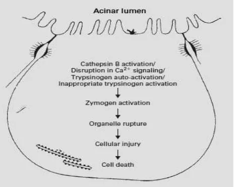
Figure 1. Digestive enzyme activation inside acinar cell
Several mechanism underlie zymogen conversion in pancreas into its active form, such as autoactivation of trypsin from trypsinogen by lysosomal hydrolase cathepsin B (CTSB), zymogen and lysosome leakage to cytoplasm and activation. This acinar cell disorder followed by pancreatic enzyme release to pancreatic tissue. Trypsinogen, chemotrypsin, and elastase in acinar cell will spontaneously activated and furthermore cause autodigestion. Trypsin will also activate complement
dysfunction would lead to increase vascular permeability that trigger proinflammatory cytokines production (IL-1,6,8, TNFĮIUHHUDGLFDODQGDUDFKLGRQDWHDFLG metabolite (prostaglandin, platelet activating factor, leukotriene) and proteolytic enzyme which induce thrombosis and tissue hemorrhage.7
In mild and moderate acute pancreatitis, the necroting segment was stil sterile. Ischemic and necrotic condition will be worsen by bacterial translocation from intestinal via lamina propria, mesenteric lymphnodes, and than into systemic circulation that cause infected necrosis. Bacterial translocation was caused by increase intestinal permeability that could be measured by increase polyethylene glycol (PEG) excretion in urine and antiendotoxin antibody (Endocab). IL-6 concentration reach its highest concentration in 36 hours after the onset of pain and organ dysfunction was found in the third day, so that ‘therapeutic window’ was between 48 to 72 hours after the onset of pain.7
Immune dysregulation was one of the factors that increase mortality in acute pancreatitis and also played an important role in SIRS and multiorgan failure incidence. Sel Th1 and Th 2 imbalance were the main mechanism of immune dysregulation. Th1 cell release
DQWLLQÀDPPDWRU\IDFWRUVVXFKDV,/DQG,/ZKLOH 7KUHOHDVHSURLQÀDPPDWRU\IDFWRUVVXFKDV,/DQG
TNF-alpha. A study by Pietruczuk showed that in acute phase, Th1 cell was suppressed so that the more active
7KZLOOWULJJHUSURIRXQGLQÀDPPDWRU\UHVSRQVH8 In acute pancreatitis with sepsis, 80% among patients was in hypermetabolic condition. Nutritional requirement was increase because of increase resting energy espenditure (REE) and protein breakdown. Negative nitrogen balance was also impact patient prognosis. In acute pancreatitis, nitrogen loss was approximately 20-40 gram each day. Several amino
DFLG GH¿FLHQF\ ZLOO ZRUVHQ LQÀDPPDWLRQ SURFHVV
The increasing endogen gluconeogenesis was a severe
LQÀDPPDWLRQPDQLIHVWDWLRQ,QSDWLHQWZLWKVHSVLVDQG
trauma, endogenous gluconeogenesis could only be suppressed partially by exogenous glucose. Oxygen consumption increase by 20-30% cause the need of more energy supply or reduce perfusion to several vital organs. Both was caused by hypovolemia and reduce
FDUGLDFIXQFWLRQEHFDXVHRILQÀDPPDWLRQSURFHVV8 In acute pancreatitis, capillary permeability will increase and lead to pancreatic enzyme leakage. This will trigger local hydrolysis process of triglyceride that comes from cylomicrone. This hydrolysis process was toxic and induce further pancreatic destruction.
An animal study showed that triglyceride cause pancreatitis and increase serum free fatty acid. This will lead to microthrombus formation that cause tissue ischemia.9 Negative nitrogen balance was correlate to worse prognosis in severe acute pancreatitis. Pancreas also need amino acid for its protein synthesis program.9
Carbohydrate was the main source of energy in acute pancreatitis because of its wide availability, its ability to suppress intrinsic gluconeogenesis, and reduce hyperlipidemia risk. A maximum of 4 mg/ kgBW/minute of glucose can be given with always monitor hyperglycemia and hypercapneu risk. Insulin secretion was also impaired in acute pancreatitis so that parenteral nutrition administration will increase hyperglycemia risk. Insulin resistance could only be corrected by exogenoud insulin administration.9
Table 1. Nutritional requirement in acute pancreatitis
Total calorie NFDONJ%:GD\
Carbohydrate Less than 5 mg/kgBW/minute
Fat /HVVWKDQJUDPNJ%:GD\
Protein JUDPNJ%:GD\
9LWDPLQWUDFHHOHPHQW 9LWDPLQ$&(DQGVHOHQLXP
KgBW: kilogram of body weight
E N T E R A L N U T R I T I O N R O L E I N A C U T E PANCREATITIS
There were no specific therapy yet for acute pancreatitis, but in general an early treatment will improve patient outcome. Fluid resuscitation, analgesic drugs, and antibiotic prophylaxis was the main treatment for acute pancreatitis. However, lower mortality seen in last decade was known to be affected by nutritional management. Enteral nutrition was improved to be the part of acute pancreatitis management.4
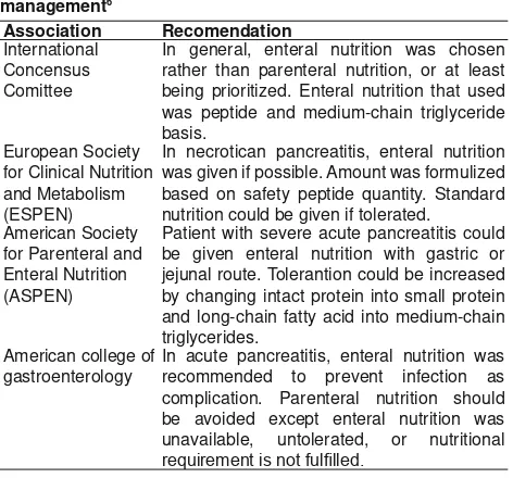
Table 2. Enteral nutrition guideline for severe acute pancreatitis management6
Association Recomendation
International Concensus Comittee
In general, enteral nutrition was chosen rather than parenteral nutrition, or at least being prioritized. Enteral nutrition that used was peptide and medium-chain triglyceride basis.
European Society for Clinical Nutrition and Metabolism (ESPEN)
In necrotican pancreatitis, enteral nutrition was given if possible. Amount was formulized based on safety peptide quantity. Standard nutrition could be given if tolerated.
American Society for Parenteral and Enteral Nutrition (ASPEN)
Patient with severe acute pancreatitis could be given enteral nutrition with gastric or jejunal route. Tolerantion could be increased by changing intact protein into small protein and long-chain fatty acid into medium-chain triglycerides.
American college of gastroenterology
In acute pancreatitis, enteral nutrition was recommended to prevent infection as complication. Parenteral nutrition should be avoided except enteral nutrition was unavailable, untolerated, or nutritional
The importance of nutrition role in acute pancreatitis has been known since 70’s era. Parenteral nutrition was a standard therapy for the last four decades based on ‘pancreatic rest’ concept. ‘Pancreatic rest’ concept told that nutrition administration above mid jejunum will stimulate pancreatic enzyme. According to this, per oral nutrition was started since acute pancreatitis sign reduced so that pancreatic enzyme will turn back to its normal level.4
The rationality of this ‘pancreatic rest’ concept is
WKDW E\ UHVWLQJ LQÀDPHG SDQFUHDV LW ZLOO VWLPXODWH
proteolytic enzyme release and reduce exocrine stimulation. Otherwise, this pancreatic enzyme secretion was known to be inverse with pancreatitis severity. Patients with severe acute pancreatitis will secrete lower level of trypsin, amylase, and lipase than mild to moderate acute pancreatitis patients because
RILWVLQVXI¿FLHQF\,QVXI¿FLHQF\VHYHULW\ZDVEDVHG
on disease severity. Unfunctional acinar cell could not respond to physiologic secretion stimulus.10
This ‘pancreatic rest’ concept was correlate to higher administration cost, increase sepsis risk because
of catheter used, and increase risk of electrolyte and metabolic disorder. Several studies also showed that it also caused gastrointestinal barrier dysfunction and increased permeability. Parenteral nutrition was also induce electrolyte imbalance, hyperglycemia, intestinal barrier dysfunction, and increase intestinal permeability so that ‘pancreatic rest’ was doubt.4
Enteral nutrition was having an immunomodulatory effect for systemic and intestinal mucosa. Intestinal epithelial cell integrity and immune cell along lymphoid tissue together with intestinal barrier played an important role in homeostasis and bacterial translocation. Intestinal epithelial cell was not only functioned as intestina mucosal defence, but also to support lymphocyte cell function and maturation. Intestinal mucosa permeability was the main parameter. Immunoglobulin M was an indirect marker for intestinal permeability. Three RCT shown an IgM reduction in patients received enteral nutrition.4,11
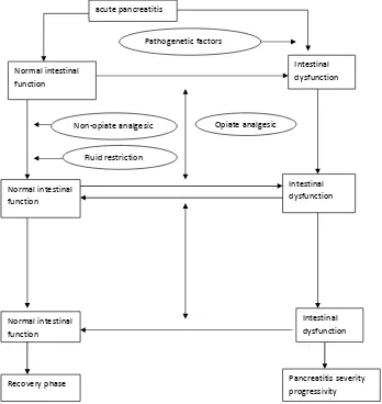
Nowadays, ‘gut rousing’ concept was slowly replace ‘pancreatic rest’. It explained that nutrition administration was needed to stimulate and generate
ĂĐƵƚĞ ƉĂŶĐƌĞĂƚŝƚŝƐ
WĂƚŚŽŐĞŶĞƚŝĐ ĨĂĐƚŽƌƐ
EŽƌŵĂů ŝŶƚĞƐƚŝŶĂů ĨƵŶĐƚŝŽŶ
/ŶƚĞƐƚŝŶĂů ĚLJƐĨƵŶĐƚŝŽŶ
EŽŶͲŽƉŝĂƚĞ ĂŶĂůŐĞƐŝĐ
&ůƵŝĚ ƌĞƐƚƌŝĐƚŝŽŶ
KƉŝĂƚĞ ĂŶĂůŐĞƐŝĐ
EŽƌŵĂů ŝŶƚĞƐƚŝŶĂů ĨƵŶĐƚŝŽŶ
/ŶƚĞƐƚŝŶĂů ĚLJƐĨƵŶĐƚŝŽŶ
/ŶƚĞƐƚŝŶĂů ĚLJƐĨƵŶĐƚŝŽŶ EŽƌŵĂů ŝŶƚĞƐƚŝŶĂů
ĨƵŶĐƚŝŽŶ
ZĞĐŽǀĞƌLJ ƉŚĂƐĞ WĂŶĐƌĞĂƚŝƚŝƐ ƐĞǀĞƌŝƚLJ
ƉƌŽŐƌĞƐƐŝǀŝƚLJ
intestinal function, contrastly differ to ‘pancreatic rest’ concept. In ‘gut rousing’ concept, there were four important phase: (1) Phase I: from onset of acute pancreatitis until hospital administration; (2) Phase II: from hospital administration until 24 hours
DIWHUÀXLGUHVXVFLWDWLRQ3KDVH,,,¿QDOSKDVHRI UHVXVFLWDWLRQWRIXO¿OOHGFDORULHQHHGVKRXUV
after resuscitation end); (4) Phase IV: after phase III until recovery stage.
There were several factors that affect intestinal function and patients outcome in each phase. Acute pancreatitis was started from local pancreas
LQÀDPPDWLRQ VR WKDW LQ 3KDVH , LQWHVWLQDO IXQFWLRQ
is normal. But, not all patients was having normal intestinal function so that therapy in the next phase will have an impact on the later phases. In phase II,
ÀXLGUHVXVFLWDWLRQDQGSDLQPDQDJHPHQWZDVGRQH
Resuscitation in acute pancreatitis should be done aggressively with positive balance, but this increased risk of intestinal submucosal edema and ileus. Opiate as analgesic have a side effect to delay gastric emptying, inhibit gastric motility, reduce pancreatic enzyme secretion and intestinal hormone so that intestinal dysfunction was mainly caused by iatrogenic etiology. Strategies to reduce this effect was giving per oral nutrition in normal intestinal patients and nasogastric tube for patients with intestinal dysfunction. If there were no enteral nutrition administration during phase III, it will worsen intestinal dysfunction and cause a progressive acute pancreatitis. In this phase, per oral nutrition should be consider, or using tube. This nutritional optimalization in this phase III will affect patient prognosis.10
Meta-analysis done by McClave, Marik, and Zalogan showed that enteral nutrition will reduce infection complication, length of stay, and organ-failure risk. However, enteral nutrition in this studies did not reduce mortality. Meta-analysis by Petrov et al involving 202 severe acute pancreatitis patients showed significant mortality difference of 4% compared to 15,9%. A study by Cao et al showed a reduced mortality and organ failure incidence among enteral nutrition intervention group while a
metaanalysis by Yi et al showed a reduced mortality, infection complication, and surgical intervention, but
QRVLJQL¿FDQWGLIIHUHQFHLQOHQJWKRIVWD\,QFOLQLFDO VWLQJV WKHUH ZHUH QR VSHFL¿F FRQWUDLQGLFDWLRQ IRU
enteral nutrition administration. This could still be
DGPLQLVWHUHGDOWKRXJKD¿VWXODDVFLWHVRUSVHXGRF\VW
present, except in severe ileus.12
ENTERAL NUTRITION ADIMINSTRATION TIME
During pancreatitis, intestinal dysfuction will
LQFUHDVH LQÀDPPDWRU\ UHVSRQVH DQG RUJDQ IDLOXUH
risk. In multicenter study, Besselink et al showed
WKDWEDFWHUHPLDZDVKDSSHQHGLQWKH¿UVWVHYHQGD\V
Bacterial translocation will triger SIRS and could lead to sepsis. Early enteral nutrition administration could improve intestinal barrier and prevent bacterial translocation.12
Unstable hemodynamic level cause intestinal ischemia so that enteral nutrition in this phase will worsen ischemia and could lead to necrosis and bacterial overgrowth. Fluid resuscitation was
QHHGHGWR¿OOGHSOHWHGLQWUDYDVFXODUÀXLGDQGUHGXFH
splanchnic vasoconstriction reflex before enteral nutrition administration. Therefore, enteral nutrition was recommended no to be given too early until
SKDVH ,,, H[DFWO\ DIWHU ÀXLG UHVXVFLWDWLRQ (QWHUDO
nutrition could be administered 24-48 hours after stable hemodynamic and intestinal function, and expected to
IXO¿OOQXWULWLRQDOWDUJHWLQKRXUV
Absolute enteral nutrition contraindication was multiple trauma with peritonitis ann retroperitoneal hematoma, intestinal obstrukction, active intestinal bleeding, and unstable hemodynamic level. Relative contraindication where parenteral nutrition was still
QHHGHGLVGLYHUWLFOHDEFHVVPDODEVRUSWLRQ¿VWXODEDG
tolerance, and malnutrition.11
A metaanalysis by Petrov et al that reviwed 11 RCT showed a reduction in organ failure risk, infection complication, and mortality in patients given enteral
QXWULWLRQ LQ WKH ¿UVW KRXUV ,I HQWHUDQO QXWULWLRQ DGPLQLVWHUHGDIWHUKRXUVWKHUHZHUHQRVLJQL¿FDQW
differences. An RCT study by Sun et al investigate
Table 3. Studies that compared enteral and parenteral nutrition12
Reference Year &RXQWU\,QVWLWXWLRQ Sample
Size Control
%HQH¿WRIHQWHUDOFRPSDUHGWR parenteral nutrition
Paraskeva et al Yunani/Pireus General Hospital 23 Parenteral nutrition Lower surgical intervention Olah et al Hungaria/Petz A. Teaching Hospital, Parenteral nutrition Lower sepsis complication
Abou-assi et al $PHULNDVHULNDW9LUJLQLD8QLY+RVS 53 Parenteral nutrition Lower sepsis complication, lower cost Gupta et al Inggris/Southampton General Hospital Parenteral nutrition Shorter length of stay, lower cost Louie et al Kanada/University of Alberta 28 Parenteral nutrition Lower complication and better glycemic
control
enteral nutrition administration in 48 hours and its relation to immune function and outcomes in 60 severe acute pancreatitis patients. Patiens received enteral
QXWULWLRQLQ¿UVWKRXUVVKRZHGDORZHUPXOWLSOH
organ failure syndrome, SIRS, and pancreatic infection compared to patients received enteral nutrition after day-8. Otherwise, mortality among both group was not signiciantly difference.12
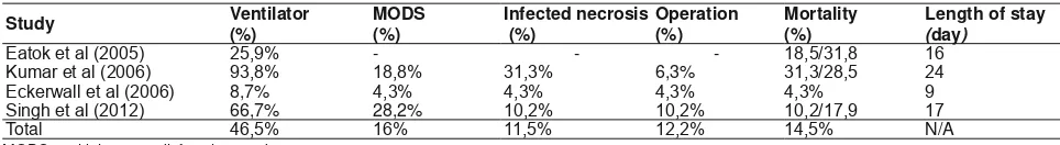
Wereszczynska-Siemiatkowska et al investigate 197 patients with severe acute pancreatitis, result in lower necrosis incidence, respiratory failure, intensive care, and mortality among patients received enteral
QXWULWLRQLQ¿UVWKRXUVFRPSDUHGWRDIWHUKRXUV
Surgical intervention and multi organ failure was also found to be lower. Enteral nutrition in this study was using nasojejunal route. All death subject (9 patients) was from group that received enteral nutrition after 48 hours. There were no difference in local complication among both groups. Infection complication predictor was SIRS > 48 hours, enteral nutrition > 48 hours, and APACHE score in day-3.12
In mild acute pancreatitis, enteral nutrition could be
DGPLQLVWHUHGHDUO\ZLWKRXWDQ\VSHFL¿FLQWHUYHQWLRQ
Study by Eckerwall et al that randomized 50 acute
SDQFUHDWLWLVSDWLHQWVWRFRPSDUHWKHHI¿FDF\DQGVDIHW\
of enteral nutrition in 24 hours to total parenteral nutrition. Enteral nutrition was given via nasogastric route. There were no difference in amilase level and SIRS between both group, but hyperglycemia was found to be lower in enteral nutrition group. Nutrition should not always be started from liquid diet, but it
LVPRUHVDIHWRJLYHORZIDWVRIW¿HW(DUO\QXWULWLRQDO
intervention will reduce length of stay.8
OPTIMAL ROUTE FOR ENTERAL NUTRITION
There were still in debate which location is the best for enteral nutrition administration. There are two options: postpyloric, especially jejenum, and prepyloric in stomach. Postpyloric tube insertion needs endoscopy specialist or radiologist, and this could lead to late eneteral nutrition administration.4
Tube insertion in jejenum was correlate to ‘pancreatic rest’ concept. Based on that concept,
nutritional administration in upper GI tract (above jejenum) will stimulate pancreatic enzyme secretion that worsen patients condition, so that gastric tube insertion was avoided. Nasogastric tube considered to increase aspiration and pancreas stimulation risk. This opinion have been investigated in several studies. 4
Several RCT and meta analysis showed that nutritional administration via nasogastric route and nasojejunal route have a better safety and tolerantion in critical patients. Acute pancreatitis patients tend to have
LOHXVFDXVHGE\LQÀDPHGSDQFUHDV7KLVDOVRVXSSRUW
the evidence that jejunal nutritional administration was better.4
Meta analysis that compare nasogastric and nasojejunal route was reviewing three RCT. In that
VWXG\WKHUHZHUHLQVLJQL¿FDQWGLIIHUHQFHLQPRUWDOLW\
p = 0.97) among both groups. In general, patients
WROHUDQWLRQIURPERWKJURXSZDVQRWVLJQL¿FDQWO\GLIIHU
and no aspiration incidence in both group. RCT that included were consistenly found no difference in safety and tolerantion of both group. 13 A study by Kumar et al showed a higher mortality rate in both group because of late nutritional administration in conservative acute pancreatitis treatment will increase mortality rate.11
Eatock et al investigate the use of gastric tube for severe acute pancreatitis patients, followed by two another RCT, concluded that enteral nutrition administration via nasogastric tube (NGT) was
well-WROHUDWHG DQG QR VLJQL¿FDQW GLIIHUHQFH LQ PRUWDOLW\
and the need of surgical correction. Two meta analysis with 131 severe acute pancreatitis concluded that there were no difference in motality, length of stay, infection complication, and multi organ failure in patients having nutrition via nasogastric tube and conventional method. However, there were still need a larger sample size study for the effectivity of NGT nutritional route tolerantion and safety.12
7DEOH6WXGLHVLQYHVWLJDWHWKHEHQH¿WRIHQWHUDOQXWULWLRQDW¿UVWKRXUVYVDIWHUKRXUV12
Study Year &RXQWU\LQVWLWXWLRQ Sample
size Control
%HQH¿WRIHDUO\KRXUVHQWHUDO nutrition
Sun et al Cina/ Nanjing Medical University
Enteral nutrition in day-8 Lower SIRS, organ failure, and complication
W e r e s z c y n s k a -Siemiatkowska et al
Polandia/ Medical University Bialystok
(QWHUDOQXWULWLRQDIWHU!KRXUV Lower mortality and complication
Sun et al China/Nanjing Medical University
From several study above, nasogastric tube location was not stimulating pancreas but less known about its effect on pancreatic secretion stimulation. A developed hypothesis told that nasogastric tube will stimulate
SDQFUHDWLFHQ]\PHEXWFOLQLFDOO\LQVLJQL¿FDQW14 This was investigated by O’keefe et al. Per oral liquid polymeric diet compared to placebo showed an
LQFUHDVH DPLODVH WU\SVLQ DQG OLSDVH VLJQL¿FDQWO\
Polymeric enteral formula administration into duodenum also stimulate amilase, trypsin, and lipase, otherwise elemental formula only slightly increase lipase. Studies also compared secretory response in nutritional administration at duodenum, mid-jejunal (40-60 cm distal ligamentum treitz) and distal jejunum (100-120 cm distal ligamentum treitz). There were
QRVLJQL¿FDQWGLIIHUHQFHDPRQJWKRVHWKUHHGLIIHUHQW
locations. Low trypsin and lipase secretion were found in elemental formula administration in mid jejenum. This response was similar to control group (fasting). 4
7KHEHQH¿WRIQDVRJDVWULFWXEHZDVQRWRQO\LQ
severe acute pancreatitis, but also in mild and moderate acute pancreatitis.5 Per oral nutritional administration via nasogastric route still stimulate pancreas but its effect was subclinical and did not affect outcomes.1 Mild to moderate acute pacreatitis early nasogastric tube feeding compared with pancreatic rest (MIMOSA) was a study that firstly investigate the safety,
WROHUDQWLRQDQGHI¿FDF\RIQXWULWLRQDODGPLQLVWUDWLRQ
via nasogastric tube in non-severe acute pancreatitis patients. Approximately in 30%, SIRS was found in the early hospital admission with median APACHE score of 6 in both group. This study compared enteral
QXWULWLRQYLDQDVRJDVWULFWXEHLQ¿UVWKRXUVDIWHU
hospital admission to non enteral feeding patients. The result showed that patients given enteral nutrition was not having progressive pancreatitis. This study was also showed that enteral nutrition reduce intensity and duration of abdominal pain, reduce opiate-analgesic needs, and reduce intolerantion risk.14 From several studies, ‘pancreatic rest’ have not been proven to improce acute pancreatitis patients and now ‘gut rousing’ concept was used as an important part of acute pancreatitis management
.
15ENTERAL NUTRITION FORMULA
In acute pancreatitis nutrition management, besides tube location, nutritional formula was also an important thing. Several composition could increase pancreatic secretion response so that in ‘pancreatic rest’ concept, nutritional formula was very important. There were
PRUHWKDQYDULHW\RIIRUPXODPDLQO\FODVVL¿HGLQWR
three types: (1) Elemental, composed of amino acid or oligopeptide, maltodextrin, and medium-chain or
long-Table 5. Studies of nasogastric tubes for acute pancreatitis patients12,13
Study Country Method Control group Average APACHE VFRUH1*71- Time of administrartion
Nutrition
formula Duration
(DWRNHWDO UK RCT Nasojejunal KRXUV Semielemental 5 days
.XPDUHWDO India RCT Nasojejunal KRXUV Semielemantal 7 days
(FNHUZDOOHWDO Sweden RCT Parenteral KRXUV Polymeric GD\V
6LQJKHWDO India RCT Nasojejunal 8,5/8 KRXUV Semielemental 7 days
NGT: nasogastric tube; NJ: nasojejunal
Table 6. Safety and tolarention of nasogastric tube12,13
Study Sample size Diarhea (%)
1*71-Pain after nutritional administration (%)
(FNHUZDOOHWDO 23 - 66% after 7 days
6LQJKHWDO
After 7 days
Total N/A
NGT: nasogastric tube; NJ: nasojejunal
Table 7. Outcome pancreatitis patients with nasogastric tubes12,13
Study Ventilator
Length of stay
(day)
chain triglyceride; (2) Polymeric, composed of non-hydrolized protein, maltodextrin, oligofructosacharide, and long-chain triglyceride; (3) Immune enhancing, composed of substrate that enhance immune activity such as glutamine, arginine, and omega-3 fatty acid,
SURELRWLFDQG¿EHUULFKIRUPXOD
In acute pancreatitis patients, elemental nutrition
VKRZHG VHYHUDO EHQH¿W FRPSDUHG WR SRO\PHULF LQ
absorption, less stimulating pancreas, and highet tolerantion. Otherwise, this elemental nutrition was high cost. Elemental nutrition and immune enhancing formula have a higher osmolarity than polymeric formula, so that it could cause diarrhea. 10 Nutritional composition could increase tolerantion and patients compliance. Patients with severe acute pancreatitis were recommended to received peptide and medium-chain triglyceride formula. Medium-medium-chain triglyceride
ZDVNQRZQWRKDYHDQWLLQÀDPPDWRU\HIIHFWEXWLWV
evidence was limited to animal studies.6
High-fiber formula with prebiotic vs igh-fiber formula
Microbiota inside intestinal lumen have an important role in infection at acute pancreatitis. In peripancreatic
ÀXLGDQDO\VLVEDFWHULDWKDWFDXVHLQIHFWLRQZDVNQRZQ
to come from intestinal. Therefore, improving intestinal bacteria balance was one of the management target that now being studied. In previous study, probiotic has a lowering effect of CRP level and also APACHE II score.6 But, a study by Besselink et al doubt the effects of prebiotic. Besselink randomized 298 patients predicted as severe acute pancreatitis into two groups. Intervention group received probiotic, compared to placebo group. Infection complication in both group
ZDVQWVLJQL¿FDQWO\GLIIHUHQW0RUWDOLW\ZDVIRXQGWR
be higher in intervention group.9
0HWDDQDO\VLVIURP5&7FRPSDULQJKLJK¿EHU
diet with no-fiber diet showed a reduce diarrhea
LQFLGHQFH7KLVVWXG\H[SODLQHGWKDW¿EHUFRXOGEH
considered clinically to reduce diarrhea.11 This enteral
IRUPXODZDVVLJQL¿FDQWO\UHGXFHVHSVLVFRPSOLFDWLRQ
but not in mortality of both group.10 Several studies
VKRZHG WKDW WKHUH ZHUH QR VLJQL¿FDQW GLIIHUHQW LQ
tolerantion, infection complication risk, and mortality between polymeric and enteric formula nutrition.
High-¿EHUGLHWZDVDOVRVDIHEXWDGGLQJLPPXQRQXWULHQRU
probiotic did not improve patients outcome.
Propratria trial showed that probiotic administration in severe acute pancreatitis patient will increase intestinal ischemia risk, organ failure, and mortality,
VLJQL¿FDQWO\ %XWLWVKRXOGEH QRWHGWKDWSURELRWLF
given in this study was widely variated so that high-fiber diet combined with probiotic will increase mortality.15
Immunonutrien in acute pancreatitis
Meta analysis by Asrani et al involving 505 patients from 12 studies showed that glutamine
VXSSOHPHQWDWLRQVLJQL¿FDQWO\UHGXFHPRUWDOLW\55
= 0.30; 95%; CI: 0.15-0.60; p0.001) and infection complication (RR = 0.58; 95% CI: 0.39-0.87; p = EXWGLGQRWUHGXFHOHQJWKRIVWD\7KHEHQH¿W
of glutamine supplementation was clearly found in enteral nutrition. Enteral nutrition supplemented by intravenous glutamine will reduce pancreatic necrosis, infection complication, and length of stay.12
PARENTERAL NUTRITION IN ACUTE PANCREATITIS
Based on ESPEN recommendation, parenteral nutrition indication in acute pancreatitis was inadequacy of enteral nutrition after 5-7 days. This period could be shorter in formerly malnourished patients. Complication
VXFKDVLOHXVSVHXGRF\VWSDQFUHDWLF¿VWXODLQWHVWLQDO
edema, pancreatic ascites, and infected necrosis impair enteral nutrition administration so that those patients was recommended to received parenteral nutrition. There were no absolute contraindication for parenteral nutrition. Parenteral nutrition could be started in 24-48 hours after resuscitation and stable hemodynamics.14 In mild acute pancreatitis study, partenteral nutrition for 5-7 days have no positive effect so that it is not recommended, except in malnourished patients. Substrate metabolism in severe acute pancreatitis was similar to severe sepsis patients. Daily nitrogen target was 0,2-0,24 gram/kgBW. This target was similar to amino acid administration of 1,2-1,5 gram/kgBW/ day. A reduction of 0,14-0,2 gram nitrogen/kgBW/day should be done in patients with chronic kidney disease or liver disease.16
Glutamine was an amino acid that have a central role in metabolism process such as nitrogen transport between organs, anabolize carbons, nucleotide and glutation precursor, and acid base regulator. An RCT study in moderate acute pancreatitis explained that parenteral nutrition with glutamine reduce overall complication and also reduce length of stay. There were no data for other amino acid administration in acute pancreatitis patients, such as BCAA, EAA, and arginine.14
but hyperglycemia as side effect should be prevented. In acute pancreatitis and other critical condition, glucose oxidation reach its maximum level, about 4-7 mg/kgBW/minute, similar to 5-6 gram/kgBW/day. if glucose consumption higher than this limit, lipogenesis, hypercapneu, and hyperglycemia will happen. This risk was increasing in severe acute pancreatitis because of
SDQFUHDWLFQHFURVLVDQGLQÀDPPDWLRQUHGXFHLQVXOLQ
secretion. Data of non-carbohydrate glucose in acute pancreatitis patients was very limited.1
OR TOLERANTION AND NUTRITIONAL ADEQUACY
Nutritional and metabolic monitor was aimed to control macronutrien (glucose, protein, fat) and micronutrient level, evaluate energy, and control glycemia. Macronutrien was evaluated intensively in 48 hours. In acute phase, blood glucose lecel was evaluated several time in a day, and reduce to once a day after acute phase. Electrolyte, ureum, creatinine, phosphate, magnesium, and liver function
Not well-tolerated or inadequate calorie intake in 3-5 days
Well-tolerated: no pain, no abdominal distension
Pancreatitis severity assessment: mild or moderate
Start enteral nutrition in 24
Increase diet progressively, evaluate tolerantion
Combine with parenteral nutrition
Normal diet in 5-7 days
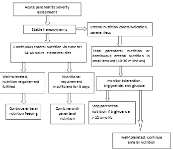
Acute pancreatitis severity assessment
Stable hemodynamic
Continuous enteral nutrition via tube for 24-48 hours, elemental diet
Enteral nutrition contraindication, severe ileus
Total parenteral nutrition or continuous enteral nutrition in small amount (10-30 ml/hours)
Nutritional requirement insufficient for 3 days
Combine with parenteral
nutrition
monitor tolerantion, triglyceride, and glucose
Stop parenteral nutrition if triglyceride > 12 umol/L
well-tolerated: continue enteral nutrition Well-tolerated,
nutrition requirement fulfilled
Continue enteral nutrition feeding
Figure 3. Nutritional management of mild to moderate acute pancreatitis
were monitor before nutritional administration. If abnormalities found, daily evaluation was done until normal, then continued to several times in a week. Albumin was not evaluated in acute phase but every week in post acute and rehabilitation phase. Body weight and muscle mass were evaluated in post acute and rehabilitation phase. 16
CONCLUSION
Acute pancreatitis management was aimed to support hemodynamic and prevent infection complication. Patients with acute pancreatitis was susceptible to be malnourished because of lower intake and higher catabolism processs so that nutritional management was
YHU\LPSRUWDQWHVSHFLDO\WRIXO¿OOQXWULWLRQDOUHTXLUHPHQW
and modulate immunity. ‘Pancreatic rest’ concept’ was now slowly replaced by ‘gut rousing’ concept. Enteral nutrition was chosen rather than parenteral nutrition or fasting because it lower length of stay, infection complication risk, and have a lower budget. Parenteral nutrition was still be used if nutritional intake was inadequate during 3-5 days or therapy, or in the condition of enteral nutrition contraindicated. Otherwise, otatl parenteral nutrition will increase infection risk, metabolic complication, electrolyte disturbance,
KLJKHUFRVWDQGKLJKHUPRUWDOLW\ULVNLQJHQHUDO)XO¿OO
nutritional requirement and prevent pancreas exocrine stimulation should not alter the main goal of therapu to increase intestinal function.
In mild acute pancreatitis, enteral nutrition could be administered as soon as possible since there were
no contraindication. Nutritional administration via nasogastric tube was better than nasojejunal, and proven not to worsen pancreatitis condition. Parenteral nutrition should be acouded except enteral nutrition
FRXOGQRWIXO¿OOGDLO\FDORULHUHTXLUHPHQW1XWULWLRQDO
and metabolic nutrition should be done regularly to control macronutrien (glucose, protein, fat) and micronutrient, evaluate total energy, control glycemia and hypertriglyceride.
REFERENCES
1. Sun E, Tharakan M, Kapoor S. Poor compliance with ACG guideline for nutrition and antibiotics in the management of acute pankreatitis: a North American survey of gastrointestinal specialists and primary care physicians. J Pancreas 2013;14:221-7.
2. Ahmad AS, Iain JD, Mc CB, Peter E, Coyne A, Keith S. Nutritional strategies in severe acute pankreatitis: a systematic review of the evidence. The surgeon 2010;8:105–10. 3. Tenner S, Baillie J, De WJ. American collage of
gastroenterology guideline: management of acute pancreatitis. Am J Gastroenterol 2013;108:1400-15.
4. 3HWURY01XWULWLRQ,QÀDPPDWLRQDQGDFXWHSDQNUHDWLWLV
,651,QÀDPPDWLRQ
5. Uomo G. Pancreatic rest or not? The debate on the nutrition in acute pankreatitis outcome. Journal of the Pancreas 2013;14(2) 6. Refaat A Hegazi, Tiffany DeWitt. Enteral nutrition and immune
modulation of acutepankreatitis. World J Gastroenterol 2014;20:16101-5.
7. Feldman M, Feldman L, Brand L. Gastrointestinal and liver disease ninth edition volume 1. Elsevier 2010
8. Sun JK, Mu XW, Li W. Effect of early enteral nutrition on immune function of severe acute pankreatitis patient. World J Gastroenterol 2013;19:917-22.
9. Petrov M. Moving beyond the ‘pancreatic rest’ ini severe and critical acute pancreatitis. Petrov Critical Care. 2013;17:161-5.
7DEOH0RQLWRULQJSDUHQWHUDODQGHQWHUDOQXWULWLRQLQVHYHUHDFXWHSDQFUHDWLWLV17
Criteria Aim Method Period
Energy target 3UHYHQWHQHUJ\GH¿FLW Calculation of energy expenditure with indirect calorimeter
,QKRXUVDWSDVWDFXWHDQG
rehabilitation phase
Protein and fat target 3UHYHQWSURWHLQDQGIDWGH¿FLW Prediction ,QKRXUVDWSRVWDFXWHDQG rehabilitation phase
Micronutrien 3UHYHQWPLFURQXWULHQWGH¿FLHQFXDQG optimize macronutrien metabolism
Monitoring sheet Daily
Glycemia Prevent overfeeding and hypoglycemia Laboratory examination In acute phase: several times In post acute phase: daily Insulin dosage Prevent overfeeding and hypoglycemia Dynamic algoritm of therapy In acute phase: several times
In post acute phase: daily Electrolyte, ureum,
creatinine, liver function
Prevent refeeding syndrome Laboratory examination At the beginning. If abnormal, evaluate daily. If normal, evaluate 2-3 times/ week
Albumin Know nutritional status while
LQÀDPPDWLRQQRWSUHVHQW Laboratory examination Post acute: weeklyNot recommended in cute phase
EHFDXVHRILQÀDPPDWLRQSUHVHQW
Body weight, BMI Nutritional status evaluation Body weight instrument In post acute and rehabilitation phase. Not in acute phase because of hidration variety
Free fat mass, intra and
H[WUDFHOOXODUÀXLG Nutritional status Bioimpedance analysis Postacute and rehabilitation phase ZKHQQRÀXLGUHWHQWLRQ
Muscle strenght Evaluate muscle strenght Dynamometer Rehabilitation phase (post ICU)
10. Maxim S. Petrov and John A. Windsor. Nutritional management of acute pankreatitis: the
11. concept of ‘gut rousing’. Curr Opin Clin Nutr Metab Care.2013;16:557–63.
12. Kumar A, Singh N, Prakash S.. Early enteral nutrition in severe acute pankreatitis: a prospective randomized controlled trial comparing nasojejunal and nasogastric routes. Journal of Clinical Gastroenterology 2006;40:431-4.
13. Olah A, Romics L. Enteral nutrition in acute pancreatitis: a review of the current evidence.World J Gastroenterol 2014;20:16123-31.
14. Yu-sui Chang, Hua-qun Fu, Yuan-mei Xiao. Nasogastric or nasojejunal feeding in predicted severe acute pancreatitis: a meta-analysis. Critical Care 2013;17:2-9.
15. Gianotti L, Meier R, Lobo DN. ESPEN guideline on parenteral nutrition: pancreas. Clinical Nutrition 2009;28:428-35. 16. Besselink MG, Van SH, Buskens E. Probiotic prophylaxis in
predicted severe acute pankreatitis: a randomised, double-blind, placebo-controlled trial. Lancet 2008;371:651-9. 17. Thibault R, Heidegger C, Bergerc. Parenteral nutrition in the
intensive care unit: cautious use improves outcome. Swiss Medical Weekly 2014;144:13997.
18. Nguyen N. Pharmacological therapy of feed intolerance in the critically ills. World J Gastrointestinal Pharmacol Ther