High-frequency transformation of oat via microprojectile
bombardment of seed-derived highly regenerative cultures
Myeong-Je Cho *, Wen Jiang
1, Peggy G. Lemaux
Department of Plant and Microbial Biology,Uni6ersity of California,Berkeley,CA94720,USA Received 23 February 1999; received in revised form 3 May 1999; accepted 5 May 1999
Abstract
A highly efficient and reproducible transformation system for oat (A6ena sati6a L. cv. GAF/Park-1) was developed using microprojectile bombardment of highly regenerative tissues derived from mature seeds. Callus was induced under dim light conditions on medium containing 2,4-dichlorophenoxyacetic acid (2,4-D), 6-benzylaminopurine (BAP) and high cupric sulfate. Highly regenerative tissues, generated from embryogenic callus, were used as a transformation target. From 327 individual explants bombarded with theb-glucuronidase gene (uidA;gus) and a hygromycin phosphotransferase gene (hpt), 84 independent transgenic events were obtained after an 8 – 12-week selection period on hygromycin. All events were regenerable, giving an effective transformation frequency of 26%; co-expression of GUS activity occurred in 70% of the independent events. Presence of the foreign genes in DNA from leaf samples of T0and T1plants was confirmed by polymerase chain reaction (PCR) amplification
and/or DNA blot hybridization. Fertility of the plants from the transgenic lines was 63% (24/38) and the transgene(s) was stably transmitted to T1and T2progeny. © 1999 Elsevier Science Ireland Ltd. All rights reserved.
Keywords:A6ena sati6aL.; Mature seed; Embryogenic callus; Highly regenerative tissue; Transformation
www.elsevier.com/locate/plantsci
1. Introduction
Introduction of genes using genetic engineering is dependent on efficient, reproducible in vitro culturing methods. Successful transformation of oat using microprojectile bombardment of cell sus-pension cultures or embryogenic calli developed from immature embryos was reported [1 – 4]. How-ever, the utility of these target tissues is limited because the regenerability decreases and so-maclonal variation increases during the prolonged culturing periods [5] necessary for transformant identification. Other in vitro-derived tissues have also been used in successful transformation
experi-ments with oat. These include mature embryo-derived callus tissue [6,7], leaf base segments of young oat seedlings [8,9] and shoot meristem cul-tures initiated from the shoot apex of germinated seedlings [10 – 12].
We have recently established an efficient in vitro system to proliferate highly regenerative tissues from immature scutellar tissues of barley [13,14]. These tissues, which yield large numbers of shoots during regeneration and can be maintained for more than a year with minimal loss in regenerabil-ity, have been used for the successful transforma-tion of previously recalcitrant commercial barley cultivars [13,14]. Genomic DNA methylation analyses showed that barley plants regenerated from the highly regenerative tissues are less vari-able with regard to those regenerated from em-bryogenic callus tissues in methylation pattern polymorphism and agronomic performance [15]. In this report we describe the development from mature oat seed of similar highly regenerative tissues to be used as transformation targets. We
Abbre6iations:2,4-D, 2,4-dichlorophenoxyacetic acid; BAP, 6-ben-zylaminopurine; CIM, callus-induction medium; GUS, b -glu-curonidase;hpt, hygromycin phosphotransferase gene.
* Corresponding author: Tel.: +1-510-6421347; fax: + 1-510-6427356.
E-mail address:[email protected] (M.-J. Cho) 1Present address: Calgene Inc., Davis, CA 95616, USA
M.-J.Cho et al./Plant Science148 (1999) 9 – 17
10
describe the generation of a large number of inde-pendently transformed lines that give rise to trans-genic plants.
2. Materials and methods
2.1. Plant material and culture of explants
Mature seeds of GAF/Park-1, a spring cultivar
of oat (A6ena sati6aL.), were surface-sterilized for
20 min in 20% (v/v) bleach (5.25% sodium
hypochlorite) followed by three washes in sterile water. Seeds were placed on three different MS-based [16] callus-induction media (CIMs)
contain-ing different combinations of
2,4-dichloro-phenoxyacetic acid (2,4-D), 6-benzylaminopurine
(BAP) and CuSO4(Table 1): (1) D%-2.0 mg/l 2,4-D
and 0.1mM CuSO4, (2) D%BC2-2.0 mg/l 2,4-D, 0.1
mg/l BAP and 5.0mM CuSO4, and (3)
DBC3-con-taining 1.0 mg/l 2,4-D, 0.5 mg/l BAP and 5.0 mM
CuSO4 [13]. Then 5 – 7 days after initiation,
germi-nating shoots and roots were completely removed by manual excision. After 3 weeks of incubation at
2491°C under dim light (approximately 10 – 30
mE, 16-h light), high-quality tissues with nodular and embryogenic structures were selected and maintained on each medium, subculturing at 3 – 4-week intervals.
2.2. Shoot regeneration test
A total of ten pieces (4 – 6 mm) of 5- and 7-month-old oat tissue, which had been main-tained on each medium, were selected and placed
on BCI-DM− media (phytohormone-free CIMs)
[13,20] for regeneration at a light intensity of
approximately 45 – 55mE; each treatment had four
replicates. After 4 weeks on regeneration medium, the numbers of pieces of highly regenerative tissue producing green sectors and the numbers of shoots per piece of highly regenerative tissue were counted. If more than one leaf arose from the same tissue base, it was counted as one shoot.
2.3. Plasmids
Plasmids, pAct1IHPT-4 and pAHC15, were used for transformation. pAct1IHPT-4 [13]
con-tains the hygromycin phosphotransferase (hpt)
gene under control of the rice actin1 promoter
(Act1), its intron (Act1I) and the nos 3% end.
pAHC15 [17] contains theuidAgene under control
of the maize ubiqutin UbiI promoter and intron
(Ubi1I) and nos.
2.4. Particle bombardment and stable transformation
Approximately 4 – 5-month-old highly
regenera-tive tissues, induced on D%BC2 medium and
main-tained on D%BC2 or DBC3 medium, were used as
targets for bombardment. Tissues (3 – 4 mm) were
transferred for osmotic pretreatment to D%BC2 or
DBC3 medium containing equimolar amounts of mannitol and sorbitol to give a final concentration of 0.4 M. After 4 h, tissues were bombarded as previously described [18] with modifications. In
summary gold particles (1.0 mm; Analytical
Scien-tific, Alameda, CA) were coated with 25 mg of a
1:1 molar ratio of pAct1IHPT-4 and pAHC15 followed by bombardment using a PDS-1000 He biolistic device (Bio-Rad, Hercules, CA) at 900 psi. Then 16 – 18 h after bombardment, tissues
were moved to D%BC2 or DBC3 medium without
osmoticum or selective agent and grown at 249
1°C under dim light.
Following the initial 10 – 14-day culturing pe-riod, each tissue was broken into one to three pieces (3 – 4 mm), depending on initial tissue size,
and transferred to D%BC2 or DBC3 medium with
20 mg/l hygromycin B (Boehringer Mannheim,
Mannheim, Germany). From the second-round selection onward, tissues were subcultured at 3 – 4-week intervals on the same medium containing 20
mg/l hygromycin B. When sufficient rapidly
grow-ing material was available, tissues were moved to
non-selective BCI-DM− or FHG regeneration
medium [19,20] and exposed to higher intensity
light (approximately 45 – 55 mE). After 4 weeks,
regenerated shoots were transferred to Magenta
boxes containing BCI-DM− without selective
agent. When the shoots reached the top of the box, plantlets were transferred to the soil.
2.5. Histochemical GUS assay
Highly regenerative, transgenic tissue and leaf
material from T0 to T2 plants transformed with a
mixture of pAct1IHPT-4 and pAHC15 were tested for GUS activity by histochemical staining [21],
.-J
.
Cho
et
al
.
/
Plant
Science
148
(1999)
9
–
17
11
Table 1
Regenerability of oat tissues grown on different mediaa
No. tissues with green sectors/total no.
Medium Composition Tissue age No. shoots/ Tissue age No. shoots/
pieces of tissue
(months) tissue piece (months) tissue piece 2,4-D (mg/l) BAP (mg/l) CuSO4(mM)
4.690.7 7 1.490.8 5
0 0.1 9.591.0/10
D% 2.0
2.0 0.1 5.0 10.090.0/10 10.891.0 7 5.791.6
D%BC2 5
5 10.090.0/10 8.290.4 7 6.191.6 DBC3b 1.0 0.5 5.0
aA total of ten pieces (4–6 mm) of 5- and 7-month-old tissue from each treatment were transferred to regeneration medium; after 4 weeks, the numbers of shoots were
counted. Values represent means9S.D. of four replicates for each treatment.
M.-J.Cho et al./Plant Science148 (1999) 9 – 17
12
acid (X-gluc) (Gold Biotechnology, St. Louis, MO). Samples were incubated overnight at 37°C in GUS assay buffer before being scored.
2.6. Genomic DNA isolation, polymerase chain reaction (PCR) and DNA blot hybridization
Total genomic DNA from leaf tissues of puta-tively transformed plants was purified as described
[22]. To test for the presence of uidA and hpt in
genomic DNA of putatively transformed lines, 500 ng of genomic DNA was amplified by PCR using
the primer set, UIDA1 (5%
-agcggccgcaTTACGTC-CTGTAGAAACC-3%) plus UID2R (5%
-agagctcT-CATTGTTTGCCTCCCTG-3%) [13] or HPT6F
(5%-AAGCCTGAACTCACCGCGACG-3%) plus
HPT5R (5%
-AAGACCAATGCGGAGCATATA-C-3%) [13], respectively. Amplifications were
per-formed in a 25-ml reaction with Taq DNA
poly-merase (Promega, Madison, WI) according to a protocol described by Cho et al. [13]. For DNA
hybridization analysis, 10 mg of total genomic
DNA from leaf tissue of each line was digested
with BamHI and EcoRI, separated on a 1.0%
agarose gel, transferred to Zeta-Probe GT mem-brane (Bio-Rad, Hercules, CA) and hybridized
with a radiolabeled uidA-specific probe following
manufacturer’s instructions. The uidA-containing
1.9-kb BamHI-SacI fragment from pAHC15 was
isolated with QIAEX gel extraction kit (QIAGEN,
Chatsworth, CA) and labeled with a-32P-dCTP
using random priming according to manufactur-er’s instructions (Promega, Madison, WI).
3. Results
3.1. Establishment of an in 6itro system
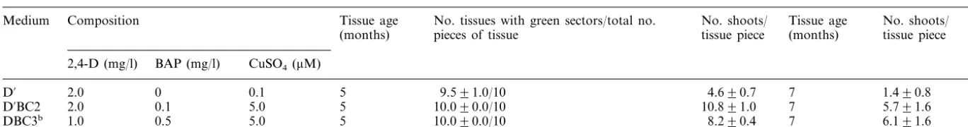
To establish a highly efficient in vitro system for culturing and regenerating oat tissue, three differ-ent media were tested (Table 1). A higher callus induction frequency and larger numbers of
em-bryogenic structures were observed on D% medium
(Fig. 1A) and D%BC2 medium than on DBC3. The
use of DBC3 at the initial callus-induction step resulted in high rates of seed germination and low frequencies of callus induction (data not shown); however, DBC3 was optimal for maintaining highly regenerative tissues derived from
embryo-genic callus initiated on D% or DBC2 medium
(Table 1; Fig. 1B).
The addition of BAP and copper to 2,4-D in
D%BC2 and DBC3 medium resulted in the
produc-tion of greater numbers of shoots per piece of tissue on regeneration medium compared to
tis-sues derived from D% medium (Table 1). The
fre-quency of shoot regeneration was increased
2.3 – 4.1-fold and 1.8 – 4.4-fold with D%BC2 and
DBC3 medium, respectively, compared to D%
medium. The extent of negative effects of the length of culture time on regenerability varied with
the three media tested (Table 1). The tissues on D%
medium resulted in a smaller percentage of regen-erated shoots at 7 months compared to 4 months than did tissues from the other two media; tissues from DBC3 yielded the highest percentage. Tissues maintained on DBC3 appeared to maintain regen-erability better than tissues on the other two media.
3.2. Bombardment and selection of transgenic tissues
For bombardment, 4 – 5-month-old highly
re-generative tissues, initiated on D%BC2 and
main-tained on D%BC2 or DBC3 medium, were used.
During the selection period on hygromycin non-transgenic tissues gradually turned brown; the presence of light-green, hygromycin-resistant tis-sues was generally observed at the third round of selection (Fig. 1C). Putative transgenic tissues were maintained and proliferated on the same medium after the fourth round of selection, until sufficient tissue was obtained for regeneration. Us-ing this transformation protocol, 84 independent transgenic lines were obtained from 327 pieces of bombarded tissue, giving a 26% transformation frequency (Table 2). Since all transformed lines were regenerable, this gives an effective
transfor-mation frequency (total no. lines regenerable/total
no. lines) [14] of 26%. To date 63% (24/38) of
independent T0 lines were fertile (Table 2), but
with varying levels of seed set (data not shown).
3.3. Analysis of T0 plants and their progeny
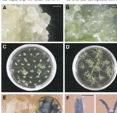
Presence of uidA in genomic DNA of
indepen-dent T0 oat lines was confirmed by DNA
hy-bridization analysis. Genomic DNA from T0
plants of ten out of 11 lines tested that were transformed with pAHC15 and positive in PCR
frag-ment. In some cases other fragments were present
after digestion with BamHI and EcoRI; line
AS-GPB%-17 produced only a larger-sized fragment
(Fig. 2). Copy numbers of uidA ranged from one
to more than ten copies per genome.
Histochemi-cal analysis for GUS activity in T0 plants derived
from the putative transgenic lines provided addi-tional evidence of stable transformation. GUS ex-pression was detected in putatively transformed cultured callus tissues, and leaf (Fig. 1E), anther, ovary and stigma (Fig. 1F) tissues. Out of 84
independent hygromycin-resistant lines examined, 70 lines contained both genes, giving an 83% cotransformation frequency; 59 were positive for GUS expression, giving a 70% coexpression fre-quency (Table 2).
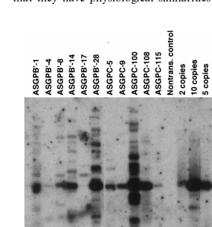
Segregation of GUS expression was analyzed in
T1 progeny of eight fertile transgenic oat lines
(ASGPB%-1, -4, -8, -10, ASGPC-5, -6, -100 and
-105); one of these lines ASGPB%-10 did not
ex-press GUS in T0 plant tissue (Table 3; Fig. 3). Of
the seven lines that expressed GUS in T0 plants,
Fig. 1. Production of transgenic oat (GAF-30/Park-1) lines and GUS expression in transgenic plants. (A) Embryogenic callus tissues of oat initiated from mature seeds on D%medium and maintained for 3 months under dim light. Bar=0.5 mm. (B) Highly regenerative tissues of oat initiated from mature seeds on D%BC2 medium and maintained for 3 months under dim light on DBC3 medium. Bar=0.5 mm. (C) Oat tissues on third round of selection on hygromycin-containing medium (photographed 10 days after transfer). (D) Plantlets regenerated from highly regenerative tissues of a transgenic oat line. (E) GUS activity in leaf tissue of non-transgenic (left) and transgenic oat (ASGPB%-4) (right). (F) GUS activity in anther (left), ovary and stigma (right) tissues
M.-J.Cho et al./Plant Science148 (1999) 9 – 17
14
Table 2
Summary of oat transformation experiments
Maintenance % Coexpression fre- % Regenerability % Fertility (no. fertile Tissue age (days % Transformation
fre-medium quency (no. indepen- (no. regenerable lines) lines/no. lines after induction) quency (no. lines with
dents/no. bombarded GUS activity/ no. regenerated) total lines)
explants)
100 (21)
18.6 (21/113) 66.7 (14/21) 58.3 (7/12) 112 D%BC2a
17.8 (8/45) 62.5 (5/8) 100 (8) 62.5 (5/8) DBC3b
73.5 (25/34) 100 (34)
133 D%BC2 33.0 (34/103) 50.0 (3/6) 100 (21)
71.4 (15/21) 75.0 (9/12) 31.8 (21/66)
DBC3
100 (84) 63.2 (24/38) Total 25.7 (84/327) 70.2 (59/84)
aD%BC2 medium is callus-induction medium containing 2.0 mg/l 2,4-D, 0.1 mg/l BAP and 5.0mM cupric sulfate. bDBC3 medium is callus-induction medium containing 1.0 mg/l 2,4-D, 0.5 mg/l BAP and 5.0mM cupric sulfate.
GUS expression was detected in T1 progeny from
six of these lines (ASGPB%-1, -4, -8, ASGPC-5, -6,
-100 and -105); one line (ASGPC-6) did not express
GUS in the T1plants (Table 3). Of the six
GUS-ex-pressing lines, only one line (ASGPB%-4) segregated
in the expected 3:1 ratio, four lines (ASGPB%-1, -8,
ASGPC-100 and -105) at lower than 3:1 and the remaining line (ASGPC-5) at higher than 3:1.
GUS expression and physical transmission of the
transgenes,uidAandhpt, were also analyzed in two
different T1segregating populations (ASGPB%-4 and
ASGPC-100). Expression of GUS in T1progeny of
ASGPB%-4 was consistent with the presence ofuidA,
as assessed by PCR. From both ASGPB%-4 and
ASGPC-100 all T1plants that were PCR-positive for
uidAwere also PCR positive forhpt. Expression of
GUS in ASGPB%-4 was strong, in ASGPC-100 weak
(Fig. 3). All seven GUS-positive T1 progeny of
ASGPB%-4 produced T2progeny with strong GUS
expression; three putative homozygous progeny
(ASGPB%-4-3, -6 and -8) were obtained (Fig. 3). A
total of five of six GUS-positive T1 progeny from
ASGPC-100 tested had weak GUS expression in T2
progeny (ASGPC-100-1, -2, -3, -4, -5, and -7); ASGPC-100-6 did not express GUS (Fig. 3).
4. Discussion
In this report, 84 transgenic lines of oat were obtained using microprojectile bombardment of 327 pieces of highly regenerative, green tissue derived from embryogenic callus induced from mature seeds. The transformation frequency (the number of independent transformants per total number of explants) was 26%, higher than that higher than that reported previously using other target tissues [1,2,6,9,11].
One of the main factors responsible for the high
transformation frequency reported in this study is likely to be the use of highly regenerative, green tissues as transformation targets. These tissues contain multiple, light-green, shoot meristem-like structures. In oat and barley the expression of a gene associated with maintenance of the
meristem-atic state, a knotted 1 homologue, was studied in
shoot meristem cultures derived from the excised shoot apex in oat [11] and barley [23] and from highly regenerative tissue (barley only) [14]. The pattern was similar in the two tissues, suggesting that they have physiological similarities [14].
Fig. 2. Hybridization analysis of DNA from T0plant tissues.
A total of 10mg of genomic DNA per lane, isolated from leaf tissues of non-transformed control and 11 uidA-positive T0
Table 3
Histochemical GUS activity in T0and T1plants
Transgenic GUS activity in GUS activity in T1
leaf tissue (+:−) T0leaf tissue
linea
+
ASGPB%-1 2:26 ASGPB%-4 + 18:4b
15:18
+
ASGPB%-8
–
ASGPB%-10 0:24
25:0 ASGPC-5 +
0:33
+
ASGPC-6
+
ASGPC-100 16:18
+
ASGPC-105 4:21
aASGPB% lines were initiated, maintained and selected on
D%BC2 medium while ASGPC lines were initiated on D%BC2,
maintained and selected on DBC3 medium.
bAnalyses using the x2-test indicated that the segregation
ratios of T1progeny were not significantly different from the
expected 3:1 (ata=0.05).
represent a high-quality target tissue for transfor-mation. The highly regenerative tissues of oat could be maintained for more than 1.5 years with minimal loss in regenerability; similar results were observed with highly regenerative, green tissues of barley [13]. All transgenic lines (100%) in this study produced multiple green shoots and were regenerable. In contrast to this situation, regenerability of trans-genic oat lines from immature embryo-derived cul-tures was 36% [2], from mature embryo-derived cultures, 58% [6] and from leaf base-derived cul-tures, 91% [9]. Of the transgenic lines derived from cultured axillary meristematic tissue, 100% were regenerable, which is not unexpected given the developmental similarities between the highly re-generative, green tissues and the shoot meristem cultures [11,14].
Despite the improvement in the in vitro culture system and regenerability of the transformed cul-tures in this study, on average only 63% of trans Highly regenerative, green tissues with a high
percentage of cells capable of sustained cell division and competent for regeneration over long periods
Fig. 3. PCR and transgene expression analysis of T1and T2plants. PCR products amplified from T1plants of two independent
lines were visualized by electrophoresis on a 0.8% agarose gel stained with ethidium bromide. (A) Lane 1, marker; lanes 2 – 16 contain the primed fragments from eight progeny of ASGPB%-4 (strong GUS-expressing line) and seven progeny of ASGPC-100
(weak GUS-expressing line) using the primers UIDA1 and UIDA2R. Lane 17 is from non-transformed tissue; lane 18 from plasmid pAHC15. (B) Lane 1, marker; lanes 2 – 16 contain the primed fragments from eight progeny of ASGPB%-4 and seven progeny of ASGPC-100 using the primers HPT6F and HPT5R. Lane 17 is from non-transformed tissue; lane 18 from plasmid pAct1IHPT-4. (C) GUS activity in T1 leaf tissue was determined as high expression (+ + +), low expression (+) or no
M.-J.Cho et al./Plant Science148 (1999) 9 – 17
16
genic lines yielded plants that were fertile; of those varying levels of seed set were observed. Tissues maintained on DBC3 appeared to have a higher level of fertility (70%) versus those maintained on
D%BC2 (56%). This might be due to the fact that
tissues maintained on DBC3 were more organized
than those on D%BC2 [13] and therefore gave rise
to more phenotypically normal plants. The phe-nomenon of reduced fertility or sterility has been
observed frequently in transgenic cereals
[13,20,24,25], especially in oat [1,2,6,9,11,12]. Pre-viously Somers et al. [1] reported that 34% (38 of 111) of transgenic lines generated from cell suspen-sion cultures or embryogenic calli derived from immature embryos gave rise to green plants; only one line was fertile. Fertility of transgenic oat plants produced from immature embryo-derived calli, mature seed-derived calli and leaf base seg-ments ranged from 19 to 64% [2,6,9]. Recently it was reported that 71% of transgenic lines pro-duced from cultured axillary meristematic tissue were fertile [11], comparable to the rate of plants maintained on DBC3 in this study. In general, the reduction in fertility in transgenic plants, relative to plants that have not undergone in vitro culture, might be related to the cytological instability that occurs during in vitro culture period associated
with transformation or to the physiological
changes in the transgenic plants in terms of the synchrony of pollen and egg development.
PCR analyses on DNA extracted from T1 leaf
tissue indicated that hpt alone or hpt and uidA
were present in all 84 transgenic lines. A total of 70 lines contained both genes, giving an 83%
cotransformation frequency; coexpression
fre-quency was 70%. DNA hybridization analysis
confirmed the presence of uidA in the genomic
DNA of all 11 transgenic lines analyzed, with copy numbers ranging from one to more than ten copies, similar to that reported in earlier studies using microprojectile bombardment [1,3].
Most GUS-positive lines in our study had strong GUS expression uniformly throughout the highly regenerative tissues; however, tissues from some transgenic lines were chimeric with respect to GUS expression (data not shown). This could be due to transgene silencing in certain parts of the tissue, survival of non-transformed tissues under the selection pressure used, or the fact that two or more transgenic lines were present in the same piece of bombarded tissue and one of the lines did
not have or express GUS. Expression of GUS was
shown to be stably inherited in T1 progeny of six
out of seven GUS-expressing T0 plants tested. In
general the level of GUS expression observed in T0
plants was stably inherited in their progeny.
T0 lines with a single site of transgene
integra-tion should give a segregaintegra-tion ratio for transgene
expression of 3:1, but one line (ASGPB%-4) with a
single copy did give this expected ratio. The cause of some lines having a lower than 3:1 ratio could be due to several factors. In some earlier reports lower segregation ratios were due to loss or low-rates of physical transmission of the transgene(s) to progeny [26 – 28]. This is not likely to be the explanation for ASGPC-100 since PCR analysis of
DNA from T1 progeny of this line indicated that
uidA was present. The more likely explanation for
the distorted segregation ratios is transgene silenc-ing, which has been reported to occur in trans-genic oat plants [11,26,27,29]. Lack of transgene expression in progeny may also be due to muta-tions, chromosomal changes and methylation changes, which have the potential to exert pleitropic effects on transgene expression and in-heritance (for review see Ref. [27]).
For skewed expression ratios higher than 3:1, it is possible that the two genes could be integrated into different chromosomes in the transgenic plants, causing them to segregate in the higher
ratio. Of the two transgenic lines studied in the T2
generation, expression of GUS was stably
trans-mitted to most T2 plants (strong GUS expression
in ASGPB%-4 plants and weak GUS expression in
ASGPC-100 plants); putative homozygous plants
were obtained from ASGPB%-4. The stability of
transgene expression in this event might be due to the fact that it existed as a single copy and that it integrated into a chromosomal location that sup-ported stable expression.
Acknowledgements
M.-J. Cho and W. Jiang were supported by funds from the BioSTAR Program of the Univer-sity of California and APCoor, a business alliance between Applied Phytologics, Inc. and the Coors Brewing Company. P.G. Lemaux was supported by the USDA Cooperative Extension Service through the University of California. The authors thank Dr D. Somers (University of Minnesota, St.
Paul, MN) for GAF/Park-1 seeds and Dr P. Quail
(Plant Gene Expression Center, USDA-ARS, Al-bany, CA) for pAHC15. We are also grateful to B. Alonso and R. Williams-Carrier for assistance in the preparation of the manuscript.
References
[1] D.A. Somers, H.W. Rines, W. Gu, H.F. Kaeppler, W.R. Bushnell, Fertile, transgenic oat plants, Bio/Technology 10 (1992) 1589 – 1594.
[2] K.A. Torbert, H.W. Rines, D.A. Somers, Use of paro-momycin as a selective agent for oat transformation, Plant Cell Rep. 14 (1995) 635 – 640.
[3] W.P. Pawlowski, D.A. Somers, Transgenic DNA inte-grated into the oat genome is frequently interspersed by host DNA, Proc. Natl. Acad. Sci. USA 95 (1998) 12106 – 12110.
[4] K.A. Torbert, M. Gopalraj, S.L. Medberry, N.E. Ol-szewski, D.A. Somers, Expression of the Commelina yellow mottle virus promoter in transgenic oat, Plant Cell Rep. 17 (1998) 284 – 287.
[5] T.J. McCoy, R.L. Phillips, H.W. Rines, Cytogenetic analysis of plants regenerated from oat (A6ena sati6a) tissue culture: high frequency of partial chromosome loss, Can. J. Genet. Cytol. 24 (1982) 37 – 50.
[6] K.A. Torbert, H.W. Rines, D.A. Somers, Transforma-tion of oat using mature embryo-derived tissue cultures, Crop Sci. 38 (1998) 226 – 231.
[7] K.A. Torbert, H.W. Rines, H.F. Kaeppler, G.K. Menon, D.A. Somers, Genetically engineering elite oat cultivars, Crop Sci. 38 (1998) 1685 – 1687.
[8] C. Gless, H. Lo¨rz, A. Jahne-Gartner, Establishment of a highly efficient regeneration system from leaf base seg-ments of oat (A6ena sati6aL.), Plant Cell Rep. 17 (1998)
441 – 445.
[9] C. Gless, H. Lo¨rz, A. Jahne-Gartner, Transgenic oat plants obtained at high frequency by microprojectile bombardment of leaf base segments, J. Plant Physiol. 152 (1998) 151 – 157.
[10] S. Zhang, H. Zhong, M.B. Sticklen, Production of multi-ple shoots from shoot apical meristems of oat (A6ena
sati6aL.), J. Plant Physiol. 148 (1996) 667 – 671.
[11] S. Zhang, M.-J. Cho, T. Koprek, R. Yun, P. Bregitzer, P.G. Lemaux, Genetic transformation of commercial cultivars of oat (A6ena sati6a L.) and barley (Hordeum 6ulgare L.) using in vitro shoot meristematic cultures
derived from germinated seedlings, Plant Cell Rep. 1999 (in press).
[12] M.-J. Cho, S. Zhang, P.G. Lemaux, Transformation of shoot meristem tissues of oat using three different
se-lectable markers, In Vitro Cell. Dev. Biol. 34 (4) (1998) 1012.
[13] M.-J. Cho, W. Jiang, P.G. Lemaux, Transformation of recalcitrant barley cultivars through improvement of re-generability and decreased albinism, Plant Sci. 138 (1998) 229 – 244.
[14] P.G. Lemaux, M.-J. Cho, S. Zhang, P. Bregitzer, Trans-genic cereals: Hordeum 6ulgare (barley), in: I.K. Vasil
(Ed.), Molecular Improvement of Cereal Crops, Kluwer, Dordrecht, 1999, pp. 255 – 316.
[15] S. Zhang, S. Zhang, M.-J. Cho, P. Bregitzer, P.G. Lemaux, Comparative analysis of genomic DNA methy-lation status and field performance of plants derived from embryogenic calli and shoot meristematic cultures, in: Proc. IX Internatl. Cong. Plant Tiss. Cell Cult., Kluwer, Dordrecht, The Netherlands, 1999 (in press). [16] T. Murashige, F. Skoog, A revised medium for rapid
growth and bioassays with tobacco tissue cultures, Phys-iol. Plant. 15 (1962) 473 – 497.
[17] A.H. Christensen, P.H. Quail, Ubiquitin promoter-based vectors for high-level expression of selectable and/or screenable marker genes in monocotyledonous plants, Transgenic Res. 5 (1996) 1 – 6.
[18] P.G. Lemaux, M.-J. Cho, J. Louwerse, R. Williams, Y. Wan, Bombardment-mediated transformation methods for barley, Bio-Rad. Bull. 2007 (1996) 1 – 6.
[19] C.P. Hunter, Plant regeneration from microspores of barley, Hordeum 6ulgare. Ph.D. thesis, Wye College,
University of London, Ashford, Kent, 1988.
[20] Y. Wan, P.G. Lemaux, Generation of large numbers of independently transformed fertile barley plants, Plant Physiol. 104 (1994) 37 – 48.
[21] R.A. Jefferson, T.A. Kavanagh, M.W. Bevan, GUS fu-sions: b-glucuronidase as a sensitive and versatile gene fusion marker in higher plants, EMBO J. 6 (1987) 3901 – 3907.
[22] S. Dellaporta, Plant DNA miniprep and microprep, in: M. Freeling, V. Walbot (Eds.), Maize Handbook, 1993, pp. 522 – 525.
[23] S. Zhang, R. Williams-Carrier, D. Jack, P.G. Lemaux, Expression of CDC2Zm and KNOTTED1 during in-vitro axillary shoot meristem proliferation and adventi-tious shoot meristem formation in maize (Zea maysL.) and barley (Hordeum 6ulgare L.), Planta 204 (1998) 542 – 549.
[24] W.J. Gordon-Kamm, T.M. Spencer, M.L. Mangano, T.R. Adams, R.J. Daines, W.G. Start, J.V. O’Brien, S.A. Chambers, W.R. Adams, N.G. Willets, T.B. Rice, C.J. Mackey, R.W. Krueger, A.P. Kausch, P.G. Lemaux, Transformation of maize cells and regeneration of fertile transgenic plants, Plant Cell 2 (1990) 603 – 618.
[25] V. Vasil, A.M. Castillo, M.E. Fromm, I.K. Vasil, Herbi-cide resistant fertile transgenic wheat plants obtained by microprojectile bombardment of regenerable embryo-genic callus, Bio/Technology 10 (1992) 667 – 674. [26] W.P. Pawlowski, K.A. Torbert, H.W. Rines, D.A.
Somers, Irregular patterns of transgene silencing in allo-hexaploid oat, Plant Mol. Biol. 38 (1998) 597 – 607. [27] W.P. Pawlowski, D.A. Somers, Transgene inheritance in
plants genetically engineered by microprojectile bom-bardment, Mol. Biotechnol. 6 (1996) 17 – 30.
[28] D.A. Somers, K.A. Torbert, W.P. Pawlowski, H.W. Rines, Genetic engineering of oat plants, in: Improve-ment of Cereal Quality by Genetic Engineering, Plenum, New York, 1994, pp. 37 – 46.
[29] D.A. Somers, Transgenic cereals: A6ena sati6a(oat), in: