Effects of White Turmeric Infusion on the Liver Cells in Carbon
Tetrachloride-Induced Mice
Fitria Oktaviani,1 R. B. Soeherman Herdiningrat,2 Herry Yulianti3
1Faculty of Medicine Universitas Padjadjaran, 2Department of Anatoy and Cell Biology Faculty
of Medicine Universitas Padjadjaran, 3Department of Anatomical Pathology Faculty of Medicine,
Universitas Padjadjaran/Dr. Hasan Sadikin General Hospital Bandung
Abstract
Background: The liver is an organ that has important functions in the body. Liver damage can be caused by oxidative stress and free radicals. White turmeric (Curcuma zedoaria L) contains antioxidants that can be used to neutralize the effects of free radicals. This study was conducted to determine the effect of white turmeric infusion on histological appearance of liver cells in male mice induced by carbon tetrachloride (CCl4).
Methods: This laboratory experimental study was conducted using male mice (Mus musculus) with Balb/c strain. Thirty-three mice were randomly divided into 3 groups i.e.,group 1 as a control group was given
standard food and drink, group 2 was given 10% of CCl4 0.1 ml by intraperitoneal injection on the first day and group 3 was given 10% of CCl4 0.1 mL by intraperitoneal injection on the first day, followed by the
administration of 50% of white turmeric infusion 0.2 mL. Kruskal-Wallis test method was used to analyse
the significant differences of the average percentage of damaged liver cells in the group given CCl4 alone
with the group given CCl4 and white turmeric infusion and control group.
Results: The percentage of damaged liver cells between groups of mice given CCl4 alone and the group given CCl4 followed by white turmeric infusion were respectively(p <0.05) compared to group1.
Conclusions: Administration of white turmeric infusion gave an influence by decreasing the percentage of damaged liver cells in CCl4-induced mice. White turmeric can serve as an alternative antioxidants that can be used to neutralize the effects of free radicals. [AMJ.2015;2(4):506–10]
Keywords: Carbon tetrachloride, liver cells, white turmeric
Correspondence: Fitria Oktaviani, Faculty of Medicine, Universitas Padjadjaran, Jalan Raya Bandung-Sumedang Km.21, Jatinangor, Sumedang, Indonesia, Phone: +62812201106627 Email: [email protected]
Introduction
Liver as an organ that bears important functions in the body is very susceptible to damage from metabolic disorders, circulation, toxic substances, microbes, and malignancy.1
One of the most serious liver diseases is hepatocellular carcinoma, with risk factors such as hepatitis B or C infections and long-term alcohol consumption.1 As many as 82%
cases of hepatocellular carcinoma are found in an endemic area of hepatitis B and C, especially in Asia and Africa.2
Hepatitis virus can cause liver tissue damage
and inflammation through the formation
of free radicals which induce mutation and transformation of normal hepatocyte cells into malignant cells.3 Other agents that can cause
liver damage are chemical agents. Chemical
agents often cause subclinical injury to the liver, which manifest only as abnormal liver enzyme tests.
Carbon tetrachloride (CCl4) is a chemical widely used as cleaning agents, solvents, as well as industrial materials.4 The CCl4 entering
the body will be metabolized in the liver to form trichloromethyl radicals and further initiate lipid peroxidation process in liver cells to cause tissue damage.5
White turmeric (Curcuma zedoaria L) is a plant of the genus Curcuma contains various
antioxidants such as phenolic and flavonoides,
and terpenoides.6 Such compounds can be
used as an analgesic, anti-inflammatory, and
may provide protection to the liver cells from damage caused by chemical agents.7 This study
Methods
This experimental study was conducted in the laboratory of the Department of Cell Biology, Faculty of Medicine Universitas Padjadjaran from August to November 2012. The total of mice used as sample was determined by using
the Federer formula (r-1)(t-1)≥15, where
r is the total of samples and t is the total of treatment groups. Based on these calculations, we determined that the value of r or number
of samples was ≥8.5. In this study, the total
of samples used in each group was 11 mice to anticipate an event of exclusion during adaptation. Inclusion criteria in this study, the
mice had an average weight of 20−25 grams
and age of 2 months old.
Mice in accordance with the inclusion and exclusion criteria were adapted for 7 days. During the adaptation period and throughout the experimental period, the mice were fed a standard pellets and water. After the adaptation period, the mice were put into 3 groups randomly. Group 1 served as control group. Mice from group 2 were given CCl4
injection on the first day only and then were
observed for 7 days. Mice from group 3 was
given CCl4 injection on the first day and then
followed by administration of white turmeric infusion 2 hours later. White turmeric infusion was given once daily until the 7th day of
experiment.
The CCl4 of 10% concentration was achieved by dissolving 0.3 mL of pure CCl4
with 0.7% of liquid paraffin. As much as 0.1
mL of CCl4 was injected intraperitoneally to induce liver tissue damage.
For making the infusion, white turmeric rhizomes were peeled and cut into small pieces, weighed up to 50 grams and then mixed with 100 mL of water in a pot. A pan
filled with water was heated until it reached a temperature of 90 ⁰C, and then placed the pot
containing the mixture of white turmeric and water on it. Heated for 15 minutes and stirred.
Then the mixture was filtered and poured into
a tube. White turmeric infusion was given to the mice using a feeding tube as much as 0.2 mL per day, each day.
By the end of the experiment, the mice were
anesthetized using chloroform and then fixed
on a surgical table. As much as 5 mL saline and formaldehyde solution were prepared in syringes. Laparotomy began with an incision on the lower abdomen up to the lower neck. A wing needle was attached in the left ventricle and connected with the syringe. A cut the
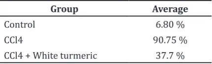
Table 1 Percentage of Damaged Liver Cells in Mice
Group Average
Control 6.80 %
CCl4 90.75 %
CCl4 + White turmeric 37.7 %
inferior vena cava was made to drain the blood while saline solution was injected, followed by liquid formalin, then the ligaments of the liver were cut and the middle third portion of the largest lobe was taken as the sample.
The tissue was fixed using 37% of
formaldehyde solution then rinsed and dehydrated with 100% of alcohol solution. Then we used xylol and toluol to cleanse the
specimen before infiltrating the paraffin. After the paraffin hardened, the specimen was cut
using microtome. Before staining procedure, specimens were cleaned with xylol and toluol followed by rehydration using alcohol solution. The staining procedure was done by using a solution of hematoxylin and eosin counterstain.
Microscopic observation was conducted to see the effect of treatment on the liver cells qualitatively and quantitatively. Qualitative assessment was done microscopically with
400x magnification, while quantitative
observation was done by counting the average percentage of injured cells.
The data were then analysed using the non-parametric Kruskal-Wallis test on SPSS.
It aimed to prove the significant differences of
the average percentage of damaged liver cells in the group given CCl4 alone with the group given CCl4 and white turmeric infusion and control group as negative control was given standard food and drink.
Results
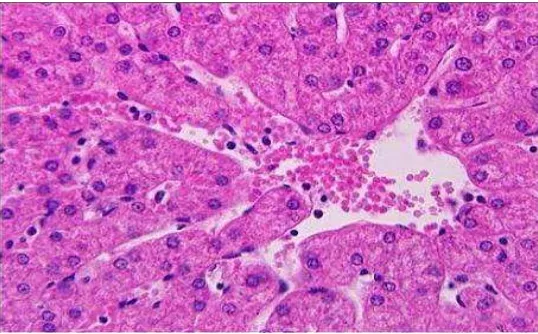
The percentage of damaged liver cells from each group was presented (Table 1). Alteration of the liver tissue architecture in the group administrated with injection of CCl4 was shown by the presence of light-colored cytoplasmic vacuoles and fusion of cell membranes.
Figure 1 Liver Cells of CCl4-induced Mice
Figure 2 Liver Cells of The Group Treated With White Turmeric Infusion Following CCl4 Induction
as in the group given CCl4 alone. There were some evidences of mitotic activity. Sinusoids
were deranged but identifiable (Figure 2).
Administration of white turmeric infusion
significantly decreased the percentage of
damaged cells in mice previously induced with CCl4 (p<0.05), 37.7 % from groups sample.
Discussions
Liver damage caused by hepatotoxic substances generally occurs in liver cells of the group treated with white turmeric
infusion following CCl4 induction, reflecting
the enzymes that play a role in the metabolism of such substances.8 CCl4 metabolism
produced an accumulation of free radicals that can damage the cell membrane or DNA directly.9 Prolonged liver damage or
cirrhosis is associated with the occurrence of hepatocellular carcinoma. Risk factors for hepatocellular carcinoma itself include chronic liver infection, metabolic liver disease, and liver cirrhosis of any cause.3 In Indonesia,
44−45% of patients with liver cirrhosis and
hepatocellular carcinoma were positive for hepatitis B detection.10 Hepatocellular
carcinoma can also occur in conditions where the liver histological structure was altered.3
In mice and rats, administration of CCl4 may cause hepatocellular carcinoma.5 CCl4
is shown to have genotoxic and mutagenic effects associated with the cytotoxicity, lipid peroxidation, and oxidative stress.5 The effect
of long-term exposure to CCl4in humans has not been widely reported, but acute exposure of CCl4 is associated with the onset of various signs and symptoms such as liver enlargement (hepatomegaly), increased aspartate aminotransferase and alanine transferase enzymes, and elevated levels of bilirubin.5
Rhizome white turmeric (Curcuma zedoaria L) has a variety of antioxidants that can be used as an analgesic and
anti-inflammatory. It also prevent the development
of cancer cells and protect liver cells from the harmful effects of toxic chemicals.7 The
effect of acetone extract and other ingredients isolated from white turmeric rhizome such as furanodiene, curdione, neocurdione, 13-hydroxygermacrone, zederone, and
curcuminoid with a dose of 12.5−50 mg/kg
through oral administration has revealed to protect liver cells from damage caused by D-GalN/LPS induction in mice, assessed by the levels ofaspartate aminotransferase (AST) and alanine transferase (ALT) enzymes in serum.11
Administration of curcumin to the
Sprague-Dawley strain rats at a dose of 200 mg/kg and 400 mg/kg has shown to provide protective and therapeutic effects on liver cells exposed
to CCl4 by inhibiting the inflammatory process
and oxidative stress.12 Curcumin alters the
transcription processes in the regulation of gene expressions that regulate the cell and
inhibits the process of inflammation, tumor
formation, angiogenesis, cell division and cellular invasion.13 Administration of curcumin
can inhibit inflammatory processes and fibrogenesis by suppressing the production of various cytokines such as TNF-α, IFN-γ,
IL-6. It also restrains the process of collagen deposition.12 Curcumin is well tolerated in
the body, thereby reducing the probability of toxicity.13 Other antioxidant substances in white turmeric also play a role in inhibiting the cytotoxicity and the production of nitric oxide (NO) produced in the process of
inflammation.11
In this study, induction of CCl4 caused damage liver cells in mice shown by the widespread histological changes. While in the group of mice given white turmeric infusion following CCl4 induction, the average percentage of damaged liver cells were
significantly lower. This is consistent with the
previous studies proving that the antioxidant contents of white turmeric rhizome can be used in the prevention and treatment of damage to the liver cells.11 Several studies
have reported that the major histopathological parameters altered with CCl4-induced damage are degeneration in hepatocytes, the presence
of necrosis and inflammatory infiltration. Our
histology analyses showed a positive effect of white turmeric in inhibiting the in the process
of inflammation.12
This laboratory experimental study has several limitations, including a small size sample. In this study, induction of CCl4 caused damage liver cells in mice shown by the widespread histological changes. Carbon tetrachloride (CCl4) is a chemical widely used as cleaning agents, solvents, as well as industrial materials Future study with large size sample seems warranted to resolve these and other related issues.
References
1. Kumar V, Abbas AK, Fausto N, Aster JC. Robbins and cotran pathologic basis of disease. 8th ed. Philadelphia: Saunders Elsevier; 2010.
3. Fattovich G, Stroffolini T, Zagni I, Donato F. Hepatocellular carcinoma in cirrhosis: incidence and risk factors. Gastroenterology. 2004;127(5 Suppl 1): S35–50.
4. United States Department of Health and
Human Services. Toxicological profile
for carbon tetrachloride. Atlanta: United States Department of Health and Human Services; 2005 [cited 2012 September 20]; Available from: http://www.atsdr.cdc.
gov/toxprofiles/tp30.pdf.
5. United States Environmental Protection Agency. Toxicological review of carbon tetrachloride. Washington, DC: United States Environmental Protection Agency; 2010 [cited 2012 September 17]; Available from: http://www.epa.gov/iris/ toxreviews/0020tr.pdf.
6. Cho WY, Kim SJ. Anti-oxidative actions of Curcuma zedoaria extract with inhibition of inducible nitric oxide synthase (iNOS) induction and lipid peroxidation. J Med Plants Res. 2012;6(22):3837–44.
7. Lobo R, Prabhu KS, Shirwaikar A. Curcuma zedoaria rosc. (white turmeric): a review of its chemical, pharmacological and ethnomedicinal properties. J Pharm Pharmacol. 2009;61(1):13–21.
8. Ramachandran R, Kakar S. Histological
patterns in drug-induced liver disease. J Clin Pathol. 2009;62(6):481–92.
9. Panjaitan RGP, Handharyani E, Chairul, Masriani, Zakiah Z, Manalu W. Pengaruh pemberian karbon tetraklorida terhadap fungsi hati dan ginjal tikus. Makara Kesehatan. 2007;11(1):11–6.
10. Merican I, Guan R, Amarapuka D, Alexander M, Chutaputti A, Chien R, et al. Chronic hepatitis B virus infection in Asian countries. J Gastroenterol Hepatol. 2000; 15(12):1356–61.
11. Morikawa T, Matsuda H, Ninomiya K, Yoshikawa M. Potent protective effects of sesquiterpenes and curcumin from zedoariae rhizoma on liver injury induced by d-galactosamine/lipopolysaccharidase or tumor necrosis factor alpha. Biol Pharm Bull. 2002;25(5):627–31.
12. Fu Y, Zheng S, Lin J, Ryerse J, Chen A. Curcumin protects the rat liver from
CCl4-caused injury and fibrogenesis
by attenuating oxidative stress and
suppressing inflammation. Mol Pharmacol.
2008;73(2):399–409.