37
1 indonesiajournalchest Vol.4 No.4 | Okt -Des. 2017
Pulmonary Hypertension in Obesity Hypoventilation Syndrome: A Case Report
Davin Takaryanto,1,2, Ade Erna1,2, Hendarsyah Suryadinata1,3
1
Faculty of Medicine, UniversitasPadjadjaran, Bandung, Indonesia 2
Department of Internal Medicine, Sumedang Regional Public Hospital, Sumedang, Indonesia 3
Respiratory Critical Disease Division, Department of Internal Medicine, HasanSadikin General Hospital, Bandung, Indonesia
Koresponensi: Dr. Davin Takaryanto
Email: [email protected]
Abstract
Introduction:The prevalence of obesity continues to increase every year across the world. Obesity has been a risk factor for developing some chronic and non-communicable diseases. Obesity hypoventilation syndrome (OHS) is one of non-communicable disease that emerges with obesity as its primary risk factor. We presented a case with severe obesity, chronic hypoventilation and multiorgan impairmentscaused by OHS, a disease that is often misdiagnosed and mistreated hence leads to higher mortality rate.
Case Report: A woman, 54 years old, was brought to hospital emergency ward with decrease of consciousness (hypersomnolent) and breathlessness. The patient had an excessive sleeping amount of time with morning or nocturnal headache, snoring, and sleeping apnea. She had been diagnosed as asthma and pulmonary hypertension and had been given therapy. She was severely obese with BMI of 44.07 kg/m2. The patient suffered multiorgan failures of respiratory failure, hepatic impairment, cardiac failure, and renal disease. She underwent positive pressure ventilatory assistance after being delayed until after fourth day of hospitalization. She suffered from cardiac arrest due to septic shock and respiratory failure. Cardiopulmonary resuscitation was carried out, but eventually the patient died.
Discussions:OHS is an alveolar hypoventilation or hypercapnia that occurs daily in obese patient without underlying disease that could lead to hypo ventilatory state. Majority of patients with OHS also experience obstructive sleep apnea hypopnea syndrome (OSAHS). Patients who complain respiratory problems are often misdiagnosed as asthma or chronic obstructive pulmonary disease even though theyshowsome obvious OHS clinical features. Chronic hypercapnia could lead to dyspnea worsened by physical activity, sleep problem, excessive daily sleeping time, delirium, myoclonus, and seizures. Along with clinical findings, further confirmatory examinations are needed to support OHS diagnosis. OHS could lead to some complications such as pulmonary hypertension and right heart failure (corpulmonale). Bariatric surgery and positive pressure ventilation with mechanical ventilator are primary therapies for OHS.
Conclusions: Diagnosis of OHS should be considered when finding obese patients who complain respiratory problems. Missed diagnosis and inadequate therapy in OHS patients could lead to high morbidity and mortality rate. Advance diagnostic tools are needed to accurately diagnose, follow up and treat OHS patients intensively.
Keywords: hypoventilation, hypercapnia, obesity, Pickwickian syndrome
Abstrak
Introduksi: Prevalensi obesitas secara global meningkat setiap tahunnya. Obesitas menjadi faktor risiko berbagai penyakit kronik dan penyakit tidak menular. Sindroma Hipoventilasi Obesitas (SHO) atau sindroma pickwickian merupakan salah satu penyakit tidak menular dengan faktor risiko utama obesitas. Kami mempersentasikan kasus obesitas berat, hipoventilasi kronik, dengan kerusakan banyak organ yang disebabkan sindroma hipoventilasi obesitas yang sering salah terdiagnosis dan mendapatkan terapi yang tidak tepat sehingga angka mortalitas tinggi.
somnolen. Pasien mengalami berbagai kegagalan organ berupa gagal napas, gangguan hati, gagal jantung, dan gagal ginjal. Pasien mengalami penundaan pemberian tekanan positif jalan napas dengan ventilator mekanik hingga hari keempat perawatan. Pasien mengalami henti jantung yang disebabkan syok sepsis dan gagal napas. Pasien meninggal setelah dilakukan resusitasi jantung paru.
Diskusi: SHO adalah hipoventilasi alveolar atau hiperkapnia pada waktu sehari-hari pada pasien obesitas tanpa adanya penyebab lain dari hipoventilasi. Mayoritas pasien dengan SHO juga memiliki gangguan napas obstruksi saat tidur. Pasien obesitas dengan gangguan napas sering salah terdiagnosis sebagai asma dan penyakit paru obstruktif kronik, meskipun gambaran klinis menunjukkan sindroma hipoventilasi obesitas. Hiperkapnia kronik dapat menunjukkan gejala sesak yang memberat dengan aktivitas, gangguan tidur, waktu tidur harian yang berlebih, delirium, mioklonus, dankejang.Dibutuhkan berbagai pemeriksaan penunjang guna menyokong diagnosis sindroma hipoventilasi obesitas selain dari gambaran klinis saja.Komplikasi hipertensi pulmonal hingga gagal jantung kanan (corpulmonale) dapat terjadi pada pasien SHO. Prosedur operasi bariatric dan pemberian alat bantu nafas tekanan positif jalan napas dengan ventilator mekanik merupakan terapi utama untuk SHO.
Simpulan: SHO harus dipertimbangkan ketika bertemu pasien obesitas datang dengan gangguan napas. Kesalahan diagnosis dan terapi yang tidak adekuat dapat menyebabkan tingginya morbiditas dan mortalitas. Sarana yang lengkap dibutuhkan untuk mendiagnosis, memantau perkembangan, serta memberikan terapi intesif pada pasien SHO.
Kata kunci: hipoventilasi, hiperkapnia, obesitas, pickwickian, sindroma
Introduction
Obesity is a condition where total energy intake exceeds total energy expenditure. Body mass index (BMI) is used to measure personal nutritional status. [1] It is calculated by dividing body weight (kg) with the square of body height (m2). Generally, World Health Organization (WHO) classifies BMI in over-nutrition as overweight (BMI 25-29,9 kg/m2) and obesity (BMI ≥ 30 kg/m2). In Asian population, overweight is defined as BMI between 23-24,9 kg/m2 and obesity as BMI more than 25 kg/m2. [1]
The global prevalence of obesity increases to 1,5 billion people in 2008. [2] In South East Asian population, the prevalence of overweight and obesity tends to increase; previously from 8% to 30% in male population, and from 8% to 52% in female population. [1] Republic of Indonesia’s Ministry of Health reported in
2013 that 15,4% of population suffered from obesity with female:male ratio of 32,9: 19,7. [2]
Obesity is the determinant risk factor for developing various chronic and non-communicable diseases. [3] According to data obtained by WHO, non-communicable diseases lead to more number of death than communicable diseases. [3] Mortality is also higher in adults aged above 65 years old with obesity. [4]
39
2 indonesiajournalchest Vol.4 No.4 | Okt -Des. 2017 (BMI ≥ 30 kg/m
3 indonesiajournalchest Vol.4 No.4 | Okt -Des. 2017 hypoventilation and multiple organ
impairments due to OHS, which is often mistakenly diagnosed and treated.
Case Report
A 54 years old woman was brought to hospital emergency ward with decrease of consciousness that had been worsened for about a month. According to her family members, she unusually had had more sleep and complained of having dizziness after waking up from sleep. She had experienced breathlessness which worsened by physical activities, snoring, and wheeze for about five years and already being treated. There were no presence of coughing, neurological disturbances, or skeletal malformations such as kyphoscoliosis.
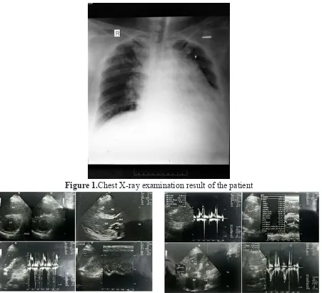
The patient had been diagnosed as asthma and heart failure. Antero-posterior chest x-ray examination was performed two months before and showed the enlargement of heart. Echocardiography was also performed and showed the presence of pulmonary hypertension (PH) (mean PH=44mmHg), trivial PR, abnormal RV contractility, dilated cardiomyopathy and mild TR. She was treated as asthma and pulmonary hypertension.
By the time of her arrival to emergency room, she was somnolent. Her nutritional status were 120 kg of weight and 165 cm of height (BMI of 44,07 kg/m2)with short neck and waist circumference of 122 cm. Her blood pressure was 120/80 mmHg, with respiratory rate of 12 times per minute, pulse rate of 100 beats per minute and
oxygen saturation of 88%. There were nasal flaring and stridor with coarse crackles in basal left lung and expiratory wheeze in both upper lungs. Swelling was observed in both of her lower extremities.
Figure 1.Chest X-ray examination result of the patient
Figure 2.Ecocardiographic examination result of the patient
The patient’s family members did not
consent her to be undergone PAP at the second day of hospitalization. She was planned to be transferred to the national referral hospital. The patient was hospitalized in emergency observational room with worsening general condition, soporous consciousness, and respiratory rate of 12 times per minute with deep breath. After revealing SGOT value of 1,467 IU/L (<31 IU/L), SGPT of 433 IU/L (<32IU/L), and random blood glucose of 360 mg/dL (100-150 mg/dL), she was considered suffering from either obesity
hypoventilation syndrome (OHS) or multiorgan damage due to sepsis.
She was eventually transferred to the intensive care unit (ICU) at the fourth day of hospitalization and was given PAP. The setting was PCV mode, 300 cc of tidal volume (TV), respiratory rate of 20 cc, PEEP of 7 mmHg, FiO2 of 95% and
41
4 indonesiajournalchest Vol.4 No.4 | Okt -Des. 2017 5 indonesiajournalchest Vol.4 No.4 | Okt -Des. 2017
At the fifth day of hospitalization, blood gas analysis (BGA) result revealed that the patient was experiencing hypercapnia, respiratory acidosis with respiratory failure (7,21 pH, 122,2 pCO2, 61 mmHg pO2, 47,9 HCO3, +20 mEq base excess, 82% SpO2, 18,0 g/dL hemoglobin, 53% hematocrite, 145,9 mEq/L sodium, 2,87 mEq/L potassium, 91,7 mEq/L chloride). Other laboratory results were 15,3 g/dL hemoglobin, 2.900/mm3 leukocytes, 64.000/mm3 thrombocytes, total cholesterol of 178 mg/dL (<200 mg/dL), 152 mg/dL LDL (<150 mg/dL), 27 mg/dL HDL (>35 mg/dL), 227 mg/dL triglycerides (<170 mg/dL), 619 IU/L SGOT, 455 IU/L SGPT, 198 mg/dL urea, 3,06 mg/dLcreatinine, 15,8 mg/dL uric acid (2,4-5,7 mg/dL), 22,6 s prothrombine time, 2,04 INR and 33,1 s APTT. The patient was considered suffering from respiratory failure with PaO2/FiO2 less than 100% (64,21%). It was revealed that she also had disseminated intravascular coagulation (64.000/mm3 thrombocytes, 2,04s INR, She was administered with norepinephrine to sustain her hemodynamics. Parenteral hypokalemia correction was also carried out.
She eventually fell into cardiopulmonary arrest due to repiratory failure and severe infection. Cardiopulmonary resuscitation was conducted, but the final ECG record showed asystole. She was declared being deceased after being hospitalized for 5 days.
Discussion
According to American Academy of Sleep Medicine, OHS is defined as the presence of daytime alveolar hypoventilation (awake, sea level, PCO2 >45 mm Hg) among obesity patients (IMT >30kg/m2) in the absence of other causes hypersomnolent state, overslept, had stridor, and dyspnea. The patient had no other clinical conditions which caused hypoventilations such as neuromuscular disturbance or mechanical and metabolic disorders which can cause hypoventilation.
Majority patients with OHS also had obstructive sleep apnea hypopnea syndrome (OSAHS). Our patient also had OSAHS, this syndrome was suspected because the patient complained that he always overslept and had stridor when she slept at night time. OHS patient with OSAHS had a > 10 mmHg higher PaCO2 in night time compared to when the patient
was awake. We couldn’t obtain a blood
gas analysis when the patient was asleep
because of the family’s economic problem.
hospital. The patient was diagnosed with asthma and cardiac enlargement based on physical examination and chest x-ray, in
spite of the patient’s clinical findings
consistent with OSAHS and OHS. Therefore, both general practitioners and specialist must have more understanding of OHS, so that the patients can have adequate therapy immediately.
Clinically, OHS patients have an excessive time of daytime sleeping or headache in the morning or night time, the same symptoms as OSAHS. Our patient also complained that she often overslept and always had a headache when she woke up. In patients with OHS, there is an unexplainable hypoxemia condition with or without hypersomnolent symptoms, dyspnea, even right heart failure, which were present in our patient. Usually, OHS patients also have other symptoms such as easily tired and mood disorder. In patients with apnea or hypopnea, there can be symptoms such as stridor, choking, or strangling sensation while sleeping.
Our patient had already shown worsening chronic hypercapnia symptoms. Usually, in the early stage of alveolar hypoventilation or hypercapnia, the patients will show no symptoms. A worsening hypercapnia is indicated with the presence of symptoms such as worsening dyspnea on exertion, sleep disturbance, excessive daytime sleepiness, delirium, myoclonic and seizure (CO2 narcosis).
We were not be able to diagnose OHS solely based on clinical manifestation, but there were some diagnostic criteria had to be met. According to Respiratory Failure Research Group set up by the Japanese Ministry of Health, the criteria diagnosis of OHS are: 1. Extreme Obesity (BMI>30), 2.
Excessive daytime sleepiness 3.Chronic daytime hypercapnia(PaCO2>45), and 4.
Severe OSAS (AHI>= 30/jam or severe oxygen desaturation). AHI is defined as the number of apnea and hypopnea per hour while the patient was sleeping. Patient with OSAHS without daily hypercapnia is diagnosed with pure OSAHS.
BGA examination must be done in patient with obesity morbidity and unexplained hypoxemia. The diagnosis of OHS need BGA result to confirm the presence of daily hypercapnia. The result of BGA examination usually shows the presence of compensated respiratory acidosis or uncompensated respiratory acidosis with hypoxemia. This patient had hypercapnia (PaCO2 122,2 mmHg) with uncompensated respiratory acidosis (pH 7,201) and hypoxemia (PO2 61mmHg). Chronic hypercapnia in this patient was indicated because there is an increase of bicarbonate serum > 27 (HCO3 = 47,9mEq/L). Daily hypercapnia is reported in 10-38% patients with OSAHS.
Polysomnography examination in patient with OHS is also needed to identify sleep disturbance and to adjust CPAP therazy or noninvasive mechanical ventilation. The limited facility is the reason why the examination was not done
in our patient, which is why we can’t
43
6 indonesiajournalchest Vol.4 No.4 | Okt -Des. 2017 7 indonesiajournalchest Vol.4 No.4 | Okt -Des. 2017
hypopnea index (AHI) which is evaluated based on American Academy of Sleep Medicine Guideline 2007. Apnea is defined as a cessation of air flow of O2
90% for >10 seconds while there is still a respiratory effort, while hypopnea is defined as a reduction of airflow amplitude of >30% for >10 seconds which is associated with oxygen desaturation >3%
OHS patients have a higher risk to have a pulmonary hypertension and mortality compared to obese population without OHS. Pulmonary hypertension is defined as mean pulmonal arterial pressure> 25 mmHg in right heart catheter. PH is found more frequently in patient with OHS compared to patient with OSAHS (50% vs 15%). Hypercapnia and hypoxemia in OHS are associated with the presence of pulmonal hypertension and right heart failure (CPC). While sleeping, there is an increase of pulmonal arterial pressure which is caused by hypoxia vasoconstriction.
Secondary factors that cause pulmonary hypertension in OHS patients are restrictive lung disease associated with severe obesity and the increasing of upper airway resistance. Airway obstruction results in profound negative intrathoracic pressure during inspiration up to -70mmHg. This negative intrathoracic pressures increases RV filling, causing the shifting of intraventricular septum to the left, which results in obstruction of left vascular beds initially response the hypoxemia with a vasoconstriction in
pulmonary arteriolar and capillary level. In the long term, hypoxemia will cause a persistent vasoconstriction. Chronic hypoxemia in OHS patients results in remodeling of pulmonary artery, which is a transition from a process of vasoconstriction to endothelial dysfunction, thickening of arterial wall and
fibrosis. Our patient’s previous
echocardiography examination revealed that pulmonary hypertension and corpulmonale were present (mean PH = 44mmHg, Right Ventricle abnormal contractility, dilated cardiomyopathy). Pulmonary hypertension and corpulmonale indicated that there was a complication from this chronic process of OHS. Patients with OHS generally had edema in lower extremities, which either associated or not with CPC. Some of these patients also had lung edema which caused by left ventricular dysfunction associated with obesity (Hypertension, cardiomyopathy obesity) or other comorbids which causing left heart failure. OHS patient usually treatment in the hospital. Adequate therapy lowering the mortality rate to 3%.In addition, OHS patient which not receiving adequate therapy need invasive mechanic ventilation with a longer hospital length of stay.
obesity which is associated with comorbid condition (including OHS) or patient with BMI >40 are recommended to receive bariatric surgery. Bariatric surgery is an operation technique which is done by cutting and making a intestinal bypass, mainly by excising the small intestine which has a primary function to absorb food. This surgery is the most effective treatment for obesity, which results in a significant body weight reduction. We
didn’t do this procedure in our patient
because of the limitation of facility and the unstable condition of our patient. Bariatric surgery in obese patient showed the
improvement of the lung’s vital capacity,
expiration peak flow, functional residual capacity, total lung capacity, and FEV1/FVC ratio. In MOHS, there is biochemical change including improvement in glucose metabolism and quickly reducing the inflammation after the surgery.
The use of respiratory aid such as mechanic ventilator with positive airway pressure, especially CPAP is an effective treatment of OHS. CPAP therapy provides continuous positive pressure in respiratory cycle. Treating apnea and hypopnea, keep the airway patency and returning the daily eucapnia. Our patient had already received PAP therapy with mechanic ventilation (PCV mode, TV 300cc RR 20 PEEP 7mmHg FiO2 95%) but after 4 days of therapy, the effectivity of PAP therapy was decreasing caused by the delayed therapy, although we already suggested this therapy in the first day. PAP therapy in OHS
patient’s actually can significantly improve
the health related quality of life, health expense, even mortality. PAP therapy can improve the physiology by increasing airway exchange and a better quality of sleep.
Conclusions
We presented severe obesity patient with a morbidity which showed the clinical manifestation of OHS. The increase of obesity prevalence is a global issue which can cause many morbidities which need intensive treatment. OHS is one of the morbidities caused by obesity which is often misdiagnosed. General practitioners and specialists had to have a deeper understanding to this morbidity so they can accurately diagnose this condition and give adequate therapy so that we can reduce the mortality rate and increase the patient’s quality of life. A complete
45
8 indonesiajournalchest Vol.4 No.4 | Okt -Des. 2017 9 indonesiajournalchest Vol.4 No.4 | Okt -Des. 2017
References
1. Rachmi C.N, Li M, Baur L.A. Overweight and obesity in Indonesia: prevalence and risk factorsda literature review. public health. 2017; 147: 20-29
2. Luglio H.F, Nisa F.Z, Penggalih M.H.S.T et al. Socioeconomic Determinants of Obesity among Women Living in Indonesian Remote Islands, Raas and Sapudi. Pak. J. Nutr. 2017. 16 (4): 279-284
3. Mihardja L, Soetrisno U. Prevalence and Determinant Factors for Overweight and Obesity and Degenerative Diseases Among Young Adults in Indonesia. JAFES. 2012;27: 77-81
4. Tatusov M, Joseph J.J, Cuneo B.M. A case report of malignant obesity hypoventilation syndrome: A weighty problem in our ICUs. Respiratory Medicine Case Report. 2017; 20: 38-41
5. Macavei V.M, Spurling K.J, Loft J, Makker H.K. Diagnostic Predictors of Obesity-Hypoventilation Syndrome in Patients Suspected of Having Sleep Disordered breathing. J Clin Sleep Med. 2013; 9(9): 879-884
6. Balachandran J.S, Masa J.F, Mokhlesi B. Obesity Hypoventilation Syndrome Epidemiology and Diagnosis. Sleep Med Clin. 2014; 9(3): 341-347
7. Akashiba T, Akahoshi T, Kawahara S. et al. Clinical Characteristics of Obesity-hypoventilation Syndrome in Japan: a Multi-center Study. Internal Medicine. 2006; 1121-1125
8. Jacob M.G, Rajesh K.R, AsokanA et al. Hypoxic hepatitis owing to obesity hypoventilation syndrome: case report. International Journal of
Medical Science and Public Health. 2016; 5: 1979-1980
9. Manthous C.A, Mokhlesi B. Avoiding Management Errors in Patients with Obesity Hypoventilation Syndrome. AnnalsATS. 2016; 13(1): 109-114 10. Liam CK. CorPulmonale due to
Obstructive Sleep Apnoea. Med J Malaysia. 1993; 48: 347-350
11. Olson A.L, Zwillich C. The obesity hypoventilation syndrome. The American Journal of Medicine. 2005; 118: 948-956
12. Bingol Z, Pihtili A, Cagatay P. et al. Clinical Predictors of Obesity Hypoventilation Syndrome in Obese Subjects With Obstructive Sleep Apnea. Respiratory Case. 2015; 60(5): 666-672
13. Dursunoglu N. Obesity Hypoventilation Syndrome and Pulmonary Hypertension Case with Excessive Daytime Sleepiness. J Sleep Disorder Ther. 2017; 6(1)
14. Pories W.J. Bariatric Surgery: Risks and Rewards. J ClinEndocrinolMetab. 2008; 93(11): 589-596