Diagnosis and Molecular Marker Analysis
of Bali’s Rabies Virus Isolates
(DIAGNOSIS DAN ANALISIS PENANDA MOLEKULER VIRUS RABIES ISOLAT BALI)
I Nyoman Dibia1, Bambang Sumiarto2, Heru Susetya2,
Anak Agung Gde Putra1, I Gusti Ngurah Kade Mahardika3, Helen Scott-Orr4
1Animal Disease Investigation Center, Denpasar,
Balai Besar Veteriner Denpasar, jln Raya Sesetan, Banjar Pegok, Denpasar, Bali 2Faculty of Veterinary Sciences, University of Gadjah Mada, Yogyakarta,
3Faculty of Veterinary Sciences, University of Udayana, Denpasar, Bali 4University of Sidney, Sidney, Australia.
E-mail : [email protected]
ABSTRACT
The direct fluorescent antibody test (dFAT) was recommended by both World Health Organization (WHO) and Office International des Epizooties (OIE) as a standard diagnostic technique for rabies. Since the outbreak of rabies in Bali, it was ascertain the importance to develop a reverse transcriptase-polymerase chain reaction (RT-PCR) technique with specific primers as an alternative diagnostic method. The aim of this study was to develop a RT-PCR technique for rabies diagnosis in animals and find out the molecular marker of Bali’s rabies virus (BRV) isolates based on the sequence of nucleoprotein (N) gene. Brain samples were obtained during 2009 from 14 suspected rabid dogs and one cattle, where rabies viruses were isolated. The dFAT was used to detect the presence of rabies viral antigen. Ribonucleic acid (RNA) of rabies viruses was extracted with TRIzol reagent. Fragment of N gene was amplified using one-step RT-PCR method with specifically-designed primer pairs and sequenced using ABI automatic sequencer. Multiple alignment of nucleotide and deduced amino acid sequences were analyzed using ClustalWof MEGA 4.0 program. This study found that twelve out of fifteen animal brain samples confirmed as rabies by dFAT. Similarly, a single band of 1215 bp PCR product for rabies virus was also detected in twelve out of twelve (100%) dFAT rabies positive samples. It is therefore evident that alternative diagnostic of rabies in animals can be established using RT-PCR technique. The results showed that the RT-PCR has a very high agreement with dFAT. Polymorphic sites of N gene of twelve BRV isolates were identified at the position 186, 501, 801, 840, 1068 and 1153. Bali’s rabies virus isolates have conserved amino acid (isoleucine) alterations at position 308 (open reading frame). Isoleucine distinguished between all Bali’s isolates and the all of isolates from other area of Indonesia and other part of the world. This finding significantly different as compared to other rabies virus isolates from other part of Indonesia or the world documented on the GenBank. Accordingly it is proposed that it can be used as molecular marker and believed to be the first study of molecular marker of rabies virus in Indonesia.
Keywords :rabies virus, diagnosis, molecular marker, nucleoprotein gene, Bali
ABSTRAK
Uji direct fluorescent antibody test (dFAT) merupakan teknik diagnostik baku internasional untuk diagnosis rabies pada hewan dan direkomendasikan oleh Office International des Epizooties (OIE). Teknik
reverse transcriptase-polymerasechain reaction (RT-PCR) dengan primer khas dapat digunakan sebagai alternatif. Penelitian ini bertujuan untuk mengembangan RT-PCR untuk diagnosis rabies pada hewan dan menemukan marka molekuler virus rabies Bali berdasarkan sekuens gen nukleoprotein (N). Otak hewan tersangka dikumpulkan dari kasus rabies di Bali tahun 2009. Sampel yang diuji adalah 14 otak anjing dan 1 otak sapi. Teknik dFAT digunakan untuk mendeteksi antigen rabies pada sampel. Total RNA diekstraksi dari otak tersebut dengan reagen TRIzol. Fragmen gen N diamplifikasi dengan metode
INTRODUCTION
Rabies is a fatal zoonotic disease caused by rabies virus. In Bali, rabies was firstly confirmed laboratorically in November 2008. The disease gives rise to a serious public health problem, with 135 human deaths reported from 2008-2011. Human rabies characteristically follows a bite from a rabid dog. Currently, free-roaming dogs play an important role as the reservoir and transmitter of the disease to humans and domestic animals. It is generally accepted that mass vaccination program has been recognized as an important tool to control the disease.
Rabies virus belongs to the genus Lyssavirus of the family Rhabdoviridae (Boldbaatar et al., 2010; Nguyen et al., 2011; Muleya et al., 2012). The rabies virus is non-segmented single-stranded RNA (Sato et al., 2005; Benedictis et al., 2011) and has a negative-sense (Metlin et al., 2007; Talbi et al., 2009). The viral genome contains five genes, approximately 12 kb in length (Wunner, 2007; Bourhy et al., 2008) and encodes five proteins namely: nucleoprotein (N), phosphoprotein (P), matrix protein (M), glycoprotein (G), and RNA polymerase or large protein (L) (Ito et al., 2001; Sugiyama and Ito, 2007; Wunner, 2007; Yousaf et al., 2012).
The N gene of rabies virus genome contains 1353 nucleotides and consists of 450 amino acids, when open reading frame (ORF) was translated (Ito et al., 2001). The antigenic sites of the N gene were identified that involved the stretches of amino acid at position 313–337 and 374–383 (Tordo, 1996). The virus nucleoprotein plays critical role in replication and transcription. Nucleoprotein is produced abundantly during viral replication and has been used as a target for diagnosis (Nicholson, 2000).
Ribonucleic acid viruses are often characterized by abundant genetic variation (Pybus et al., 2007). Each RNA virus genome exhibits a high degree of sequence variation (Khawplod et al., 2006). Even, Worobey and Holmes (1999) declared that RNA viruses
deserve their reputation as Nature’s swiftest evolvers. According to Murphy et al., (2007) nearly every progeny genome in infected cell will be different from the parental genome at least one nucleotide. Point mutation is the main force
in lyssavirus evolution and it is showed no
evidence of recombination (Badrane and Tordo, 2001). The changes found were distributed unevenly in the genome, which means that genes coding for different proteins evolved at different rates (Fenner et al., 1993; Murphy et al., 2007). The amino acid sequence of N gene was highly conserved, with homologies 99% (Ito et al., 2001). Kouznetzoff et al., (1998) identified that the most conserved region of the N gene was at amino acid position 298–352. Therefore, molecular characterization of the new-rabies virus isolates is important to perform.
The direct flourescent antibody test (FAT) is one of main diagnostic tool for rabies in Indonesia. The method was recommended by both World Health Organization (WHO) and Office International des Epizooties (OIE) as a standard technique. In order to be able to undertake molecular characterization, it is a necessity to develop a reliable molecular assays both rabies virus diagnosis and characterization such as reverse transcription-polymerase chain reaction/RT-PCR with specific primers. In this article we present the diagnosis and molecular marker of Bali’s rabies virus isolates in order to investigate dynamic of rabies virus and its spreading.
RESEARCH METHODS
Samples
Fifteen brain samples (14 dogs and one cattle) were obtained during 2009 in this study. The dog brain samples were collected in Denpasar, Badung, Tabanan, Gianyar, Bangli, Karangasem, and Buleleng districts of the Bali province. Meanwhile, brain samples of cattle suspected infected with rabies virus was collected in Tabanan district.
tunggal dapat divisualisasikan dalam 12 dari 12 sampel yang positif itu. Hasil tersebut merupakan bukti bahwa peneguhan diagnosis rabies pada hewan dapat dilakukan dengan teknik RT-PCR sebagai alternative. RT-PCR menunjukkan kesesuaian dengan FAT sampai 100%. Situs polimorfik gen N dari 12 isolat tampak pada posisi 186, 501, 801, 840, 1068, dan 1153. Yang unik, isolat rabies Bali mempunyai asam amino khas pada posisi 308 yaitu isoleusin. I308 membedakan isolat Bali dengan isolat lain di Indonesia dan dunia. Dengan hasil tersebut, I308 dapat diguanakan sebagai marka molekuler virus isolat Bali.
anti-sense primer NR1251, 0.5 µL SuperScript III RT/Platinum Taq Mix (containing SuperScriptTM III Reverse Transcriptase and Platinum Taq DNA Polymerase) and 5.1 µL ultrapure aquabidest. Conditions of the thermal cycler for cDNA synthesis followed immediately by PCR amplification were as follows: one cycle cDNA synthesis at 50°C for one hour, one cycle initial denaturation at 95°C for 45 seconds and followed by 40 cycles PCR amplification of denaturation at 94°C for 45 seconds, annealing at 50°C for 45 seconds, and extension at 72°C for one minute. The final steps were extension at 72°C for five minutes. The PCR products were visualized using electrophoresis on 1% ultrapure agarose gel (Invitrogen) containing ethidium bromide. Expected amplification product was a 1215-bp DNA fragment.
Nucleotide Sequencing and Genetic Analysis
The RT-PCR products were excised from the gel and purified using QIAquick PCR Purification Kit (QIAGEN). The purified products (amplified cDNAs) were employed for direct sequencing with the N gene–specific primers NF36Y, NF303R, NF587, and NR1251. Cycle sequencing reaction was carried out with a Big Dye Terminator v3.1 Cycle Sequencing Kit (Applied Biosystems, USA). Sequencing products were obtained using the ABI PRISM 3100 Genetic Analyzer (Applied Biosystems, USA) at the Eijkman Institute, Jakarta. The sequencing results were analyzed and rabies virus was determined by BLAST analysis (http:// www.ncbi.nlm.nih.gov/blast/Blast.cgi) The N gene target sequence was 1125-b corresponding to the nucleotides at position 40 to 1164.
Genetic analysis was performed to determine the molecular marker of Bali rabies virus isolates based on the N gene. A nucleotide sequence of the N gene of the isolates from Bali and other regions of Indonesia were compared genetically with those available in the GenBank data base. Multiple alignment of nucleotide and deduced amino acid sequences were analyzed using ClustalW of MEGA 4.0 software (Tamura et al., 2007).
RESULTS AND DISCUSSION
Flourescent Antibody Test
The direct flourescent antibody test was conducted on 15 animal brain samples that rabid Rabies Antigen Detection and Virus
Inactivation
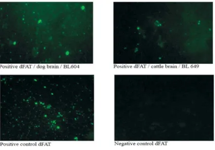
The direct fluorescent antibody test was used to confirm the presence of rabies viral antigen. Smears were prepared from composite sample of original brain tissue, that includes the hippocampus. The brain smear slide was then stained with specific conjugate (fluorescein isothiocyanate-labeled rabbit anti-rabies nucleocapsid immunoglobulins) (BioRad). In the dFAT, the specific aggregates of nucleocapsid protein are identified by their fluorescence. The antigen reacting with antibodies tagged with fluorescein isothiocyanate, appears under ultraviolet light a brightly coloured apple-green or greenish-yellow objects against a dark background (Dean et al., 1996). A negative control using a healthy dog brain smear and a positive control using a rabid dog brain smear were performed for an accurate diagnosis. dFAT-positive samples were made up as a 20% (wt/ vol) suspension with phosphate-buffered saline (PBS) and inactivated with sodium dodecylsulphate (500 µL of 20% original brain suspension containing 50 µL of 10% SDS). The inactivated virus samples were stored at –80oC for further study.
Primer Design
The specific primers were developed in-house using Primer 3 software (http://biotool. umass-med.edu/bioapps/Primer3-www.cgi). They were designed based on nucleotide sequences for the nucleoprotein (N) gene that are available in GenBank. The primers are NF36Y (5’-TCAGGTGGTCTCYTTGAAGCC-3’) at positions 36 to 56, NF303R (5’-CCGATGTRGAAGGGAGT TGG-3’) at positions 303 to 322, NF587 (5’-ACTCACAAGATGTGTGCCAA-3’) at positions 587 to 606, and NR1251 (5’-CTTTAGTCGACCT CCGTTCA-3’) at positions 1232 to 1251. RNA Extraction and RT-PCR
Figure 2. Electrophoresis result of PCR products. M = DNA ladder 100bp (Marker), (+) = positive control, (-) = negative control, and lanes 1-12 = Bali’s isolates. Spesific PCR products at position 1215-bp are indicated by the arrow.
suspected. Of 15 brain samples tested, 12 samples were rabies positive. Some examples of dFAT shown in Figure 1.
RT-PCR and Nucleotide Sequence of N Gene (nt 40 -1164)
All of the dFAT-positive samples from the twelve brain samples obtained from the seven
districts of Bali province (Table 1) were amplified successfully using RT-PCR method. An expected PCR product was 1215 bp (Figure 2).
The specific PCR products were used for direct sequencing analysis. Nucleotide sequences (1125 bp) of the N genes (nt 40 – 1164) of 12 rabies isolates were determined. BLAST analysis (http://www.ncbi.nlm.nih.gov/blast/Blast/cgi) in
this study showed that genetic information obtained was spesific for N gene of rabies virus (data not shown). Nucleotide and amino acid sequences among isolates of Bali rabies were 99.6 to 100% and 99.7 to 100% homologous, respectively. All sequences have been submitted to GenBank and assigned accession numbers (Table 1).
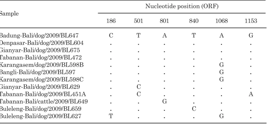
Polymorphic sites of nucleoprotein gen of twelve Bali’s rabies virus isolates were identified at the position 186, 501, 801, 840, 1068, and 1153 (Table 2). At these six polymorphic sites, all substitution occurrences are transition subs-titution, whilst none for transversion substi-tution. The occurrence of transition substitution at the third base of codon is nine times, at the
first base is one times, and none at the second base.
Based on sequence of this 1125 nucleoide, it is obtained 375 amino acids with the following details, one amino acid having non-synonymous (nucleotide and amino acid-altering) and nine amino acids having synonymous (nucleotide altering but amino acid) (data not shown). Alignment of deduced amino acid sequences of Bali’s rabies isolates, other area of Indonesia (non-Bali) and other countries in the world accessed at GenBank was analyzed. The result of the analysis found unique amino acid in all Bali’s rabies isolates, where they have isoleucine amino acid at the position 308, while other isolates outside Bali have valine amino acid at Table 1. Rabies field isolates in 2009 from the seven districts of Bali used in this study.
District Host Isolate GenBank Accession No.
Badung dog BL647 JX448340
Denpasar dog BL604 JX462616
Tabanan dog BL472 JX462617
Tabanan dog BL451A JX462618
Tabanan cattle BL649 JX462619
Karangasem dog BL598B JX462620
Karangasem dog BL598C JX462621
Buleleng dog BL627 JX462622
Buleleng dog BL659 JX462623
Gianyar dog BL629 JX462624
Gianyar dog BL675 JX462625
Bangli dog BL597 JX462626
Table 2. Polymorphic sites of twelve sequences of N gene of Bali’ rabies viruses. Nucleotide position (ORF) Sample
186 501 801 840 1068 1153
Badung-Bali/dog/2009/BL647 C T A T A G
Denpasar-Bali/dog/2009/BL604 . . . . . .
Gianyar-Bali/dog/2009/BL675 . . . . . .
Tabanan-Bali/dog/2009/BL472 . . . . . .
Karangasem/dog/2009/BL598B . . . . G .
Bangli-Bali/dog/2009/BL597 . . . . G .
Karangasem/dog/2009/BL598C . . . . G .
Gianyar-Bali/dog/2009/BL629 . C . . . .
Tabanan-Bali/dog/2009/BL451A . C . . . A
Tabanan-Bali/cattle/2009/BL649 . . G . . .
Buleleng-Bali/dog/2009/BL659 . . . C . .
Buleleng-Bali/dog/2009/BL627 T . . . G .
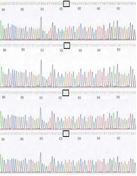
Figure 3. Electropherogram of direct sequencing result of PCR products. Three nucleotides at position 922 (A), 923 (T) and 924 (A) in box show codon for Isoleucine (I) at position 308 of N gene of Bali’s isolates.
same position (Figure 4). Codon encoding specific amino acid (isoleucine) of N gene for Bali’s rabies virus isolates were shown in eletropherogram (Figure 3).
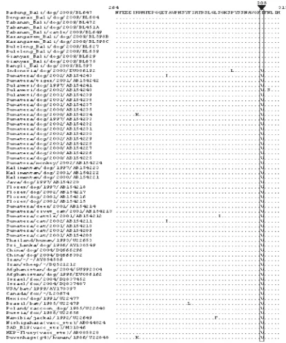
non-synonymous substitutions at position amino acid 308 (Isoleucine) whilst all rabies viruses in Indonesia and all over the world have valine at the same position. Amino acid alignment of N gene of rabies field isolates in Bali obtained in this study and accessed at GenBank shown in Figure 4.
Rabies diagnosis in animal brains is based on the FAT as the standard technique (Wacharapluesadee et al., 2008) and it provides a reliable diagnosis in 98-100% of cases (OIE, 2008). Rabies diagnostic methods based on molecular biology approach have been developed one of them is RT-PCR. This method is also Figure 4. Alignment of the deduced amino acid sequences of the rabies N protein from rabies field
much beneficial in further analyzing on genetic characterization of rabies viruses.
Twelve of twelve (100%) dFAT-positive samples in this study were detected succesfully using one step RT-PCR (Figure 2). This condition showed that RT-PCR developed has a high agreement with dFAT, so it can be used as a reliable confirmative diagnostic tool. The result of this study supported similar study result conducted by previous researchers. Superiority of using RT-PCR is this method still able to detect viral antigen in decomposed brain specimen, which is not detected by using dFAT, as reported David et al., (2002). Benedictis et al., (2011) reported that the one-step RT-PCR showed high relative specificity 98.94% (CI of 97.55 to 99.65), sensitivity 99.71% (CI of 98.40 to 99.99) and accuracy 98.90% values in comparison with those obtained with the FAT used as a gold-standard method. It is also reported that the agreement between the one-step RT-PCR developed and the gold-standard method (FAT) was calculated as 98.91% with a Cohen’s kappa coefficient of 0.977, which corresponds to nearly perfect agreement between the two methods. It is concluded that RT-PCR could be used as an adjunct to dFAT, especially to suspected rabid dog that died more than eight hours before specimen is taken.
Polymorphic sites of nucleoprotein gen of twelve Bali’s rabies virus isolates were identified at the position 186, 501, 801, 840, 1068, and 1153 (Table 2). Nei and Kumar (2000) and Cann (1993) described that a transition substitution is the substitution of a purine (adenine or guanine) for another purine or the substitution of a pyrimidine (thymine or cytosine) for another pyrimidine. The occurrence of substitution toward six polymorphic sites in this study is transition substitution whist non transversion substitution. The happening of transition substitution at the third base of codon is nine times, at the first base is one times, and none at the second base. The result of this study refers to opinion of Murphy et al., (2007) who stated that the most common mutation are single nucleotide substitution that called point mutation. Point mutation in third nucleotide of a codon are often silent, so do not result an altered amino acid because of redundancy in the genetic code. According to Yang et al. (2000) that comparison of relative fixation rates of synonymous (silent) and non-synonymous (amino acid-altering) mutations provides a means for understanding the mechanisms of molecular
sequence evolution. The event of mutation of Bali’s rabies virus isolates corresponds to RNA viruses character. Murphy et al., (2007) explained that RNA viruses are characterized by a high mutation rate during replication, because of the absence of a cellular proof reading mechanism and post replication error correction by RNA polymerase. Ming et al., (2010) also reported that rabies virus evoluted continously through genetic mutation. Hughes et al., (2005) showed that the evolutionary rate for N gene of rabies virus in bats in North America was estimated to be 2.32 x 10⁻⁴ substitutions per site per year, meanwhile Talbi et al., (2009) reported that the mean rates of nucleotide substitution for the N gene of rabies virus isolates belonging to the Africa was 3.82 x 10⁻⁴ substitutions per site per year.
The deduced amino acid sequences of N gene of Bali rabies virus isolates were compared with those of rabies virus isolates from other part of Indonesia, Asia, the America, Erope, Africa, and related rabies virus isolates (Figure 4). A unique amino acid substitution between Bali’s rabies virus isolates (BRV) and non-BRV isolates (accessed in GenBank) was found in the most conserved region at amino acid position 308 of nucleoprotein gene and distinguished all of isolates in Bali from the isolates in other part of Indonesia and the world.
This study is believed to be the first study to detect molecular marker of rabies virus in Indonesia. It was found unique amino acid (isoleucine) at the position 308 from all Bali isolates, which has not been possessed by rabies isolates in all Indonesia and some other countries all over the world, accessed at GeneBank (Figure 4). In addition, this specific amino acid (isoleucine) is precise to most conserve region of Ngene of rabies virus. Thus, it can be said that isoleucineat position 308 (open reading frame) of N gene is molecular marker of Bali’s rabies virus isolates and it can be used as an epidemiological marker in other to investigate dynamic and its spreading. The data obtained in this study support opinion of Nagarajan et al., (2006) who stated that amino acid mutations of gene might specifically carry a molecular marker that can be used as an epidemiological marker.
and vampire bat related rabies virus isolates (DRRV and VRRV, respectively) in Brazil using strain-spesific (SS) primers. All the DRRV and VRRV were successfully distinguished by RT-PCR with SS primers developed. Theoretically and referring to success of RT-PCR development conducted by Ito et al., (2003) that it is really possible and necessary to do. Strain-specific primers for detection of Bali’s rabies virus isolates were designed namely: Primer1 (5’-TACTCATCTAATGCAGTTGGTCACA-3’ at position 898 to 922) and Primer2 (5’-TCCAACAAAGTGAATGAGATTGAATAT-3’ at position 922 to 948).
CONCLUSION
This research finding showed that confirmative diagnostic of rabid animals can be established using RT-PCR technique with specific primers and isoleucine at position 308 of N gene of Bali’s rabies virus isolates identified as molecular marker.
ACKNOWLEDGMENT
This study was funded by ACIAR AH 2006-166 project. The authors would like to thank Heads of DIC Denpasar, for having provided some of the animal brain specimens.
REFERENCES
Badrane H, Tordo N. 2001. Host Switching in
Lyssavirus History from the Chiroptera to
the Carnivora Orders. J Virol 75(17) : 8096-8104.
Benedictis PD, Battisti CD, Dacheux L, Marciano S, Ormelli S, Salomoni A, Caenazzo ST, Lepelletier A, Bourhy H, Capua I, Cattoli G. 2011. Lyssavirus Detection and Typing Using Pyrosequencing. J Clin Microbiol 49(5) : 1932-1938.
Boldbaatar B, Inoue S, Tuya N, Dulam P, Batchuluun D, Sugiura N, Okutani A, Kaku Y, Noguchi A, Kotaki A, Yamada A. 2010. Molecular Epidemiology of Rabies Virus in Mongolia, 2005-2008. Jpn J Infect Dis 63 : 358-363.
Bourhy H, Reynes J, Dunham EJ, Dacheux L, Larrous F, Huong VTQ, Xu G, Yan J, Miranda MEG, Holmes EC. 2008. The origin and phylogeography of dog rabies virus. J
Gen Virol 89 : 2673-2681.
Cann AJ. 1993. Principles of molecular virology. San Diego, CA: Academic Press Inc. David D, Yakobson B, Rotenberg D, Dveres N,
Davidson I, Stram Y. 2002. Rabies virus detection by RT-PCR in decomposed naturally infected brains. Vet Microbiol 87(2) : 111-118.
Dean DJ, Abelseth MK, Anatasiu P. 1996. The fluorescent antibody test. In Meslin FX, Kaplan MM, Koprowski H. (Ed). Laboratory
techniques in rabies. 4th ed. Geneva: World
Health Organization. Pp. 88-95.
Fenner FJ, Gibbs EPJ, Murphy FA, Rott R, Studdert MJ, White DO. 1993. Veterinary Virology. 2nd ed. California: Academic Press Inc.
Hughes GJ, Orciari LA, Rupprecht CE. 2005. Evolutionary timescale of rabies virus adaptation to North American bats inferred from the substitution rate of the nucleoprotein gene. J Gen Virol 86 : 1467-1474.
Ito M, Itou T, Shoji Y, Sakai T, Ito FH, Arai YT, Takasaki T, Kurane I. 2003. Discrimination between dog-related and vampire bat-related rabies viruses in Brazil by strain-specific reverse transcriptase-polymerase chain reaction and restriction fragment length polymorphism analysis. J Clin Virol 26(3) : 317-330.
Ito N, Kakemizu M, Ito KA, Yamamoto A, Yoshida Y, Sugiyama M, Minamoto N. 2001. A comparison of complete genome sequences of the attenuated RC-HL strain of rabies virus used for production of animal vaccine in Japan, and the parentral Nishigahara strain. Microbiol Immunol 45 : 51-58.
Khawplod P, Shoji Y, Ubol S, Mitmoonpitak C, Wilde H, Nishizono A, Kurane I, Morimoto K. 2006. Genetic analysis of dog rabies viruses circulating in Bangkok. Infect Gen
Kouznetzoff A, Buckle M, Tordo N. 1998. Identification of a region of the rabies virus N protein involved indirect binding to the viral RNA. J Gen Virol 79 : 1005-1013. Metlin AE, Rybakov S, Grusdev K, Neuvonen
E, Huovilainen A. 2007. Genetic heterogeneity of Russian, Estonian and Finnish filed rabies viruses. Arch Virol DOI 10.1007/s00705-007-1001-6.
Ming P, Yan J, Rayner S, Meng S, Xu G, Tang Q, Wu J, Luo J, Yang X. 2010. A history estimate and evolutionary analysis of rabies virus variants in China. J Gen Virol 91: 759-764.
Muleya W, Namangala B, Mweene A, Zulu L, Fandamu P, Banda D, Kimura T, Sawa H. Ishii A. 2012. Molecular epidemiology and a loop-mediated isothermal amplification method for diagnosis of infection with rabies virus in Zambia. Virus Res 163 : 160-168. Murphy FA, Gibbs EPJ, Horzinek MC, Studdert
MJ. 2007. Veterinary Virology. 3rd ed. USA: Elsevier, Academic Press.
Nagarajan T, Mohanasubramanian B, Seshagiri EV, Nagendrakumar SB, Saseendranath MR, Satyanarayana ML, Thiagarajan D, Rangarajan PN, Srinivasan VA. 2006. Molecular Epidemiology of Rabies Virus Isolates in India. J Clin Microbiol 44(9) : 3218–3224.
Nei M, Kumar S. 2000. Molecular Evolution
and Phylogenetics. New York: Oxford
University Press.
Nguyen AKT, Nguyen DV, Ngo GC, Nguyen TT, Inoue S, Yadama A, Dinh XK, Nguyen DV, Phan TX, Pham BQ, Nguyen HT, Nguyen HTH. 2011. Molecular Epidemiology of Rabies Virus in Vietnam (2006-2009). Jpn
J Infect Dis 64 : 391-396.
Nicholson KG. 2000. Rabies. In: Zukerman AJ, Banatvala JE, Pattison JR, (Ed). Principles
and Practice of Clinical Virology. Fourth
edition. USA: Jhon Wiley & Sons Ltd. Pp. 583-606.
OIE (Office International des Epizooties). 2008. Manual of Standards for Diagnostic tests
and Vaccines. Chapter 2.1.13. Pp.304-323.
Pybus OG, Rambaut A, Belshaw R, Freckleton RP, Drummond AJ, Holmes EC. 2007. Phylogenetic Evidence for Deleterious Mutation Load in RNA Viruses and Its Contribution to Viral Evolution. Mol Biol Evol 24(3) : 845-852.
Sato G, Tanabe H, Shoji Y, Itou T, Ito FH, Sato T, Sakai T. 2005. Rapid discrimination of rabies viruses isolated from various host species in Brazil by multiplex reverse transcriptionpolymerase chain reaction. J
Clin Virol 33: 267-273.
Sugiyama M, Ito N. 2007. Control of rabies: epidemiology of rabies in Asia and development of new generation vaccines for rabies. Comp Immunol Microbiol Inf Dis 30:273-286.
Talbi C, Holmes EC, Benedictis PD, Faye O, Nakoune E, Gamatie D, Diarra A, Elmamy BO, Sow A, Adjogua EV, Sangare O, Dundon WG, Capua I, Sall AA, Bourhy H. 2009. Evolutionary history and dinamics of dog rabies virus in western and central Africa.
J Gen Virol 90 : 783-791.
Tamura K, Dudley J, Nei M, Kumar S. 2007. MEGA4: Molecular Evolutionary Genetics Analysis (MEGA) Software Version 4.0. Mol
Biol Evol 42(8) : 1596-1599.
Tordo N. 1996. Chracteristic and molecular biology of the rabies virus. In: Meslin FX, Kaplan MM, Koprowski H. (Ed). Laboratory
techniques in rabies, 4th ed. Geneva. World
Health Organization. Pp. 28-51.
Wacharapluesadee S, Sutipanya J, Damrong-watanapokin S, Phumesin P, Chamnanpood P, Leowijuk C, Hemachudha T. 2008. Development of a TaqMan real-time RT-PCR assay for the detection of rabies virus. J Virol
Methods 151 : 317-320.
Worobey M, Holmes EC. 1999. Evolutionary aspects of recombination in RNA viruses. J
Gen Virol 80 : 2535-2543.
Wunner WH. 2007. Rabies Virus. In: Jackson AC, Wunner WH. (Ed.). Rabies. 2nd ed. USA: Elsevier Inc. Pp. 23-68.
Yang Z, Nielsen R, Goldman N, Pedersen AMK. 2000. Codon-substitution models for heterogeneous selection pressure at amino acid sites. Gen 155 : 431-449.