Feature Extraction For Application of Heart Abnormalities
Detection Through Iris Based on Mobile Devices
Entin Martiana Kusumaningtyas, Aliridho Barakbah, Aditya Afgan Hermawan
Politeknik Elektronika Negeri Surabaya Jl. Raya ITS Sukolilo Surabaya 60111, Indonesia
Telp: 6231 5947280 Fax : 6231 5946114
E-mail: {entin, ridho}@pens.ac.id, [email protected]
Abstract
World Health Organization (WHO) data in 2012 showed 17.5 million people worldwide died from heart disease or 31% of 56.5 million deaths and examining it by current methods in hospitals is not cheap. Iridology is an alternative medicine technique whose proponents claim that patterns, colors, and other characteristics of the iris can be examined to determine information about a patient's systemic health. One research on computer iridology is about the computer's iridology system to detect heart conditions. There are several stages such as capture eye base on target, pre-processing, cropping, segmentation, feature extraction and classification using Thresholding algorithms. In this study, feature extraction process performed using binarization method by transforming the image into black and white. In this process we compare the two approaches of binarization method, binarization based on grayscale images and binarization based on proximity. The system we proposed was tested at Mugi Barokah Clinic Surabaya. We conclude that the image grayscale approach performs better classification than using proximity.
Keywords: Iridology, Feature Extraction, Grayscale, Binarization, Thresholding.
1. INTRODUCTION
2014 in Indonesia showed, Coronary Heart Disease (CHD) became the highest cause of death at all age after stroke, that is equal to 12,9%. [3].
Most people think the elderly suffer from heart disease. But in fact, any ages can suffer hearti disease, including infants. It happens because unhealthy lifestyle, stress, and heredity factors. When the patient's situation becomes severe usually newly detected heart disease. It is dangerous for patient if they get helped to late or not immediately dealt with. To detect the abnormalities immediately the patient should go to the doctor. Patient must go to the hospital to check their heart condition. They usually use Echocardiogram (Heart USG), Electrocardiography (ECG), CT Scans, and etc [4]. Checking with the devices need a lot cost.
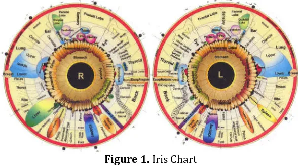
But now, in addition to medical treatment there are alternatives. It is quite popular nowadays. With this alternative method people believe the results are the same as medical and the cost is cheaper. One of the most popular alternative medicine is Iridology. Iridology is an alternative medicine technique whose proponents claim that patterns, colors, and other characteristics of the iris can be examined to determine information about a patient's systemic health. This method believes that pattern on the iris reflects body condition [5]. Ignatz Von Peczely is Hungarian physicist who discovered iridology by examining changes of iris in some patients who are recovering from illness. He created the first Chart of Iris. Dr. Bernard Jensen revised the chart and it is used internationally as shown on Figure 1.
Figure 1. Iris Chart
With Iridology, iris can represent organ conditions of human [6].
The result can be seen from the comparison of before and after the removal process wavelet noise variations in quality ECG significant gesture. The parameters of the wave Q, R, S and T cue ECG recordings can be known. [8]. Using the Bayes algorithm to generate high accuracy values to classify liver abnormalities [9]. Riyanto et.al create healthcare kiosk is used as an alternative to the health care facilities [10].
There are several studies that analyze the heart detection using a computer. Combining image processing and iridology to produce a system of body health detection through computerized iris. Using Visual C # and KNN algorithm to analyzed the condition of the kidneys through the iris of eye [11]. Present the difference auto and manual crop by dividing the iris image into 3 parts to get the iris area. The system using Visual C# to process and analysis the data [12]. And an application that has a method to cuts image automatically. Imagery series to get the iris image from the original image by automated process using desktop-based histogram projection. [13].
After presented study of automatic cropping [14], in this paper we presented feature extraction process performed using binarization method by transforming the image into black and white. In this process we compare the two approaches of binarization method, binarization based on grayscale images and binarization based on proximity.
2. RELATED WORKS
In iridology it is believed that certain positions has very close relationship with every organ in the body. Iris image segmentation is one of the most important steps in the process of the iris diagnosis, and the iris localization is a very critical step for the iris image segmentation. The quality of the iris localization directly affects the accuracy of the following operations. Different approaches have already been reported in the literature to locate the iris region and feature extraction. A 2-D Gabor filter based texture analysis and a texture fractal dimension estimation method are proposed for pathological feature extraction; and at last support vector machines are constructed to recognize[15].
This article proposed feature extraction for the heart location in Iris using binarization method by transforming the image into black and white. We will try to compare the two approaches of binarization method, binarization based on grayscale images and binarization based on proximity.
3. ORIGINALITY
4. SYSTEM DESIGN
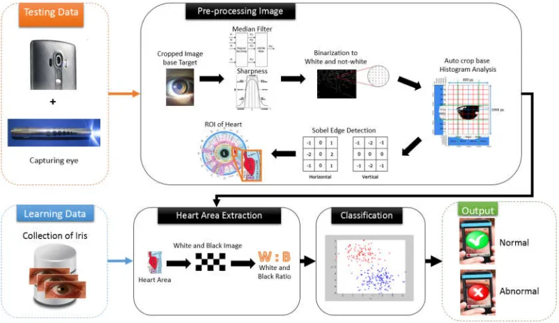
The General Design System is illustrated in Figure 2. The important phase is Pre-processing to get area of iris that required to get the position of heart. This result is impact on iris recognition process. This phase will make high quality of the original image and determine the success result of auto cropping. In this research, we use twice cropping, there are cropping image based on target and auto cropping based on histogram analysis. Then, the image will be segmented to get ROI of heart. This part is important to determine the abnormalities of heart.
Figure 2. General Design System
4.1 Image Data
Figure 3. (a) Normal Heart image (b) Abnormal Heart Image as Data Training from Mugi Barokah Clinic
And the image data that used for data test obtained from mobile device camera. The image is taken using an application that built on android devices. This application has a camera custom activity to take the iris. In the center of it, there is a target which always appears that helps us to focus on the acquisition of iris image. The iris must be exact in the center.
Figure 4. Illustration of cropping based on target
After getting the image, the images will be cropped using automatic cropping based on target according to Figure 4. The cropping is dividing the width and height into three. The taken image is divided into nine sections. The image will be cropped into 1/3 to 2/3 of width image and 1/3 to 2/3 of height image. The result is taken from the fifth section. After the image cropped, it will be resized into 800x1066 pixels.
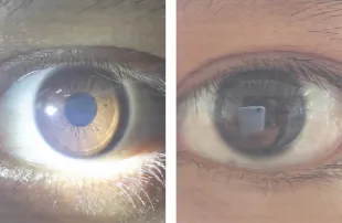
When taking image need to use an additional light from health flashlight and the best lighting for the acquisition of iris image is white color. By using this health flashlight can show the details of the iris as described in Figure 5.
Figure 5. Capture eyes using additional light and not
illustrated in Figure 6 (b). The third image shows an image with properly lighting but the light is too slant. The lighting makes a shadow beside the iris area. It will make the shadow turn into black when it converted to biner. The shadow looks like the addition in iris area as Figure 6 (c).
(a) (b)
(c)
Figure 6 (a) Ideal, (b) Over, and (c) Slant of Lighting
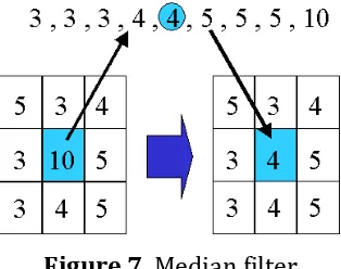
4.2 Median Filter
We use median filter to soften and remove noise in the eye image. This filter replaces the center value in the window with the median of all the pixel values in the window such illustrated in Figure 7.
4.3 Sharpness
Sharpness is a technique for increasing the apparent sharpness of an
image. The High Pass Filter and Low Pass Filter is the most method to sum up. We used Median Filter for the Low Pass Filter and Sobel Operator for High Pass Filter.
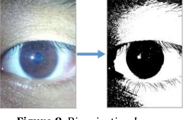
4.4 Binarization Image
Before performing the auto-cropping to get iris image, the sharpened image should be converted to a binary image. The binary image has only two possible values for each pixel. Typically, the two colors used for a binary image are black and white. In this study image converted into white and not white. Proximity is using for conversion of the original image into a binary image. Euclidian distance formula is using for Distance calculations.
dist((x, y), (a, b)) = √(x - a)² + (y - b)² (1)
A threshold is used to determine whether the pixel is included black or white and the value that used in this study is 175. With this threshold if the value exceeds then changed to black color, and if less than or equal then changed to white. Threshold value is derived from the average learning data and searched for each first threshold value.
Figure 8. Binarization Image
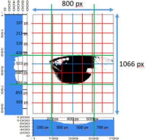
4.5 Auto Cropping based on Histogram Analysis
After getting binary image, it will be analyzed to perform the auto cropping. This phase aims to get the area of iris. The cropping method performed by observing the pattern of the histogram. The image width will be divided into 8 sections and the image height will be decided into 10 sections.
4.5.1 Histogram
pixel. The left and right point is taken from the highest changed value on the horizontal histogram. The value is seen from 1/8 to 3/8 of the total width of the image pixel for left point and from 5/8 to 7/8 of the total width of the image pixel for right point. The top and bottom point is taken from the highest changed value on the vertical histogram. It illustrates in Figure 8.
Figure 9. Histogram vertical and horizontal
The value is seen from 3/10 to 5/10 of the total width of the image pixel to get the left point and from 6/10 to 8/10 of the total width of the image pixel to get the bottom point. Then the image will be cropped in accordance with the obtained boundary before.
4.6 Edge Detection
After getting the image cropped, it will be processed in feature extraction and applying edge detection. Edge detection makes extract the lines on iris and produces a detailed and clearly iris image. Sobel method is using to perform edge detection in this study, which ia applying Sobel operator uses 3x3 kernel to calculate the gradient x and y.
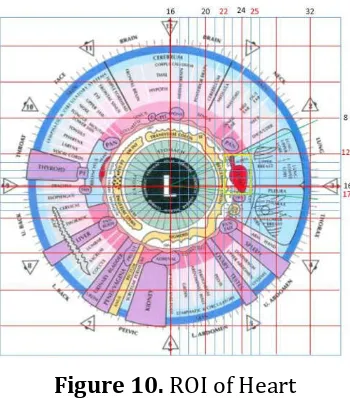
4.7 Segmentation on ROI of Heart
Figure 10. ROI of Heart
Based on the division of Iris in order to get the heart area, it can be seen that the boundary region as follows:
left : 22/32 of width image
right : 25/32 of width image
top : 12/32 of height image
bottom : 27/32 of height image
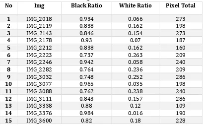
4.8 Feature Extraction
Aimed of Feature extraction to obtain the critical information of ROI heart. This method calculate the number of black and white pixels divide with the pixel total of the image. The ratio of white and black can be calculated by:
Ratio of White = (2)
Ratio of Black = (3)
In this study, feature extraction process performed using binarization method by transforming the image into black and white. In this section will try to compare the two approaches of binarization method, binarization based on grayscale images and binarization based on proximity.
4.9 Classification
examine the data captured by the camera smartphone. Here is a classification thresholding algorithm.
1. Determine threshold for analyze the test data.
2. Categorize label for the data that is less than the threshold and over
threshold.
3. Label the data according to its category, namely normal and abnormal.
5. EXPERIMENT AND ANALYSIS
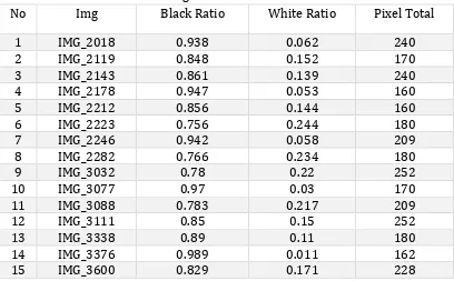
Binarization Based on Grayscale Images changes the grayscale image into a binary image with a threshold. The threshold used in this study is 50. According to the Table 1 and 2, it can be taken the average ratio of black and white pixels in the images normal and abnormal heart. The average ratio of black and white pixels to the normal heart image are 0.855 and 0.145. While the average ratio of black and white pixels to abnormal heart image are 0.594 and 0.406.
Table 2. Results of Feature Extraction Binarization with Grayscale Image to The Training Data of Abnormal Heart
No Img Black Ratio White Ratio Pixel Total
1 IMG_2235 0.649 0.351 308
2 IMG_2364 0.601 0.399 228
3 IMG_2409 0.627 0.373 209
4 IMG_2862 0.679 0.321 209
5 IMG_2873 0.574 0.426 209
6 IMG_2921 0.659 0.341 273
7 IMG_3044 0.667 0.333 336
8 IMG_3099 0.506 0.494 170
9 IMG_3122 0.519 0.481 160
10 IMG_3178 0.556 0.444 198
11 IMG_3543 0.393 0.607 308
12 IMG_3633 0.596 0.404 198
13 IMG_3872 0.542 0.458 286
14 IMG_3998 0.702 0.298 198
15 IMG_4164 0.711 0.298 273
16 IMG_4839 0.612 0.388 209
17 IMG_7833 0.506 0.494 405
From the graph shown in Figure 11, white and black ratio of the normal and abnormal image plotted and it could be seen that between them are separated. For normal is in the lower-right axis and the value of the black ratio is more than white ratio. While the abnormal is in the upper-left axis and the value of the white ratio is more than black ratio.
Figure 11. Graph Feature of Black and White Pixel Ratio
image will calculate with the white color (255,255,255). The proximity that used as the threshold is 350.
Table 3. Results of Feature Extraction Binarization with Proximity to The Training Data of Normal Heart
16 IMG_4839 0.632 0.368 209
17 IMG_7833 0.523 0.477 405
From the graph shown in Figure 12, white and black ratio of the normal and abnormal image plotted and it could be seen that between them are separated. For normal is in the lower-right axis and the value of the black ratio is more than white ratio. While the abnormal is in the upper-left axis and the value of the white ratio is more than black ratio.
Figure12. Graph Feature of Black & White Pixel Ratio
Table 5. Result of Classification to Training Data
Img Target Classification Result Grayscale Proximity
IMG_2018 Normal N N
IMG_2119 Normal N N
IMG_2143 Normal N N
IMG_2178 Normal N N
IMG_2212 Normal N N
IMG_2223 Normal N N
IMG_2246 Normal N N
IMG_2282 Normal N N
IMG_3032 Normal N N
IMG_3077 Normal N N
IMG_3088 Normal N N
IMG_3111 Normal N N
IMG_3338 Normal N N
IMG_3376 Normal N N
IMG_3600 Normal N N
IMG_2235 Abnormal AN AN
IMG_2409 Abnormal AN AN
Table 6. Result of Classification to Testing Data
approach. The performance of those approaches are calculated using formula in Figure 11 and 12, here is the calculation detail:
Performance of Training Data disease leading to death. It can affect everyone, both young and old. Iridology
is an alternative medicine technique to detect heart abnormalities. Needs a
process in feature extraction in an iris image to perform iridology and computation process. The mobile device is one of the applications platforms which is very popular right now. It could do a lot of computing that used to be only on the computer. So we can perform every computation in anywhere and anytime. This research is proposed a new method of iridology and computation process in order to check the heart condition. By applying the mobile devices, everyone can check their heart regularly.
The feature extraction method produces a high result of right classification while it performs on success image cropping. From that experiment, the black threshold used is 0.725 and the white threshold used is 0.275.
Acknowledgements
This research would have been possible with the financial of Applied Research from Ministry Research, Technology and Higher Education. We were especially indebted to Mr. Abdul Hamid from Mugi Barokah Clinic who provided training data for this research.
REFERENCES
[1] https://en.wikipedia.org,, 2017, Heart. Retrieved 26 11 16, from
[2] https://id.wikipedia.org, 2008, Jantung. Retrieved 26 11 16, from https://id.wikipedia.org/wiki/Jantung#Penyakit_jantung
[3] http://www.who.int, 2017, The top 10 causes of death. Retrieved 30 01
17, from http://www.who.int/mediacentre/factsheets/fs310/en/ [4] https://health.detik.com, 2013, Mau Cek Kesehatan Jantung? Ini Dia
Jenis-jenisnya,. Retrieved 26 11 16, from
https://health.detik.com/read/2013/03/20/132737/2198898/775/m au-cek-kesehatan-jantung-ini-dia-jenis-jenisnya.
[5] E. Ernst, M.H Cohen, J. Stone, “Ethical problems arising in evidence
based complementary and alternative medicine”; J Med Ethics 2004,30:156-159
[6] B. Jensen, Scienceand Practice of Iridology, 2005.
[7] Saparudin, Edvin Ramadhan, Identifikasi Kelainan Jantung
Menggunakan Pola Citra Digital Electrocardiagram, Sriwijaya University, 2012.
[8] B. S. Widodo, “Wavelet-Based Treatment for Heart Gesture
Detection of Myocardial Abnormalities Method Using High-Speed QRS Detection,” 2009.
[9] Aulia Fitriana Sari, 2014, Iris Recognition For Detection Of Liver Organ
Disorders, EEPIS.
[10] Sigit, Riyanto et al, “Development Healthcare Kiosk for Checking Heart Healt”, EMITTER International Journal of Engineering Technology, Vol 3, No 2, pp 99-114, 2015.
[11] N. Fauziah, "Iridology Application for Kidney Condition Analysis through Iris Eye Imagery," 2014.
[12] Martiana, Entin K. et al, “Auto Cropping On Iris Image For Iridology
Using Histogram Analysis”, Knowledge Creation and Intelligent Computing, pp 42-46, 2016.
[13] Martiana, Entin K. et al,“Application For Heart Abnormalities
Detection Through Iris”, International Electronics Symposium, pp 319-326, 2016.
[14] Martiana, Entin K. et al,“Auto Cropping For Application of Heart Abnormalities Detection Through Iris Based on Mobile Devices ”, International Electronics Symposium, pp 319-326, 2017.
[15] Lin M, Naimin Li, Texture Feature Extraction and Classification for