Inhibins in female and male reproductive physiology: role
in gametogenesis, conception, implantation and
early pregnancy
Stefano Luisi
1, Pasquale Florio
1, Fernando M.Reis
2and Felice Petraglia
1,31Department of Pediatrics, Obstetrics and Reproductive Medicine, University of Siena, Italy and2Department of Obstetrics
and Gynecology, University of Minas Gerais, Belo Horizonte, Brazil
3To whom correspondence should be addressed at: Department of Pediatrics, Obstetrics and Reproductive Medicine, University
of Siena, Policlinico ‘Le Scotte’, viale Bracci, 53100 Siena, Italy. E-mail: petraglia@unisi.it
A great deal of new information has arisen in the recent years concerning inhibin physiology and clinical relevance in reproductive medicine. It is now recognized that the two inhibin isoforms, named inhibin A and inhibin B, are produced by the gonads in the course of gamete maturation and in women have a different pattern of secretion throughout the menstrual cycle. Since inhibins are also produced by placenta and fetal membranes, it has been suggested that there is an involvement in physiological adaptation of pregnancy. Evidence from several sources has underlined the clinical usefulness of the measurement of inhibin-related proteins in the diagnosis and follow-up of different fertility disturbances and early pregnancy viability. In the male, inhibin B is produced in the testis, prin-cipally by the Sertoli cells. Inhibin B expression and secretion are positively correlated with Sertoli cell function, sperm number, and spermatogenic status and are negatively correlated with FSH. This review covers the most recent advances on the role of inhibins in human reproductive function. Considerable progress in the understand-ing of inhibin physiology has resulted from selective measurement of the two inhibin molecular forms, named inhi-bin A and B. Newly recognized alterations of inhiinhi-bin levels in gynaecological diseases as well as in normal and pathological pregnancy are discussed, with particular emphasis on the potential clinical usefulness of assessing inhibin levels in serum and other biological fluids.
Key words: implantation/inhibin/ovary/pregnancy/testis
Introduction
Inhibins are multifunctional molecules involved in the control of pituitary FSH secretion. A body of observational and experimen-tal evidence from several species, including the human, supports the concept that inhibins are gonadal messengers that exert a physiological negative feedback control on FSH release at the pituitary gland. Apart from their essential role in the selective control of FSH secretion, inhibins are currently recognized as paracrine ovarian and testicular regulators and have multiple paracrine effects in the utero-placental unit, representing a prom-ising marker for male and female infertility, gynaecological and gestational diseases.
Structure and receptor
Inhibins are glycoproteins produced by the granulosa and theca cells of the ovary and by the Sertoli cells of the testis. They are of great importance for the negative feedback control of pituitary
gonadotrophin secretion. There are at least two active molecular forms in circulation, inhibin A and inhibin B, which are hetero-dimers made by an a subunit and either bA(inhibin A) or bB (inhibin B) subunits (Hayeset al., 1998).
Activins (dimers composed of bAand/orbBinhibin subunits) act on activin type I and type II receptors which contain an extracellular domain (where the hormone binds), a transmem-brane region and a serine/threonine kinase intracellular domain. Although most inhibin effects are associated with its antagonism to activin, there are cells that bind inhibin with much higher affi-nity than activin due to the existence of specific inhibin-binding molecules (Lebrun and Vale, 1997). One of these molecules was recently discovered to be betaglycan, a membrane-anchored pro-teoglycan that operates as an inhibin co-receptor. Betaglycan binds inhibina subunit with high affinity and enhances inhibin binding to activin type II receptor through its b subunit, thus preventing activin access to the type II receptor (Lewis et al., 2000).
Human Reproduction Update Page 1 of 13 doi:10.1093/humupd/dmh057
by guest on May 19, 2016
http://humupd.oxfordjournals.org/
Assays for inhibins
Radioimmunoassays were the first assay developed and ‘the Monash assay’ (McLachlan et al., 1986, 1987; Schneyer et al., 1990) provided a wealth of insight into reproductive physi-ology. However, the ‘Monash’ assay, which used a polyclonal antibody raised against 31 kDa bovine inhibin with epitopes for the antibody on the inhibinasubunit, is unable to discriminate between dimeric bioactive inhibin forms and various forms of freeasubunit which circulate in 20-fold excess. Several groups prepared monoclonal and polyclonal antibodies with the inten-tion of producing two-site immunoassays to measure specifi-cally the bioactive dimeric forms of inhibin (Illingworth et al., 1991; Baly et al., 1993; Poncelet and Franchimont, 1994). However, due to the high degree of structural conservation of inhibin between species, it is a poor immunogen and the anti-bodies raised had limited affinity. More recently, a panel of monoclonal antibodies to synthetic peptide immunogens was used to construct ultrasensitive enzyme-linked immunosorbent assays (ELISA) for dimeric inhibin A, inhibin B, and the inhi-bin precursor pro-alphaC (Groome et al., 1994, 1995, 1996; Knight and Muttukrishna, 1994; Evans et al., 1997). The inhi-bin A and inhiinhi-bin B assays make use of a sample pre-treatment process with hydrogen peroxide (Knight and Muttukrishna, 1994) which oxidizes the methionine residues in thebsubunits and significantly increases the sensitivity of the assays. An additional specificity-enhancing step involves heating samples for these assays with a sodium dodecyl sulphate solution, which irreversibly disrupts activin – follistatin complexes and, in the inhibin B assay, removes the effect of heterophil antibodies. A very high specificity and sensitivity is required for measuring concentrations of the various members of the inhibin family in serum where physiological concentrations may be detected at concentrations as low as 5 pg/ml. The Groome ELISA, which are now available commercially (Oxford Bioinnovation, UK or from Diagnostic System Laboratories, DSL, USA), provide pre-cise and replicable results for use in clinical and physiological research.
Pituitary
A decade before the isolation and biochemical characterization of inhibins, their essential biological activity had been demon-strated in testicular extracts that selectively inhibited FSH release (Keoghet al., 1976). Since then, the negative feedback control of pituitary FSH secretion has been the most recognized physiological role of inhibin. The selective inhibition of FSH synthesis and release by circulating inhibins have been demon-strated in several animal species including rodents, other mammals, and non-human primates, and in well-controlled observational and intervention studies in humans.
The physiological role of inhibin has been evaluated by immunoneutralization experiments in which the infusion of anti-inhibin antisera at different phases of the rat estrous cycle caused a robust increase in plasma FSH concentrations, whereas LH remained unchanged (Rivieret al., 1986). Moreover, the admin-istration of inhibin antiserum to female rats (Attardiet al., 1992) or hamsters (Kishi et al., 1996) led to a substantial increase of FSH-b mRNA and protein levels in the pituitary gland,
contrasting with no change in the transcription rate of LH-bor their commonasubunit (Attardiet al., 1992).
Another body of evidence for the inhibin effects on gonado-trophin release came from the injection of recombinant human inhibin A into various animal species under different experimen-tal conditions. The administration of recombinant inhibin to female rats prevents the pro-estrous evening FSH surge but does not inhibit ovulation (Rivieret al., 1991a), probably because LH secretion is not affected (Rivieret al., 1991b). Inhibin adminis-tration disrupts the FSH pulse frequency, amplitude, and peak levels without changing LH pulsatility, suggesting that inhibin alters mostly the pituitary sensitivity to GnRH, not the hypo-thalamic GnRH release (Rivier et al., 1991b). Inhibin adminis-tration to cycling rhesus monkeys during the early follicular phase produces a rapid inhibition of FSH and thereby delays ovulation (Molskness et al., 1996). Inhibin infusion during the luteal phase does not change LH release, progesterone concen-trations nor the duration of the luteal phase, whereas the phy-siological FSH rise during the luteal – follicular transition is inhibited (Stoufferet al., 1994).
Both observational and experimental evidence in women suggests that inhibins are physiologically important regulators of FSH secretion. The prospective evaluation of women undergoing bilateral oophorectomy showed that serum FSH concentrations begin to increase shortly after surgery in both menstrual cycle phases and, notably, there is a negative correlation between FSH and inhibin A in both phases or between FSH and inhibin B in the follicular phase (Muttukrishna et al., 2002a). At late repro-ductive years, regularly cycling women with elevated day 3 FSH levels have lower inhibin A and inhibin B levels compared to age-matched controls with normal FSH levels, while activin A, progesterone and estradiol concentrations do not differ (Muttukrishna et al., 2000). In normal cycling, as well as in GnRH deficient women with appropriate GnRH replacement, the suppression of estradiol negative feedback by continuous admin-istration of tamoxifen produces an increase in FSH levels during the luteal – follicular transition, which testifies to the importance of estradiol feedback at this period; nevertheless, the FSH levels remain distant from the post-menopause range, probably because the inhibin feedback is intact. During the late follicular phase, tamoxifen does not induce any significant change in FSH levels, suggesting that at this phase it is the inhibin rather than the estradiol feedback that plays a critical role in the control of FSH secretion (Weltet al., 2003).
Ovary
In the human ovary, inhibin subunit gene expression has been demonstrated in granulosa, theca, and lutein cells with a chan-ging pattern during menstrual cycle (Hayeset al., 1998).
Inhibin peptide localization, however, is essentially restricted to the granulosa layer of antral and pre-ovulatory follicles, as well as granulosa luteal cells of active corpora lutea, being scarce or absent from theca and interstitial cells (Yamotoet al., 1991, 1992). Inhibin subunits have also been localized in the corpus luteum of pregnancy (Minami et al., 1995). Inhibin a
subunit is already expressed during fetal life in antral follicles of sheep ovaries, whereas the bAsubunit is not (Engelhardt et al., 1995).
by guest on May 19, 2016
http://humupd.oxfordjournals.org/
Although the major importance of inhibins in reproductive function resides on endocrine regulation of pituitary gonado-trophin secretion, some paracrine effects within the ovary have also been described. For example, in granulosa and theca cells isolated from sheep ovaries, inhibin mediation appears to be required for a physiological steroid production in response to gonadotrophin stimulation (Campbell and Baird, 2001).
In the human ovary, inhibin has been shown to increase androgen production by theca cells. Produced by granulosa cells in increasing amounts during follicular development and stored in the antral fluid, inhibin may reach the adjacent thecal layer and positively modulate the LH-induced androgen synthesis. This assumption is supported by observation that recombinant inhibin A enhances LH-stimulated androgen release by cultured human thecal cells (Hillier et al., 1991). Because activin A induces FSH receptors and enhances the proliferation of granu-losa cells in vitro (Mather et al., 1997), inhibin might also antagonize this activin/FSH effect by competing with activin, characterizing an autocrine regulation of granulosa cell prolifer-ation. However, this is a difficult hypothesis to test in vitro because the absence of natural follicular microenvironment would preclude all the physiological conditions (local concen-tration of inhibins, receptor modulation, other regulatory factors) in which this putative autocrine mechanism may take place.
The paracrine action of inhibins within the ovary is presum-ably mediated by inhibin/activin receptors in the target cells. Indeed, binding sites for inhibin and activin have been found in human ovary byin situligand binding (Krummenet al., 1994). Although this methodology does not exclude the influence of soluble activin-binding proteins, like follistatin, the expression of mRNA encoding both type I and type II activin receptors was subsequently shown in human granulosa cells (Eramaa et al., 1995). Furthermore, activin type II receptor has been immunolo-calized in ovarian follicles at various developmental stages, since the secondary follicle stage. Inhibin/activin subunits, their receptors, and intracellular signalling proteins from the Smad family have been identified in granulosa and theca cells of small antral follicles as well as granulosa cells of early atretic follicles (Pangaset al., 2002).
Follicular and peritoneal fluid
Both inhibin A and inhibin B are secreted into follicular fluid. An interesting comparison between serum and follicular fluid inhibin levels in matched samples collected from spontaneously cycling women showed that, in the late stage of dominant follicle development, inhibin A is 4-fold more concentrated in follicular fluid than in serum, whereas inhibin B is present in similar concentration in both fluids (Klein et al., 1996). The amount of both inhibins secreted into follicular fluid seems to increase along the process of follicle development, but current data are still conflicting. Based on cross-sectional evaluation of follicular fluid samples collected in different stages of follicle development (4.8 – 20 mm in diameter) from normal ovulatory women, Magoffin and Jakimiuk (1997) found higher inhibin B levels in follicles of intermediate diameter (13 mm) and higher inhibin B than inhibin A levels in all follicular fluid samples. However, the relatively lower inhibin B concentration in larger follicles seems to result from dilution of the hormone in greater
volumes of follicular fluid rather than a decrease in total hor-mone release. Similar patterns have been observed in peritoneal fluid of healthy women: inhibin A and inhibin B levels are very high and mimic the cyclic changes of serum inhibin levels (Florio et al., 1998). The amount of both inhibins secreted into follicular fluid seems to increase with follicle development, although their concentrations may slightly decrease in the largest follicles due to dilution in a greater fluid volume (Magoffin and Jakimiuk, 1997). In the sheep, inhibin subunit mRNA are expressed in greater amounts in large than in small follicles, which probably reflects the greater granulosa cell population of large follicles rather than an augmented inhibin production by each individual cell (Campbellet al., 1996; Campbell and Baird, 2001).
Changes of circulating levels during menstrual cycle
Puberty
Associated with the postnatal activation of gonadotrophin secretion, the inhibin B and inhibin A secretion is sustained until the age of 18 – 24 months. Serum inhibin B levels correlate posi-tively with age several years before the clinical onset of puberty, suggesting increasing follicular activity in late pre-puberty. During female puberty, the inhibin B level increases from Tan-ner stage 1 to stage 3, suggesting high follicular activity before the development of ovulatory menstrual cycles, but serum inhi-bin A levels become measurable later in puberty, in agreement with the idea that inhibin A is mainly produced by the corpus luteum (Raivio and Dunkel, 2002).
Menstrual cycle
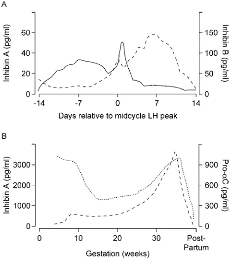
The major form of inhibin secreted during follicular phase of the menstrual cycle is inhibin B: serum levels rise sharply from early follicular phase of the menstrual cycle, with a peak follow-ing the FSH rise and a progressive fall durfollow-ing the remainder of the follicular phase (Figure 1A). Another peak of serum inhibin B is observed 2 days after the midcycle LH peak, followed by rapid decrease and constant low levels during luteal involution (Groome et al., 1996). The circulating levels of inhibin A are low in the early stage of follicular phase and rise from late fol-licular phase to peak at midluteal phase, after a short indentation coinciding with LH peak. Inhibin B levels change within a range above inhibin A levels during the whole follicular phase of men-strual cycle (Groome et al., 1996; Klein et al., 1996). These different menstrual cycle patterns suggest that the two inhibin forms may have different physiological roles. Whereas inhibin B seems to be the major marker of follicular growth in response to exogenous FSH administration, inhibin A is secreted mostly by the corpus luteum and may be involved in the gradual release of ovarian negative feedback on FSH secretion during the luteal – follicular transition (Weltet al., 1997).
Ageing cycling women have increasing FSH levels during the follicular phase in spite of normal estrogen and LH levels and regular menstrual cycles. This FSH rise occurs some years before menopause and accompanies the decline of ovarian fol-licular reserve and fertility rate. The selective FSH rise of late reproductive years might be a consequence of declining inhibin secretion by a reduced pool of ovarian follicules. This was
by guest on May 19, 2016
http://humupd.oxfordjournals.org/
suggested by early studies performed when assay methods did not discriminate between the two inhibin forms (Lenton et al., 1991). After the development of specific two-site ELISA for both inhibin A and inhibin B, the age-associated changes of ovarian – gonadotrophin interaction have become clearer. It was observed that: (i) ageing women with elevated FSH and normal estradiol levels have markedly lower inhibin B levels during the follicular phase (Klein et al., 1996; Reame et al., 1998); (ii) luteal phase inhibin A levels are also low in ageing cycling women (Danforth et al., 1998); (iii) follicular phase inhibin B levels decrease earlier than inhibin A during the process of ovar-ian ageing (Burger et al., 1998); (iv) both serum inhibin A and inhibin B levels are nearly undetectable in normal post-menopausal women (Burgeret al., 1998; Petragliaet al., 1998).
Changes of inhibin secretion in ovarian disorders
Hypothalamic amenorrhoea
Amenorrhoeic women with deficiency of GnRH secretion due to hypothalamic lesions are an interesting model to study the role of hypothalamic – pituitary axis on inhibin secretion. When trea-ted with exogenous GnRH at regular, physiological pulses, these patients display a normal increase of inhibin B levels during the luteal – follicular transition of induced menstrual cycles. In con-trast, abnormally low frequency pulses of GnRH administered during luteal – follicular transition result in sub-physiological levels of FSH and inhibin B (Weltet al., 1997). These findings indicate that the release of inhibin B is dependent on adequate FSH stimulation and may serve to confirm the effectiveness of GnRH replacement therapy on ovarian follicular development.
Conversely, inhibin A and inhibin B levels are not altered in patients with functional hypothalamic amenorrhoea (Petraglia
et al., 1998; Casper et al., 2000). These findings indicate that
basal inhibin secretion does not require a normal hypothalamus – pituitary function, but may be suddenly suppressed by primary ovarian failure, regardless of senescence.
Polycystic ovarian syndrome
Polycystic ovarian syndrome (PCOS) is characterized by increased inhibin levels due to the persistence of a cohort of small follicles that contribute to the pool of circulating inhibins, but the pulsatile rhythm of inhibin B secretion is blunted (Lockwoodet al., 1998a). Treatment of PCOS patients with low doses of FSH may be sufficient to induce the development of a single dominant follicle which grows in parallel with estradiol and inhibin A levels (Anderson et al., 1998a). Overall, these findings indicate that elevated circulating levels of inhibin B lead to the relative deficit of FSH in PCOS, while the disruption of inhibin B pulsatility may be an additional marker of multiple, incomplete follicular growth. Additionally, the paracrine action of inhibins on stimulating androstenedione production by theca cells may reinforce the hyperandrogenism of PCOS (Pignyet al., 1997, 2000).
Premature ovarian failure
Inhibins A and B are dramatically reduced in women with pre-mature ovarian failure (POF) (Petraglia et al., 1998). Serum levels of both inhibins are as low as in normal post-menopausal women matched for time elapsed since last menstrual period, and do not correlate with patient age, length of amenorrhoea, or serum gonadotrophin levels (Petraglia et al., 1998). In addition, circulating levels of inhibin A, inhibin B and pro-alphaC are reduced after oophorectomy. Women with amenorrhoea induced by GnRH analogue treatment or by antineoplastic chemotherapy still produce inhibin A and pro-alphaC. This probably reflects a residual ovarian function and hormone synthesis (Cobelliset al., 2002).
The GnRH analogue treatment should be considered in every woman of reproductive age receiving chemotherapy, in addition to assisted reproductive technologies and the investigational attempts of ovarian cryopreservation for future in vitro matu-ration or reimplantation. In addition, inhibin A concentmatu-rations may serve as a prognostic factor for predicting the resumption of ovarian function in addition to the levels of FSH, LH and estra-diol (Blumenfeldet al., 1999).
The testis
Sites and targets of inhibin
Inhibin subunits are expressed in the human testis from fetal life. At second trimester gestation, both a andbB subunits are present in Sertoli and Leydig cells, whereas gonocytes express only thebBsubunit and Leydig cells also express thebAsubunit (Rabinovici et al., 1991; Anderson et al., 2002). However, the
bA subunit serves mainly for the synthesis of its homodi-mer, activin A (Buzzard et al., 2003), whereas inhibin A is produced to a much lesser extent and remains undetectable in
Figure 1. (A) Changes of serum inhibin A and inhibin B levels during the menstrual cycle. Redrawn from Groomeet al. (1996). (B) Maternal serum inhibin A and pro-alphaC levels during pregnancy and post-partum. Accord-ing to Fowleret al.(1998).
by guest on May 19, 2016
http://humupd.oxfordjournals.org/
the peripheral serum of adult males (Anawaltet al., 1996; Illing-worthet al., 1996).
Sertoli cells are able to produce both aand bB inhibin sub-units before puberty, as suggested by immunolocalization and by the finding of normal serum inhibin B levels in pre-pubertal boys with Sertoli cell-only syndrome. After puberty, onlya sub-unit continues to be expressed by Sertoli cells whereas thebB
subunit is predominantly detected in maturating germ cells and with less intensity in Leydig cells (Andersson et al., 1998). Coherently, the acute administration of FSH to healthy adult men does not induce any significant change in serum inhibin B, while pro-alphaC levels do increase (Kinniburgh and Anderson, 2001). This suggests that Sertoli cells produce predominantly a
subunit in response to FSH stimulation, whereasbBsubunit and dimeric inhibin B require a more complex process to be released.
Testicular targets of inhibins
The receptors, co-receptors and intracellular signalling molecules thus far implicated in the inhibin mechanism of action are all expressed in the testis. Type II activin receptor and Smad pro-teins have been localized in Sertoli, Leydig, and germ cells (de Kretser et al., 2001; Hu et al., 2003), whereas the inhibin co-receptors betaglycan and inhibin-binding protein seem to be restricted to Leydig cells (Bernard et al., 2002). However, the physiological role of paracrine/autocrine inhibin effects within the testis has not been clarified. While the a subunit knockout mouse model suggests that this protein protects against the development of testicular tumours, there is no evidence for a physiological role of paracrine/autocrine inhibin signalling on spermatogenesis or steroidogenesis (reviewed by de Kretser
et al., 2001).
Physiology
Mutual regulation between inhibin and gonadotrophins in men
Physiological inhibin production by the adult testis requires a normal population of Sertoli cells, FSH stimulation, and sperma-togenesis to be present. The two later factors are not absolutely necessary for a basal inhibin B release, which is seen in some forms of hypogonadism, impaired spermatogenesis (Foresta
et al., 1999), and in men submitted to pharmacological
gona-dotrophin suppression by testosterone treatment (reviewed by Anderson, 2001). Spermatic vein blood sampling has revealed a pulsatile pattern of immunoreactive inhibin release and the pulses coincided with those of testosterone, suggesting that both hormones may respond to the same pulses of gonadotrophins (Winters, 1990). However, experimental evidence in non-human primates and clinical observations show unequivocally that the gonadotrophic effect of the pituitary gland on testicular inhibin secretion comes from FSH action on Sertoli cells (Ramaswamy and Plant, 2001). Not only is the contribution of Leydig cells to the circulating inhibin negligible (Andersson et al., 1998), but the LH effect on inhibin release also appears to be inhibitory rather than stimulatory (Ramaswamy and Plant, 2001).
Serum inhibin B concentrations are substantially decreased or undetectable in men with either hypo- or hypergonadotrophic hypogonadism (Anawaltet al., 1996). Among infertile men with
elevated FSH levels, the FSH concentration is inversely corre-lated with inhibin B but not with pro-alphaC, suggesting that the physiologically important hormone that exerts tonic negative feedback upon FSH secretion in men is inhibin B (Illingworth
et al., 1996).
Inhibin B as a marker of spermatogenesis
The functional relationship between spermatogenesis and serum inhibin B concentrations is nicely demonstrated by the study of patients with variable degrees of spermatogenesis impairment due to a single genetic disorder, the presence of microdeletions in the AZFc region of the Y chromosome (Frydelund-Larsen
et al., 2002). In this selected population, serum inhibin B
con-centrations are absolutely normal in the individuals with bilateral spermatocytic arrest but undetectable in patients with a predomi-nant Sertoli cell-only histological pattern (Frydelund-Larsen
et al., 2002).
Among the germ cells, spermatids appear to play a critical role in the mechanism of testicular inhibin B secretion in adult-hood. Men with obstructive azoospermia and spermatidic arrest show normal inhibin B concentrations, whereas those with Ser-toli cell-only syndrome or spermatogenesis arrest at pre-sperma-tid phases have considerably lower, often undetectable, inhibin B concentrations (Andersson et al., 1998; Foresta et al., 1999; Frydelund-Larsen et al., 2002). Testicular irradiation for treat-ment of carcinoma in situ leads to a Sertoli cell-only pattern with undetectable inhibin B levels in men with previously nor-mal inhibin B concentrations (Petersen et al., 1999). Neverthe-less, a minority of individuals with Sertoli cell-only syndrome are still capable of producing some inhibin B, the source of which has not been determined (Forestaet al., 1999).
Sertoli cells also release inhibin B from its apical side into the seminiferous tubules, thus contributing to the seminal plasma inhibin B content. Seminal plasma inhibin B concentrations are substantially reduced in azoospermic men regardless of obstruc-tive, non-obstructive or post-vasectomy azoospermia (Anderson
et al., 1998b; Garem et al., 2002), and correlate directly with
sperm count (Andersonet al., 1998b). Seminal plasma inhibin B is inversely related with serum FSH but their correlation is not strong, probably because the regulation of peripheral serum inhi-bin B (which exerts negative feedback on FSH secretion) differs from that of seminal plasma inhibin B (Andersonet al., 1998b; Garemet al., 2002).
Time-course from fetal to adult life and senescence
Inhibins A and B become detectable in male fetal serum at 14 – 16 weeks of gestation (Muttukrishna et al., 2004). In male fetuses, inhibin B concentrations at mid-trimester correlate directly with testosterone (Muttukrishnaet al., 2004) and inver-sely with FSH (Debieve et al., 2000), suggesting that the pitu-itary – testicular axis, and in particular the mutual regulation of inhibin B and FSH release, is already operative during fetal life.
Testicular cells collected from newborns produce more inhibin
B in vitrothan cells obtained from older children (Berensztein
et al., 2000). In the peripheral serum, inhibin B concentrations
are detectable from birth (Florio et al., 1999). In neonates, serum inhibin B concentrations are higher in males than in females, probably reflecting a testicular production, but there
by guest on May 19, 2016
http://humupd.oxfordjournals.org/
appears to be no correlation with FSH levels at this stage of development (De Schepper et al., 2000). There is a rapid post-natal increment which parallels the rise of FSH, then a peak at
,3 – 4 months and a rapid decrease to low but detectable
con-centrations during the following years until the pubertal rebound (Chada et al., 2003). The most pronounced increase in serum inhibin B levels during pubertal transition occurs between Tan-ner stages G1 and G2, coinciding with the LH and testosterone surges (Raivio et al., 1998; Chada et al., 2003). The decline in testicular function in older men is accompanied by a slight reduction in inhibin B concentrations, the mechanism of which has not been established (Mahmoud et al., 2000; Anderson, 2001).
Pathophysiology
Pubertal delay and hypogonadism
In boys with constitutional delay of puberty, as well as men with hypogonadotrophic hypogonadism, serum inhibin B concen-trations are low and dissociated from FSH. Upon GnRH replace-ment, the testis begins to respond to gonadotrophin stimulation by releasing inhibin B, which then reaches the physiological concentration range and establishes the negative feedback control of FSH release, as evidenced by an inverse correlation between inhibin B and FSH levels (Seminaraet al., 1996; Raivio
et al., 2000).
Cryptorchidism
Inhibin B can be used as a sensitive marker of testicular function in boys with various testicular disorders (Kubini et al., 2000). Basal inhibin B strongly correlates with the hCG-induced testos-terone increment and helps in the differential diagnosis between anorchia, abdominal testis with or without testicular damage, gonadal dysgenesis, and androgen insensitivity (Kubini et al., 2000). There is a direct correlation between inhibin B levels and the number of spermatogonia in testicular biopsies (Longuiet al., 1998). During early childhood, it is the basal inhibin B concen-tration that better detects testicular function, whereas in older children some increment of inhibin B following hCG injection may also be observed (Raivio and Dunkel, 1999).
Adults with cryptorchidism and subfertility have lower inhibin B levels than fertile men or even subfertile men without cryp-torchidism (Gouveia Brazaoet al., 2003).
Germ cell depletion by cancer therapy
Inhibin B is a reliable marker of spermatogenesis soon after pub-erty in cancer survivors who had received chemotherapy and/or radiation therapy in the past. These treatments cause selective depletion of germ cells without affecting Leydig cell function or steroidogenesis. Consequently, at puberty these boys have low inhibin B levels which correlate inversely with FSH and testicu-lar size but not with testosterone or LH (Lahteenmaki et al., 1999; Cicognaniet al., 2000). In adult men, the decrease of inhi-bin B and the consequent rise in FSH concentrations have been documented a few months after testicular irradiation for the treatment of carcinoma in situ (Petersen et al., 1999). Interest-ingly, these patients continue to produce freeasubunits that can
be detected by total inhibin assays (Tsatsoulis et al., 1990), while functional, dimeric inhibin B is suppressed.
Clinical usefulness in reproductive medicine
Monitoring of ovulation induction cycles
During controlled ovarian stimulation for assisted reproduction treatment, inhibin B can be used in the same way as estradiol to monitor the follicular growth. In a cohort of women undergoing ovulation induction for assisted reproduction treatment, a peak of inhibin B has been characterized at midfollicular phase, pre-ceding the pre-ovulatory estradiol peak, that correlated with the number of oocytes retrieved (Dokras et al., 2000) and fertilized (Fawzy et al., 2002). Higher serum inhibin A and inhibin B levels on the day of hCG administration to trigger ovulation are associated with a greater probability of pregnancy in assisted reproduction treatment cycles, but these markers do not add sub-stantial predictive power to classical parameters such as age and number of oocytes retrieved (Hall et al., 1999; Engel et al., 2003).
Both inhibin A and inhibin B are produced in greater amounts during IVF cycles complicated by ovarian hyperstimulation syn-drome (Enskog et al., 2000). Higher inhibin levels are already present during follicular growth and before the onset of the syn-drome, but the usefulness of inhibin determinations to predict this complication and guide cycle cancellation has not been established.
Evaluation of ovarian reserve and prediction of response to ovulation induction
Women with abnormally high FSH response to clomiphene citrate challenge also have lower serum inhibin B levels during the follicular phase of the menstrual cycle, which explains the exaggerated basal and/or post-test FSH levels (Hofmann et al., 1998). This occurs because normal ovulatory women in their late reproductive years have lower follicular phase inhibin B levels compared to young women, which reflects their reduced pool of growing follicles (Kleinet al., 1996). In addition to the number of follicles, inhibin B possibly reflects their quality as well. Thus, luteinized granulosa cells collected from women with elevated FSH levels have a reduced inhibin production rate
in vitro(Seiferet al., 1996).
As the physiological principle of the clomiphene citrate test is to unmask an exaggerated FSH release due to insufficient inhibin feedback, the recent improvement of inhibin assays has inspired the use of direct inhibin measurements to assess ovarian reserve instead of, or in addition to, FSH. As expected, low inhibin B levels at early follicular phase of spontaneous cycles predict an increased risk of poor response to controlled ovulation induction. Importantly, inhibin B offered significant prognostic information even after data correction for confounding variables such as the FSH concentration (Balaschet al., 1996; Seiferet al., 1997; Hall
et al., 1999; Tinkanen et al., 1999; Ficicioglu et al., 2003).
Whether this information is likely to influence the clinical decision as to justify the inclusion of inhibin B measurement in the work-up of assisted reproduction treatment patients is still uncertain. The latest clinical guideline released by the National Collaborating Centre for Women’s and Children’s Health (2004)
by guest on May 19, 2016
http://humupd.oxfordjournals.org/
states that the role of inhibin B for this purpose needs further evaluation.
Inhibin B has also been evaluated as an additional marker to predict the response to ovulation induction in women whose main infertility factor was ovulatory dysfunction but, in such cases, it does not appear to be of clinical relevance (Imaniet al., 2000, 2002). This is not surprising because anovulatory women who fail to respond to ovulation induction might have this resist-ance explained by a number of alternative mechanisms, apart from a diminished ovarian reserve.
Prediction of sperm retrieval in men with non-obstructive azoospermia
Serum inhibin B concentrations may be useful to predict the chances of successful sperm recovery among men with non-obstructive azoospermia submitted to testicular sperm extraction (TESE) (Ballescaet al., 2000; Baillyet al., 2003). There appears to be an association between lower inhibin B levels and unsuccessful sperm retrieval by TESE (Ballesca et al., 2000; Brugo-Olmedo et al., 2001; Bailly et al., 2003). This outcome cannot be predicted by FSH, probably because multiple factors other than spermatogenesis influence its peripheral concen-tration, whereas inhibin B reflects more faithfully the sperm pro-duction (Ballesca et al., 2000; Bailly et al., 2003). Despite this evidence, the precise accuracy of inhibin B test to predict the outcome of TESE – ICSI is still unknown. Whereas some studies suggested that inhibin B is a sensitive and specific marker of sperm recovery (Ballesca et al., 2000; Brugo-Olmedo et al., 2001), others have found it less effective (von Eckardsteinet al., 1999; Vernaeveet al., 2002). Only one study has stratified the population according to the different histopathologic patterns encompassed by the common denomination of non-obstructive azoospermia, and the sensitivity of inhibin B was much better if the pattern was maturation arrest compared to germ cell aplasia (Vernaeve et al., 2002). There is no evidence that combining inhibin B with another serum marker (e.g. FSH) will be advan-tageous over inhibin B alone to predict the outcome of TESE. Finally, it should be remembered that undetectable serum inhibin B does not exclude the possibility of successful pregnancy and delivery using TESE and ICSI (Guthauseret al., 2002). In con-trast to serum measurements, seminal plasma inhibin B does not distinguish patients with positive or negative outcome of TESE (Garemet al., 2002).
Early pregnancy
Physiology
Inhibins are also involved in the control of the feto-maternal communication required to maintain pregnancy. Human pla-centa, decidua, and fetal membranes are the major sites of pro-duction and secretion of inhibin A and inhibin B in maternal serum, amniotic fluid, and cord blood. During the first trimester of pregnancy, the feto-placental unit is the main source of circu-lating inhibin A and inhibin B. In fact, inhibin subunit mRNA (a, bA, bB) were localized in placental trophoblast (Petraglia
et al., 1987, 1991). Inhibin A is secreted into the maternal
circu-lation (Petraglia et al., 1993; Muttukrishna et al., 1995, 1997), and coelomic fluid (Riley et al., 1996; Wallace et al., 1997a;
Luisiet al., 1998). From 6 to 12 weeks of gestation, a precursor of inhibin A, pro-alphaC peptide, is mainly secreted by the cor-pus luteum (Muttukrishnaet al., 1997; Lockwoodet al., 1998b). During early pregnancy, pro-alphaC peptide levels increase to reach high concentrations at 5 weeks, and then fall after week 9 in coincidence with a small rise in inhibin A and with the peak in hCG (Fowleret al., 1998). Therefore, the profile of first trime-ster circulating maternal pro-alphaC is markedly different from inhibin A, with an excess of pro-alphaC over inhibin A, indicat-ing that a shift occurs in the tissue source and/or processindicat-ing pattern of the a subunit precursor as pregnancy advances (Figure 1B). It is likely that these events reflect the switch from corpus luteum to placental support for the pregnancy. Further-more, pro-alphaC concentrations in week 4 of pregnancy are higher in singleton pregnancy resulting from IVF treatment cycles (i.e. with multiple corpora lutea) than in spontaneous sin-gleton pregnancies (i.e. with single corpus luteum), but extre-mely low in pregnancies arising from frozen – thawed embryo (i.e. established without corpus luteum), suggesting that the cor-pus luteum is the main source of the amonomer during early pregnancy (Lockwoodet al., 1997).
Contrasting findings have been reported by Birdsall et al. (1997) as inhibin A concentrations in the first trimester of preg-nancy are not significantly different in women with or without corpora lutea, and multiple donor oocyte pregnancies were found to have higher concentrations of inhibin A compared with singleton donor oocyte pregnancies, supporting a placental source (Birdsall et al., 1997). It has also been reported that while maternal circulating concentrations of inhibin A after the removal of feto-placental unit gradually decreased within the first hour and decreased to even lower concentrations within the next 3 hours, maternal pro-alphaC levels decreased only within the first hour and then remained unaltered with levels similar to the luteal phase (Muttukrishna et al., 1997), indicating a significant contribution by a viable corpus luteum. Taken together, these findings suggest that in early pregnancy the feto-placental unit is the major source of inhibin A; and that pro-alphaC is mainly secreted by the corpus luteum.
During the first trimester of pregnancy, maternal serum levels of inhibin A are higher than in non-pregnancy, while inhibin B (Petraglia, 1997) does not significantly differ from levels detected during the menstrual cycle. At this gestational period, coelomic fluid inhibin B levels are higher than in maternal serum and amniotic fluid (Luisi et al., 1998). In amniotic fluid, inhibin A is not detectable (Rileyet al., 1996).
With respect to the possible clinical implications, the measurement of inhibin-related proteins in early pregnancy could be useful in the diagnosis of impending abortion, hydatidi-form mole and Down’s syndrome.
Pathologies
Impending abortion
Early diagnosis of a complicated or poor pregnancy outcome, particularly in patients undergoing assisted reproduction treat-ment, could aid their counselling and managetreat-ment, and the role for the inhibin family as markers of early pregnancy viability has been investigated in this respect. Initially low levels and a very rapid decline of inhibin A in non-viable clinical pregnancies
by guest on May 19, 2016
http://humupd.oxfordjournals.org/
with embryonic failure has been reported; and non-viable clinical pregnancies have very low levels of inhibin A and the initially low levels and very rapid decline in inhibin A in pregnancies with embryonic failure suggest a role for this glycoprotein as a monitor of early pregnancy viability (Lockwood et al., 1997, 1998a). Furthermore, in IVF pregnancies following inadvertent periconceptual exposure to GnRH analogue, the levels of pro-alphaC are found to be higher in successful pregnancies than in early pregnancy failure, being indicative of lytic damage, and herald pregnancy failure despite luteal supplementation with pro-gesterone (Lockwoodet al., 1998b).
However, all those studies have evaluated the early pregnancy outcome in women who became pregnant after assisted repro-duction treatment. Recently, inhibin A concentrations were measured in maternal circulation of healthy spontaneously preg-nant women progressing to deliver a healthy term singleton baby, in patients with incomplete miscarriage [carrying a non-viable (absence of heart beat activity) embryo or an anembryonic gestational sac in utero] and with complete miscarriage (an empty uterus with a history of passage of products of con-ception), in order to ascertain whether their measurement, in comparison to hCG, might provide a rapid and useful marker of early pregnancy viable placentation (Luisiet al., 2003).
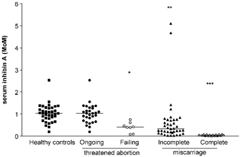
Patients with complete miscarriage had the lowest hCG, and inhibin A levels, that were lower in complete than in incomplete abortion (Figure 2). Thus, in the presence of complete miscar-riage, a condition associated with trophoblast dismissal, maternal serum levels of hCG and inhibin A are the lowest, indicating the presence of a failed trophoblast. In fact, in patients who under-went voluntary pregnancy interruption, maternal inhibin A con-centrations quickly decreased to low levels after the removal of the placenta (Muttukrishnaet al., 1997), reinforcing the concept that the feto-placental unit significantly contributes to the maternal levels. Thus, when evaluated in a larger population, according to different gestational ages (Luisi et al., 2003), or longitudinally in the same patients (Muttukrishna et al., 1997) inhibin A and hCG levels were significantly lower in miscarriage than in healthy patients.
The measurement of inhibin A during the first trimester of pregnancy could be useful in the diagnosis of trophoblast dys-functions, and therefore be helpful in the management of early pregnancy problems, to predict the first trimester pregnancy out-come in patients with early pregnancy vaginal bleeding due to threatened abortion. In fact, patients with early pregnancy bleed-ing and an intrauterine sac with fetal cardiac activity (threatened abortion) progressing till term have higher inhibin A than those with threatened abortion but whose pregnancy failed later (Florio
et al., 2004), This finding may add significant prognostic
infor-mation for predicting the pregnancy outcome. In fact, using the best thresholds indicated by the receiver operating characteristic (ROC) analysis, only inhibin A at the threshold 0.553 MoM achieved the best accuracy for prediction of failing pregnancy, showing a sensitivity of 90.6% and a specificity of 99.5% (Florio
et al., 2004) (Figure 2).
Inhibin A concentrations were also evaluated in women with a history of unexplained recurrent miscarriages: levels were lower in those patients that had a subsequent miscarriage com-pared with those who had a live birth (Muttukrishna et al., 2002b).
Taken together, all these findings would support the utility of serum inhibin A measurement in the prediction of poor outcome of pregnancy, or perhaps even as a marker of placental dysfunc-tion and damage both in the presence and before the onset of the clinical symptoms of recurrent miscarriage.
Hydatidiform mole
Partial hydatidiform mole is a triploid gestational trophoblastic tumour, occurring in about three per 1000 pregnancies (Seckl
et al., 2000). Inhibin subunits are localized in trophoblastic
tumours (McCluggageet al., 1998; Pelkeyet al., 1999; Shih and Kurman, 1999; McCluggage, 2001) and the detection by immunohistochemistry of the inhibin subunits has been recently proposed as a useful immunohistochemical marker of tropho-blastic neoplasia to be included in the diagnostic antibody panel in association with b-hCG (Pelkey et al., 1999; McCluggage, 2001). In addition, serum immunoreactive inhibin levels were found to be high in women with hydatidiform mole (Yohkaichiyaet al., 1989; Badonnelet al., 1994), and suggested as a reliable tumour marker. However, that assay was never available on a large scale, it was of poor reliability and did not distinguish specifically between the various forms of inhibin-related proteins.
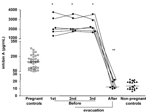
Inhibin A levels are consistently elevated in molar pregnancy without any considerable overlap with normal pregnancy values at the same stage of pregnancy (Florioet al., 2002), suggesting a role for inhibin A measurement in diagnosing molar pregnancies (Figure 3). In addition, inhibin A levels after molar evacuation decline significantly to values similar to those measured in non-pregnant women, whereas hCG levels, despite decreasing, remain far higher than in non-pregnant women, suggesting that inhibin A is more sensitive than hCG in identifying patients with spontaneous remission after molar evacuation.
Down’s syndrome
Inhibinasubunit is overexpressed in second trimester placental tissue of pregnancy affected by fetal Down’s syndrome
Figure 2.Maternal serum inhibin A levels in healthy pregnant women (con-trol), patients with threatened abortion with ongoing and failing pregnancy, incomplete and complete miscarriage. Individual values are plotted (expressed as mean of mean) and horizontal bars represent the group medians. *P,0.05, **P,0.001, ***P,0.001 versus healthy controls and threatened abortion with ongoing pregnancy.
by guest on May 19, 2016
http://humupd.oxfordjournals.org/
(Lambert-Messerlian et al., 1998), suggesting that increased a
subunit expression is one of the mechanisms leading to increased levels of inhibin A in serum. Indeed, during the second trimester of gestation, inhibin A levels were significantly elevated in maternal serum of women carrying a Down’s syndrome preg-nancy (Van Lithet al., 1992; Spenceret al., 1993; Cuckleet al., 1994), even in the presence of unchanged inhibin B (Wallace
et al., 1998a,b) and activin A levels (Lambert-Messerlianet al.,
1996, 1998), at least between 16 and 19 weeks of gestation. Amniotic fluid inhibin A levels are abnormally reduced in this syndrome, contrarily to what happens with maternal serum levels, indicating that the two compartments receive inhibin A from different sources: whereas the fetal membranes are the main source of inhibin A for the amniotic cavity, the placenta is responsible for most of that in the maternal circulation (Wallace
et al., 1997b, 1999).
The finding of high maternal serum inhibin A levels in associ-ation with Down’s syndrome may be used as a prenatal screen-ing marker. Although not sensitive enough to be used as a sscreen-ingle marker (Wenstrom et al., 1999), inhibin A may be usefully introduced into a multiple marker screening test for Down’s syndrome. The Serum Urine and Ultrasound Screening Study (SURUSS) conducted on 47 507 women recruited between September 1996 and April 2000 who attended 25 maternity centres (24 in the UK and one in Austria) has provided the lar-gest data set reported on women seen in both the first and second trimesters of pregnancy without intervention in the first trimester (Waldet al., 2003, 2004). The Quadruple test, an early second trimester (14 – 20 completed weeks) test based on the measure-ment of a-fetoprotein, unconjugated estriol (uE3), free b-hCG (or total hCG) and inhibin A together with maternal age, is now widely used in the USA and UK, and it shows a 6.2% false-posi-tive rate for an 85% detection rate, and the corresponding odds of being affected given a positive result were 1:32 (Waldet al., 2003, 2004). The integration of nuchal translucency measure-ment and pregnancy-associated plasma protein A (PAPP-A) in the first trimester with the Quadruple test in the second trimester
seems to be the most effective screening test, with an estimated 85% detection rate for a 0.9% false-positive rate, that increases to 1.3% without measuring inhibin A. The use of serum markers only (PAPP-A in the first trimester and the Quadruple test in the second trimester) has a an 85% detection rate for a 3.9% false-positive rate: however, the false-false-positive rate for the test without inhibin A measurement is 6.0% (Waldet al., 2003, 2004).
Conclusion
Based on the findings of the present review, it can be concluded that:
. Inhibin A and inhibin B are made at different times in human males and females and from several different cell types. They have a demonstrable role in suppressing FSH production and there is some evidence for local paracrine effects in the gonads. . Ovary and testis are the main inhibin A- and inhibin B-produ-cing organs from puberty.
. Inhibin A is secreted mainly by the corpus luteum, and is involved in the negative feedback control of FSH secretion during luteal – follicular transition.
. Inhibin A concentrations may serve as a prognostic factor for predicting the resumption of ovarian function.
. Inhibin B is secreted by antral follicles in response to FSH, and is the major marker of follicular growth. Thus, inhibin B level is a marker of ovarian function, which may become a useful tool to identify the potential ovarian responsiveness to ovulation induction.
. Inhibin B is a marker of spermatogenesis, and the decline in testicular function in older men is accompanied by a slight reduction in inhibin B concentrations, the mechanism of which has not been established.
. During pregnancy, the placenta becomes the predominant secretory organ for inhibin A, which is also produced by the corpus luteum.
. The measurement of inhibin A has clinical value in women at early pregnancy with impending abortion or hydatiform mole. . The increase of serum inhibin A in Down’s syndrome preg-nancies may have a clinical value, because it improves the prenatal detection of the disease.
References
Anawalt BD, Bebb RA, Matsumoto AM, Groome NP, Illingworth PJ, McNeilly AS and Bremner WJ (1996) Serum inhibin B levels reflect Sertoli cell function in normal men and men with testicular dysfunction. J Clin Endocrinol Metab 81,3341– 3345.
Anderson RA (2001) Clinical studies: inhibin in the adult male. Mol Cell Endocrinol 180,109 – 116.
Anderson RA, Groome NP and Baird DT (1998a) Inhibin A and inhibin B in women with polycystic ovarian syndrome during treatment with FSH to induce mono-ovulation. Clin Endocrinol 48,577– 584.
Anderson RA, Irvine DS, Balfour C, Groome NP and Riley SC (1998b) Inhi-bin B in seminal plasma: testicular origin and relationship to spermato-genesis. Hum Reprod 13,920– 926.
Anderson RA, Cambray N, Hartley PS and McNeilly AS (2002) Expression and localization of inhibin alpha, inhibin/activin betaA and betaB and the activin type II and inhibin beta-glycan receptors in the developing human testis. Reproduction 123,779– 788.
Andersson AM, Muller J and Skakkebaek NE (1998) Different roles of pre-pubertal and postpre-pubertal germ cells and Sertoli cells in the regulation of serum inhibin B levels. J Clin Endocrinol Metab 83,4451 – 4458.
Figure 3. Maternal serum inhibin A levels in healthy pregnant women (W), partial hydatidiform mole (†) before and after evacuation, and in non-preg-nant controls (K) during the follicular phase of the menstrual cycle. Medians are indicated by horizontal bars. *P,0.001 versus molar pregnancy before evacuation; **P,0.001 versus values before evacuation.
by guest on May 19, 2016
http://humupd.oxfordjournals.org/
Attardi B, Vaughan J and Vale W (1992) Regulation of FSH beta messenger ribonucleic acid levels in the rat by endogenous inhibin. Endocrinology 130,557 – 559.
Badonnel Y, Barbe F, Legagneur H, Poncelet E and Schweitzer M (1994) Inhibin as a marker for hydatidiform mole: a comparative study with the determinations of intact human chorionic gonadotropin and its free beta-subunit. Clin Endocrinol 41,155 – 162.
Bailly M, Guthauser B, Bergere M, Wainer R, Lombroso R, Ville Y and Selva J (2003) Effects of low concentrations of inhibin B on the out-comes of testicular sperm extraction and intracytoplasmic sperm injec-tion. Fertil Steril 79,905 – 908.
Balasch J, Creus M, Fabregues F, Carmona F, Casamitjana R, Ascaso C and Vanrell JA (1996) Inhibin, follicle-stimulating hormone, and age as pre-dictors of ovarian response in in vitro fertilization cycles stimulated with gonadotropin-releasing hormone agonist-gonadotropin treatment. Am J Obstet Gynecol 175,1226 – 1230.
Ballesca JL, Balasch J, Calafell JM, Alvarez R, Fabregues F, de Osaba MJ, Ascaso C and Vanrell JA (2000) Serum inhibin B determination is pre-dictive of successful testicular sperm extraction in men with non-obstructive azoospermia. Hum Reprod 15,1734– 1738.
Baly DL, Allison DE, Krummen LA, Woodruff TK, Soules MR, Chen SA, Fendly BM, Bald LN, Mathers JP and Lucas C (1993) Development of a specific and sensitive two-site enzyme-linked immunosorbent assay for measurement of inhibin-A in serum. Endocrinology 132,2099– 2108. Berensztein E, Saraco N, Belgorosky A and Rivarola MA (2000) Secretion
of inhibin B by human prepubertal testicular cells in culture. Eur J Endocrinol 142,481 – 485.
Bernard DJ, Chapman SC and Woodruff TK (2002) Inhibin binding protein (InhBP/p120), betaglycan, and the continuing search for the inhibin receptor. Mol Endocrinol 16,207– 212.
Birdsall M, Ledger W, Groome N, Abdalla H and Muttukrishna S (1997) Inhibin A, activin A in the first trimester of human pregnancy. J Clin Endocrinol Metab 82,1557– 1560.
Blumenfeld Z, Avivi I, Ritter M and Rowe JM (1999) Preservation of ferti-lity and ovarian function and minimizing chemotherapy-induced gona-dotoxicity in young women. J Soc Gynecol Invest 6,229 – 239.
Brugo-Olmedo S, De Vincentiis S, Calamera JC, Urrutia F, Nodar F and Acosta AA (2001) Serum inhibin B may be a reliable marker of the pre-sence of testicular spermatozoa in patients with nonobstructive azoosper-mia. Fertil Steril 76,1124 – 1129.
Burger HG, Cachir N, Robertson DM, Groome NP, Dudley E, Green A and Dennerstein L (1998) Serum inhibins A and B fall differentially as FSH rises in perimenopausal women. Clin Endocrinol 48,809 – 813.
Buzzard JJ, Farnworth PG, de Kretser DM, O’Connor AE, Wreford NG and Morrison JR (2003) Proliferative phase sertoli cells display a develop-mentally regulated response to activin in vitro. Endocrinology 144, 474 – 483.
Campbell BK and Baird DT (2001) Inhibin A is a follicle stimulating hor-mone-responsive marker of granulosa cell differentiation, which has both autocrine and paracrine actions in sheep. J Endocrinol 169, 333 – 345.
Campbell BK, Scaramuzzi RJ and Webb R (1996) Induction and mainten-ance of oestradiol and immunoreactive inhibin production with FSH by ovine granulosa cells cultured in serum-free media. J Reprod Fertil 106,7 – 16.
Casper FW, Seufert RJ and Pollow K (2000) Inverse correlation between baseline inhibin B and FSH after stimulation with GnRH: a study of serum levels of inhibin A and B, pro alpha-C and activin A in women with ovulatory disturbances before and after stimulation with GnRH. Eur J Endocrinol 143,77– 84.
Chada M, Prusa R, Bronsky J, Kotaska K, Sidlova K, Pechova M and Lisa L (2003) Inhibin B, follicle stimulating hormone, luteinizing hormone and testosterone during childhood and puberty in males: changes in serum concentrations in relation to age and stage of puberty. Physiol Res 52,45 – 51.
Cicognani A, Cacciari E, Pasini A, Burnelli R, De Iasio R, Pirazzoli P and Paolucci G (2000) Low serum inhibin B levels as a marker of testicular damage after treatment for a childhood malignancy. Eur J Pediatr 159,103 – 107.
Cobellis L, Luisi S, Pezzani I, Reis FM, De Leo V and Petraglia F (2002) Serum inhibin A, inhibin B, and pro-alphaC levels are altered after sur-gically or pharmacolosur-gically induced menopause. Fertil Steril 77, 745 – 749.
Cuckle HS, Holding S and Jones R (1994) Maternal serum inhibin levels in second-trimester Down’s syndrome pregnancies. Prenat Diagn 14, 387 – 390.
Danforth DR, Arbogast LK, Mroueh J, Kim MH, Kennard EA, Seifer DB and Friedman CI (1998) Dimeric inhibin: a direct marker of ovarian aging. Fertil Steril 70,119 – 123.
Debieve F, Beerlandt S, Hubinont C and Thomas K (2000) Gonadotropins, prolactin, inhibin A, inhibin B, and activin A in human fetal serum from midpregnancy and term pregnancy. J Clin Endocrinol Metab 85, 270 – 274.
De Schepper J, Verlinde F, Cortvrindt R, Callewaert M and Smitz J (2000) Serum inhibin B in normal term-born male and female neonates during the first week of life. Eur J Pediatr 159,465– 469.
de Kretser DM, Loveland KL, Meehan T, O’Bryan MK, Phillips DJ and Wreford NG (2001) Inhibins, activins and follistatin: actions on the tes-tis. Mol Cell Endocrinol 180,87 – 92.
Dokras A, Habana A, Giraldo J and Jones E (2000) Secretion of inhibin B during ovarian stimulation is decreased in infertile women with endome-triosis. Fertil Steril 74,35– 40.
Eramaa M, Hilden K, Tuuri T and Ritvos O (1995) Regulation of inhibin/ activin subunit messenger ribonucleic acids (mRNAs) by activin A and expression of activin receptor mRNAs in cultured human granulosa-luteal cells. Endocrinology 136,4382 – 4389.
Engel JB, Felberbaum RE, Junge K, Reissmann T and Diedrich K (2003) Inhibin B on the day of HCG-administration is a predictive factor for failure in assisted reproduction cycles. Arch Gynecol Obstet 268, 278 – 280.
Engelhardt H, Harkness LM, Thomas GB, Brooks AN, McNeilly AS and Baird DT (1995) Expression of inhibin alpha- and beta A-subunit mRNA and protein inthe fetal sheep ovary throughout gestation. Mol Cell Endocrinol 107,141 – 147.
Enskog A, Nilsson L and Brannstrom M (2000) Peripheral blood concen-trations of inhibin B are elevated during gonadotropin stimulation in patients who later develop ovarian OHSS and inhibin A concentrations are elevated after OHSS onset. Hum Reprod 15,532 – 538.
Evans LW, Muttukrishna S, Knight PG and Groome NP (1997) Develop-ment, validation and application of a two-site enzyme-linked immuno-sorbent assay for activin-AB. J Endocrinol 153,221– 230.
Fawzy M, Lambert A, Harrison RF, Knight PG, Groome N, Hennelly B and Robertson WR (2002) Day 5 inhibin B levels in a treatment cycle are predictive of IVF outcome. Hum Reprod 17,1535– 1543.
Ficicioglu C, Kutlu T, Demirbasoglu S and Mulayim B (2003) The role of inhibin B as a basal determinant of ovarian reserve. Gynecol Endocrinol 17,287 – 293.
Florio P, Luisi S, Vigano` P, Busacca M, Fadalti M, Genazzani AR and Petraglia F (1998) Healthy women and patients with endometriosis show high concentrations of inhibin A, inhibin B, and activin A in peritoneal fluid throughout menstrual cycle. Hum Reprod 13,2606– 2611.
Florio P, Benedetto C, Luisi S, Santuz M, Di Carlo C, Marozio L, Genazzani AR and Petraglia F (1999) Activin A, inhibin A, inhibin B, parturition: changes of maternal and cord serum levels according to the mode of delivery. Br J Obstet Gynaecol 106,1061 – 1065.
Florio P, Severi FM, Cobellis L, Danero S, Bome A, Luisi S and Petraglia F (2002) Serum activin A and inhibin A. New clinical markers for hydati-diform mole. Cancer 94,2618– 2622.
Florio P, Luisi S, D’Antona D, Severi FM, Rago G and Petraglia F (2004) Maternal serum inhibin A levels may predict pregnancy outcome in women with threatened abortion. Fertil Steril 81,468 – 470.
Foresta C, Bettella A, Petraglia F, Pistorello M, Luisi S and Rossato M (1999) Inhibin B levels in azoospermic subjects with cytologically characterized testicular pathology. Clin Endocrinol 50,695 – 701. Fowler PA, Evans TW, Groome NP, Templeton A and Knight PG (1998) A
longitudinal study of maternal serum inhibin A, inhibin B, activin A, pro-aC and follistatin during pregnancy. Hum Reprod 12,3530 – 3536. Frydelund-Larsen L, Krausz C, Leffers H, Andersson AM, Carlsen E,
Bangsboell S, McElreavey K, Skakkebaek NE and Rajpert-De Meyts E (2002) Inhibin B: a marker for the functional state of the seminiferous epithelium in patients with azoospermia factor C microdeletions. J Clin Endocrinol Metab 87,5618 – 5624.
Garem YF, Arini AF, Beheiry AH, Zeid SA and Comhaire FH (2002) Poss-ible relationship between seminal plasma inhibin B and spermatogenesis in patients with azoospermia. J Androl 23,825– 829.
Gouveia Brazao CA, Pierik FH, Erenpreiss Y, de Jong FH, Dohle GR and Weber RF (2003) The effect of cryptorchidism on inhibin B in a subfer-tile population. Clin Endocrinol 59,136– 141.
by guest on May 19, 2016
http://humupd.oxfordjournals.org/
Groome NP, Illingworth PJ, O’Brien M, Cooke I, Ganesan TS, Baird DT and McNeilly AS (1994) Detection of dimeric inhibin throughout the human menstrual cycle by two-site enzyme immunoassay. Clin Endocri-nol 40,717 – 723.
Groome NP, Illingworth PJ, O’Brien M, Priddle J, Weaver K and McNeilly AS (1995) Quantification of inhibin pro-alpha C-containing forms in human serum by a new ultrasensitive two-site enzyme-linked immuno-sorbent assay. J Clin Endocrinol Metab 80,2926 – 2932.
Groome NP, Illingworth PJ, O’Brien M, Pai R, Rodger FE, Mether JP and McNeilly AS (1996) Measurement of dimeric inhibin B throughout the human menstrual cycle. J Clin Endocrinol Metab 81,1401– 1405. Guthauser B, Bailly M, Bergere M, Wainer R, Ville Y and Selva J (2002)
Successful pregnancy and delivery after testicular sperm extraction despite an undetectable concentration of serum inhibin B in a patient with nonobstructive azoospermia. Fertil Steril 77,1077 – 1078.
Hall JE, Welt CK and Cramer DW (1999) Inhibin A and inhibin B reflect ovarian function in assisted reproduction but are less useful at predicting outcome. Hum Reprod 14,409– 415.
Hayes FJ, Hall JE, Boepple PA and Crowley WF Jr (1998) Differential con-trol of gonadotropin secretion in the human: endocrine role of inhibin. J Clin Endocrinol Metab 83,1835– 1841.
Hillier SG, Yong EL, Illingworth PJ, Baird DT, Schwall RH and Mason AJ (1991) Effect of recombinant inhibin on androgen synthesis in cultured human thecal cells. Mol Cell Endocrinol 75,R1 – R6.
Hofmann GE, Danforth DR and Seifer DB (1998) Inhibin-B: the physiologic basis of the clomiphene citrate challenge test for ovarian reserve screen-ing. Fertil Steril 69,474 – 477.
Hu J, Zhang YQ, Liu XP, Wang RA, Jin Y and Xu RJ (2003) Expression and localization of Smad1, Smad2 and Smad4 proteins in rat testis during postnatal development. Asian J Androl 5,51 – 55.
Illingworth PJ, Reddi K, Smith KB and Baird DT (1991) The source of inhi-bin secretion during the human menstrual cycle. J Clin Endocrinol Metab 73,667– 673.
Illingworth PJ, Groome NP, Byrd W, Rainey WE, McNeilly AS, Mather JP and Bremner WJ (1996) Inhibin-B: a likely candidate for the physiologi-cally important form of inhibin in men. J Clin Endocrinol Metab 81,1321– 1325.
Imani B, Eijkemans MJ, de Jong FH, Payne NN, Bouchard P, Giudice LC and Fauser BC (2000) Free androgen index and leptin are the most pro-minent endocrine predictors of ovarian response during clomiphene citrate induction of ovulation in normogonadotropic oligoamenorrheic infertility. J Clin Endocrinol Metab 85,676 – 682.
Imani B, Eijkemans MJ, Faessen GH, Bouchard P, Giudice LC and Fauser BC (2002) Prediction of the individual follicle-stimulating hormone threshold for gonadotropin induction of ovulation in normogonadotropic anovulatory infertility: an approach to increase safety and efficiency. Fertil Steril 77,83 – 90.
Kinniburgh D and Anderson RA (2001) Differential patterns of inhibin secretion in response to gonadotropin stimulation in normal men. Int J Androl 24,95– 101.
Keogh EJ, Lee VLWK, Rennie GC, Burger HG, Hudson B and de Kretser DM (1976) Selective suppression of FSH by testicular extracts. Endo-crinology 98,997– 1004.
Kishi H, Okada T, Otsuka M, Watanabe G, Taya K and Sasamoto S (1996) Induction of superovulation by immunoneutralization of endogenous inhibin through the increase in the secretion of follicle-stimulating hor-mone in the cyclic golden hamster. J Endocrinol 151,65– 75.
Klein NA, Illingworth PJ, Groome NP, McNeilly AS, Battaglia DE and Soules MR (1996) Decreased inhibin B secretion is associated with the monotropic FSH rise in older, ovulatory women: a study of serum and follicular fluid levels of dimeric inhibin A and B in spontaneous men-strual cycles. J Clin Endocrinol Metab 81,2742– 2745.
Knight PG and Muttukrishna S (1994) Measurement of inhibin using a modi-fied two-site immunoradiometric assay specific for oxidized (Met O) inhibin. J Endocrinol 141,417– 425.
Krummen LA, Moore A, Woodruff TK, Rice GC and Phillips DM (1994) Localization of inhibin and activin binding sites in the testis during development by in situ ligant binding. Biol Reprod 50,734 – 744. Kubini K, Zachmann M, Albers N, Hiort O, Bettendorf M, Wolfle J,
Bidlingmaier F and Klingmuller D (2000) Basal inhibin B and the tes-tosterone response to human chorionic gonadotropin correlate in prepu-bertal boys. J Clin Endocrinol Metab 85,134– 138.
Lahteenmaki PM, Toppari J, Ruokonen A, Laitinen P and Salmi TT (1999) Low serum inhibin B concentrations in male survivors of childhood malignancy. Eur J Cancer 35,612 – 619.
Lambert-Messerlian GM, Canick JA, Palomaki GE and Scneyer AL (1996) Second trimester levels of maternal serum inhibin A, a inhibin precursor, and activin in Down syndrome pregnancy. J Med Screen 3,58 – 62. Lambert-Messerlian GM, Luisi S, Florio P, Mazza V, Canick JA and
Petraglia F (1998) Second trimester levels of maternal serum total activin A and placental inhibin/activin a and bA subunit messenger ribonucleic acids in Down syndrome pregnancy. Eur J Endocrinol 138,425 – 429.
Lebrun JJ and Vale WW (1997) Activin and inhibin have antagonistic effects on ligand-dependent heteromerization of the type I and type II activin receptors and human erythroid differentiation. Mol Cell Biol 17, 1682 – 1691.
Lenton EA, de Kretser DM, Woodward AJ and Robertson DM (1991) Inhi-bin concentrations throughout the menstrual cycles of normal, infertile, and older women compared with those during spontaneous conception cycles. J Clin Endocrinol Metab 73,1180– 1190.
Lewis KA, Gray PC, Blount AL, MacConell LA, Wiater E, Bilezikjian LM and Vale W (2000) Betaglycan binds inhibin and can mediate functional antagonism of activin signalling. Nature 404,411– 414.
Lockwood GM, Ledger WL, Barlow DH, Groome NP and Muttukrishna S (1997) Measurement of inhibin and activin in early human pregnancy: demonstration of fetoplacental origin and role in prediction of early-pregnancy outcome. Biol Reprod 57,1490 – 1494.
Lockwood GM, Muttukrishna S, Groome NP, Matthews DR and Ledger WL (1998a) Mid-follicular phase pulses of inhibin B are absent in polycystic ovarian syndrome and are initiated by successful laparoscopic ovarian diathermy: a possible mechanism regulating emergence of the dominant follicle. J Clin Endocrinol Metab 83,1730 – 1735.
Lockwood GM, Ledger WL, Barlow DH, Groome NP and Muttukrishna S (1998b) Identification of the source of inhibins at the time of conception provides a diagnostic role for them in very early pregnancy. Am J Reprod Immunol 40,303 – 308.
Longui CA, Arnhold IJ, Mendonca BB, D’Osvaldo AF and Bloise W (1998) Serum inhibin levels before and after gonadotropin stimulation in cryp-torchid boys under age 4 years. J Pediatr Endocrinol Metab 11, 687 – 692.
Luisi S, Battaglia C, Florio P, D’Ambrogio G, Taponeco F, Santuz M, Genazzani AR and Petraglia F (1998) Activin A and inhibin B in extra-embryonic coelomic and amniotic fluids, maternal serum in early preg-nancy. Placenta 19,435 – 438.
Luisi S, Florio P, D’Antona D, Severi FM, Sanseverino F, Danero S and Petraglia F (2003) Maternal serum inhibin A levels are a marker of a viable trophoblast in incomplete and complete miscarriage. Eur J Endo-crinol 148,233 – 236.
Magoffin DA and Jakimiuk AJ (1997) Inhibin A, inhibina B and activin A in the follicular fluid of regularly cycling women. Hum Reprod 12, 1714 – 1719.
Mahmoud AM, Goemaere S, De Bacquer D, Comhaire FH and Kaufman JM (2000) Serum inhibin B levels in community-dwelling elderly men. Clin Endocrinol 53,141 – 147.
Mather JP, Moore A and Li RH (1997) Activins, inhibins, and follistatins: further thoughts on a growing family of regulators. Proc Soc Exp Biol Med 215,209 – 222.
McCluggage WG (2001) Value of inhibin staining in gynecological pathol-ogy. Int J Gynecol Pathol 20,79– 85.
McCluggage WG, Ashe P, McBride H, Maxwell P and Sloan JM (1998) Localization of the cellular expression of inhibin in trophoblastic tissue. Histopathology 32,252 – 256.
McLachlan RI, Robertson DM, Burger HG and de Kretser DM (1986) The radio-immunoassay of bovine and human follicular fluid and serum inhi-bin. Mol Cell Endocrinol 46,175 – 185.
McLachlan RI, Healy DL, Robertson DM et al. (1987) Circulating immuno-reactive inhibin in the luteal phase and early gestation in women under-going ovulation induction. Fertil Steril 48,1011 – 1015.
Minami S, Yamoto M and Nakano R (1995) Sources of inhibin in early preg-nancy. Early Pregn 1,62 – 66.
Molskness TA, Woodruff TK, Hess DL, Dahl KD and Stouffer RL (1996) Recombinant human inhibin-A administered early in the menstrual cycle alters concurrent pituitary and follicular, plus subsequent luteal, function in rhesus monkeys. J Clin Endocrinol Metab 81,4002 – 4006.
Muttukrishna S, George L, Fowler PA, Groome NP and Knight PG (1995) Measurement of serum concentrations of inhibin A (a-bA dimer) during human pregnancy. Clin Endocrinol 2,391 – 397.
by guest on May 19, 2016
http://humupd.oxfordjournals.org/