Enzymatic Cellulose Palmitate Synthesis Using Immobilized Lipase
Anna Roosdiana 1), Diah Mardiana 1), Ellya Indahyanti 1), Dyah Ayu Oktavianie 2) 1) Faculty of Mathematics and Natural Sciences, University of Brawijaya, Indonesia
2) Faculty of Veterinary Medicine, University of Brawijaya, Indonesia
E-mail: [email protected]
ABSTRACT
Bacterial cellulose can be modified by esterification using palmitic acid and Mucor miehei lipase as catalysts. The purpose of this research was to determine the optimum conditions of esterification reaction of cellulose and palmitic acid. The esterification reaction was carried out at the time variation of 6, 12, 18, 24 and 30 hours and the mass ratio of cellulose: palmitic acid (1:1, 1: 2, 1: 3, 1:4, 1:5, 1:6) at 50°C. The cellulose palmitate was examined its physical and chemical properties by using FTIR spectrophotometer, XRD, bubble point test and saponification apparatus. The results showed that the optimum reaction time of esterification reaction of cellulose and palmitic acid occurred within 24 hours and the mass ratio of cellulose: palmitic acid was 1:3 resulting in DS of 0.376 with swelling index of 187%, crystallinity index of 61.95%, and Φ porous of 2.40 μm. Identification of functional groups using FTIR spectrophotometer showed that C=O ester group was observed at 1737.74 cm-1 and strengthened by the appearance of C-O ester peak at 1280 cm-1. The conclusion of this study is reaction time and reactant ratio influence significantly the DS of cellulose ester.
Keywords: bacterial cellulose, esterification, lipase, palmitic acid.
INTRODUCTION
Cellulose is a high molecular weight
linear polymer comprised of
D-glucopyranose units linked by β-1.4 glycosidic bonds. Cellulose is the main structural cell
wall component of all major plant and apart from plant, it can be derived from source as certain strains Gluconacetobacter xylinus bacteria (Romling, 2012). Bacterial cellulose can be obtained in high purity, whether plant cellulose is usually mixture with lignocelluloses. Therefore, modification of cellulose is more advantageous using bacterial cellulose. Modification of cellulose can be carried out by etherification, esterification and grafting. Modified cellulose, especially cellulose acetate can be function as raw material for membrane filtration such as microfiltration, ultrafiltration and reverse osmosis (Puspitasari dan Radiman, 2006).
of substitution (Puspitasari dan Radiman, 2006).
Chemicals used for esterification are anhydride carboxylic acid such as anhydride acetic acid or trifluoacetic acid, however the use of those chemical and solvents are carcinogenic agents, it is necessary to look for another esterification method. One of the alternatives of esterification is enzymatic esterification using immobilized lipase. Lipase can be produced from Mucor miehei, followed by purification and dialysis. Purification of lipase was carried out through precipitation using ammonium sulfate saturation of 20-60% to produce a specific activity of 5.4 g/mL.minute (Roosdiana et al., 2009).
The esterification of cellulose and carboxylic acid is influenced by temperature, the ratio of reactants and reaction time. Bacterial cellulose is enzymatically esterified with citric acid for 24 hours and the weight ratio of 1:4 produce cellulose citrate with a value of DS: 0.34 and IS: 33.90% (Roosdiana et al ,2015).
This research aims to determine the influence of reaction time and mass ratio of reactants on degree of substitution of cellulose palmitate. The resulted ester is identified its physical and chemical properties.
MATERIALS AND METHODS
Bacterial cellulose was purchased from Gandaria Street 11 Malang. Lipase (102 U/ g) from Mucor miehei was purchased from Sigma-Aldrich, distilled water, chitosan, calcium hypochlorite, palmitic acid, NaOH, HCl, KBr, n-butanol (Merck), ethanol 96%, sodium tripolyphosphate, acetic acid glacial and phenolftalein indicator. The instruments used were Bubble point apparatus, FTIR spectrophotometer (Shimadzu 1601) and XRD (PANalytical Xpert PRO).
Preparation of Bacterial Cellulose Powder. Bacterial cellulose, nata de coco were washed thoroughly with running water, then treated with 0.1 M NaOH and 0.5% calcium hypochlorite solution at room temperature and kept overnight respectively. Nata de coco was washed again with running water. Nata de coco was pressed at 110-120°C for 5 minutes using a hot hydraulic press. The dried nata de coco was ground using dry grinder. The powder was sieved using 100-120 mesh of sieve.
Preparation Immobilized Lipase.
The matrix was used 1.25 g chitosan and dissolved in 50 ml of 3% glacial acetic acid solution. Then, 0.5 g of lipase was mixed with 5 mL of chitosan solution. The mixture was dropped into 10 ml of 3% sodium tripolyphosphate solution and allowed to stand for 75 minutes, then the immobilized lipase was put in the refrigerator.
Determination of Optimum Mass Ratio of Reactant.
The procedure was similar to
determination of the optimum reaction time The reaction was carried out at optimum reaction time within various mass ratio of cellulose and palmitic acid (1:1, 1:2, 1:3, 1:4, 1:5 and 1:6).
Determination of physical and chemical properties of cellulose palmitate.
Analysis of cellulose palmitate esters using FTIR was conducted to determine its functional groups. Sample was ground with KBr. Transmission was measured at the wave number range 4000-400 cm-1.
Degree of Substitution (DS).
The values 162 is the molecular weight of the anhydrous glucose unit and 239.5 is
the molecular weight of substituent from palmitic acid.
The hydrophilicity of ester was observed by determining the swelling index of ester in distilled water as the diffusing agent. The swelling index was calculated by following equation (Caillol, et al., 2012):
Swelling index (%) = (Ws-Wd)/Wd x 100
Where, Ws and Wd are the weights of the swollen bar and the dried bar, respectively.
RESULTS AND DISCUSSION
Optimum Condition of Esterification Bacterial Cellulose with Palmitic Acid Using Immobilized Lipase.
There are some factors affecting the activity of lipase in catalyzing esterification reaction, number of enzyme, temperature, reaction time, amount ratio of reactants and water activity. In this study, the esterification of bacterial cellulose with palmitic acid was conducted at optimum lipase activity of 50°C.
Figure 1. pH Value from Three Formulations.
Figure 3. Flux of Diclofenac Sodium Penetrated
For the calculation of permeability, mean permeability of every formula is (Figure 4): formula I of 1.0320. 10-4±5.92. 10-6, formula II of 1.0289. 10-4±3.69. 10-6 and formula III of 1.3637. 10-4±6.15. 10-6. The permeability value of formula III was higher than others and differ significantly with formula II. It pointed out that niosomal system increased membran permeability.
The data of pH, flux and permeability showed that the usage of niosomal system (diclofenac sodium-span 60-cholesterol= 1:6:6) on HPC gel base increase pH value, membrane permeability and penetration rate of diclofenac sodium in HPC gel base. Those elevations were due to good dispersion of drugs by niosomal system. It made the drugs release easily from gel base. Increasing of flux and permeability due to niosomal system prepared dispersion well so the drugs release easily from gel base. The impact was sufficient of drugs availability for optimal penetration. The next mechanism was niosome decreasing water losing at trans-epidermal layer and affecting conformation structure skin lipid bilayer.
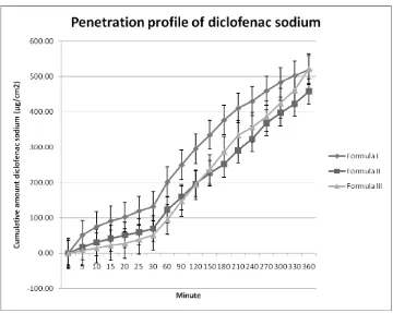
For the first minutes, formula II gave higher line than formula III. It could be seen that penetration of formula II was faster. This could be due to the effect of propylenglycol in formula II. Propylenglycol, at first, it was used as a preservative. But, it obviously acted as a wetting agent. Propylenglycol also dissolved diclofenac sodium so the drug dispersion was better.
The difference of mixing with propylenglycol was the cause of higher cumulative amount of diclofenac sodium in formula II at the first minutes. In formula II, propylenglycol wet diclofenac sodium. Meanwhile, propylenglycol had been mixed all in the gel base in formula III. The direct contact between diclofenac sodium and propylenglycol made a better dispersion. Unfortunately, this effect could not make high cumulative amount of diclofenac sodium. As we could see in Figure 2, the cumulative amount of formula II was less than formula III.
In formula II, span 60 and cholesterol were only mixed with the other component. On that way, they made the gel was at hydrophobic ambiance. Diclofenac sodium’s
affinity with span 60 and cholesterol would be stronger so the drug was less capable to release optimally. It had an effect in availability and penetration. Even though span 60 had hydration effect which could help penetration effect, but its ability was not supporting significantly. Moreover, the drug availability was less. Overall, formula III had increasing penetration rate of 19.93% and permeability 32.54% compared with formula II.
CONCLUSION
It can be concluded that the niosomal system (diclofenac sodium-span 60-cholesterol=1:6:6) with its entrapment efficiency of 74.33%; it had influence in increasing pH value and penetration (based on flux value and permeability) of diclofenac sodium in HPC gel base. That formulation also gave white milky, odorless,
smooth and watery consistency
characteristics. The pH value of formula III was high, 7.34. We suggest further research to give the formulation with normal pH value for human skin. In addition, the morphology evaluation of niosomes was very important. Optimizing SEM method preparation was needed for niosomes evaluation.
ACKNOWLEDGEMENT
Authors are grateful to Dexa Medica, Corp, Palembang, Indonesia for providing gift sample of diclofenac sodium.
REFERENCES
Australian Government Department of Health. 2014. Safety Review of Diclofenac. Commonwealth of Australia. Allen, L. V. Popovich, N. G., and Ansel, HC.
Lippincott Williams & Wilkins.
Barry, B. W. 1983. Dermatological Formulation Percutaneous Absorption. New York:Marcel Dekker Inc.
Biju, S. S., Sushama,T., Mishra, P. R., & Khar, R. K. 2006. Vesicular Systems: An Overview. Indian J Pharma Sci. 68 (2): 141-153.
Choi, M. J., and Maibach, H. I. 2005. Liposomes and Niosomes as Topical
Drug Delivery Systems. Skin
Pharmacology and Physiology. 18 (5): 209-219.
Florey, K. 1986. Analytical Profiles of Drug Substance. Orlando: Academic Press Inc.
Hardman, J. G., and Limbird, L. E. 2001. The Pharmacological Basis of Therapeutics. 10th edition. United States: The McGraw Hill. p. 709 -710.
Hendradi, E. 1995. Kinetika dan Mekanisme Transport Beberapa Antihistamin Melewati Membran Lipid. Thesis. Yogyakarta.
Jufri, M., Anwar, E., and Djajadisastra, J. 2004. Pembuatan Niosom Berbasis Maltodekstrin DE 5-10 dari Pati Singkong (Manihot utilissima). Majalah Ilmu Kefarmasian, I (1): 10-20.
Mura, S., Pirot, F., Manconi, M., Falson, F., and Fadda, A. M. 2007. Liposomes and niosomes as potential carriers for dermal delivery of minoxidil. Journal of Drug Targeting, February 15(2): 101-108.
Naresh, R. A. R., Pillai, G. K,. Udupa, K,. and
Chandrashekar, G. 1994.
Antiinflammatory Activity of Niosome Encapsulated Diclofenac Sodium in Arthritic Rats. Indian J Pharmacol. 26: 46-48.
Patel, R. 2008. Niosome: An Unique Drug Delivery System. Retrieved From the website: Stinchcomb, A. L. 2010. Challenges and opportunities in dermal/transdermal delivery. Their Deliv. July, 1(1): 109-131.
Priprem, A., Janpim, K., Nualkaew, S., and Mahakunakorn, P. (2016). Topical Niosome Gel of Zingiber cassumunar Roxb. Extract for Anti-inflammatory Activity Enhanced Skin Permeation and Stability of Compound D. AAPS PharmSciTech 17 (3): 631–639.
Rowe, R. C., Sheskey, P. J., and Quinn, M. E. (Eds). 2009. Handbook of Pharmaceutical Excipients. 6th edition. London: Pharmaceutical Press.
Shahiwala, A., and Misra, A. 2002. Studies in Topical Application of Niosomally Entrapped Nimesulide. J Pharm Pharmaceut Sci., 5 (3):220 –225. Sinko, P. J., and Singh, Y. (2011). Martin's
Physical Pharmacy and Pharmaceutical Sciences. 6th edition. Baltimore: Lippincott Williams & Wilkins.
Suresh, R. V., and Kerunath, K. P. 2015. Formulation and Evaluation of Novel Anti-Bacterial Ciprofloxacin Loaded Niosomal Cream. Int. Res. J. Pharm. 6 (8). 519-527.