U. S. ARMY MEDICAL DEPARTMENT CENTER AND SCHOOL
FORT SAM HOUSTON, TEXAS 78234
MD0006
BASIC HUMAN ANATOMY
DEVELOPMENT
This subcourse reflects the current thought of the Academy of Health Sciences and conforms to printed Department of the Army doctrine as closely as currently possible. Development and progress render such doctrine continuously subject to change.
When used in this publication, words such as "he," "him," "his," and "men" are intended to include both the masculine and feminine genders, unless specifically stated
otherwise or when obvious in context.
ADMINISTRATION
Students who desire credit hours for this correspondence subcourse must meet eligibility requirements and must enroll through the Nonresident Instruction Branch of the U.S. Army Medical Department Center and School (AMEDDC&S).
Application for enrollment should be made at the Internet website:
http://www.atrrs.army.mil. You can access the course catalog in the upper right corner. Enter School Code 555 for medical correspondence courses. Copy down the course number and title. To apply for enrollment, return to the main ATRRS screen and scroll down the right side for ATRRS Channels. Click on SELF DEVELOPMENT to open the application and then follow the on screen instructions.
In general, eligible personnel include enlisted personnel of all components of the U.S. Army who hold an AMEDD MOS or MOS 18D. Officer personnel, members of other branches of the Armed Forces, and civilian employees will be considered eligible based upon their AOC, NEC, AFSC or Job Series which will verify job relevance. Applicants who wish to be considered for a waiver should submit justification to the Nonresident Instruction Branch at e-mail address: [email protected].
For comments or questions regarding enrollment, student records, or shipments, contact the Nonresident Instruction Branch at DSN 471-5877, commercial (210) 221-5877, toll-free 1-800-344-2380; fax: 210-221-4012 or DSN 471-4012, e-mail
5 THE HUMAN MUSCULAR SYSTEM
Section I. The Skeletal Muscle ... 5-1--5-4 5-2 II. Some Elementary
Skeleto-Section I. The Respiratory System ... 7-1--7-5 7-2 II. Breathing and Breathing
Mechanisms in Humans ... 7-6--7-8 7-9
Exercises ... 7-11
8 THE HUMAN UROGENITAL SYSTEMS
9 THE HUMAN CARDIOVASCULAR AND LYMPHATIC SYSTEMS
Section I. Introduction... 9-1--9-3 9-3 II. The Human Cardiovascular
System... 9-4--9-8 9-4 III. The Human Lymphatic System ... 9-9--9-10 9-16
XII. Controls in the Human
Nervous System ... 11-38--11-39 11-37
LIST OF ILLUSTRATIONS
Figure Page
LIST OF TABLES
Table Page
4-1 The tissues and functions of structures of
CORRESPONDENCE COURSE OF THE
U.S. ARMY MEDICAL DEPARTMENT CENTER AND SCHOOL
SUBCOURSE MD0006
BASIC HUMAN ANATOMY
INTRODUCTION
In this subcourse, you will study basic human anatomy. Anatomy is the study of body structure. Physiology is the study of body functions. Anatomy and physiology are two subject matter areas that are vitally important to most medical MOSs. Do your best to achieve the objectives of this subcourse. As a result, you will be better able to
perform your job or medical MOS.
Subcourse Components:
This subcourse consists of 11 lessons and an examination. The lessons are:
Lesson 1, Introduction to Basic Human Anatomy.
Lesson 2, Tissues of the Body.
Lesson 3, The Human Integumentary and Fascial Systems.
Lesson 4, The Human Skeletal System.
Lesson 5, The Human Muscular System.
Lesson 6, The Human Digestive System.
Lesson 7, The Human Respiratory System and Breathing.
Lesson 8, The Human Urogenital Systems.
Lesson 9, The Human Cardiovascular and Lymphatic Systems.
Lesson 10, The Human Endocrine System.
Lesson 11, The Human Nervous System.
Credit Awarded:
Material Furnished:
In addition to this subcourse booklet, you are furnished an examination answer sheet and an envelope. Answer sheets are not provided for individual lessons in this subcourse because you are to grade your own lessons. Exercises and solutions for all lessons are contained in this booklet.
You must furnish a #2 pencil to be used when marking the examination answer sheet.
You may keep the subcourse.
Procedures for Subcourse completion:
You are encouraged to complete the subcourse lesson by lesson. When you have completed all of the lessons to your satisfaction, fill out the examination answer sheet and mail it to the AMEDDC&S along with the Student Comment Sheet in the envelope provided. Be sure that your name, rank, social security number, and address is on all correspondence sent to the AMEDDC&S. You will be notified by return mail of the examination results. Your grade on the examination will be your rating for the subcourse.
Study Suggestions:
Here are some suggestions that may be helpful to you in completing this subcourse:
Read and study each lesson assignment carefully.
After reading and studying the first lesson assignment, work the lesson exercises for the first lesson, marking your answers in the lesson booklet. Refer to the text material as needed.
When you have completed the exercises to your satisfaction, compare your answers with the solution sheet located at the end of the lesson. Reread the referenced material for any questions answered incorrectly.
After you have successfully completed one lesson, go to the next lesson and repeat the above procedures.
Student Comment Sheet:
LESSON ASSIGNMENT
LESSON 1 Introduction to Basic Human Anatomy.
TEXT ASSIGNMENT Paragraphs 1-1 through 1-15.
LESSON OBJECTIVES After completing this lesson, you should be able to:
1-1. Define anatomy.
1-2. Characterize individuals according to body type and state clinical significance.
1-3. Identify kinds of anatomical studies.
1-4. Trace the organization of the human body into cells, tissues, organs, organ systems, and the total organism.
1-5. List the parts of an upper member and the parts of a lower member.
1-6. Identify a reason for studying terminology.
1-7. Define the anatomical position.
1-8. Given drawings illustrating planes and directions, name the planes and directions.
1-9. Define the cell and match names of major components with drawings representing them.
LESSON 1
INTRODUCTION TO BASIC HUMAN ANATOMY
Section I. GENERAL
1-1. DEFINITIONS
a. Anatomy is the study of the structure of the body. Often, you may be more interested in functions of the body. Functions include digestion, respiration, circulation, and reproduction. Physiology is the study of the functions of the body.
b. The body is a chemical and physical machine. As such, it is subject to certain laws. These are sometimes called natural laws. Each part of the body is engineered to do a particular job. These jobs are functions. For each job or body function, there is a particular structure engineered to do it.
c. In the laboratory, anatomy is studied by dissection (SECT = cut, DIS = apart).
1-2. BODY TYPES
No two human beings are built exactly alike, but we can group individuals into three major categories. These groups represent basic body shapes.
MORPH = body, body form
MESOMORPH = body type between the two others, "muscular" type
Ectomorphs, slim persons, are more susceptible to lung infections. Endomorphs are more susceptible to heart disease.
1-3. NOTE ON TERMINOLOGY
terminology. Accountants have debits, credits, and balance sheets. Physicists have quantums and quarks. Mathematicians have integrals and differentials. Mechanics have carburetors and alternators. Educators have objectives, domains, and curricula.
b. To work in a legal field, you should know the meaning of quid pro quo. To work in a medical field, you should know the meanings of terms such as proximal, distal, sagittal, femur, humerus, thorax, and cerebellum.
1-4. KINDS OF ANATOMICAL STUDIES
a. Microscopic anatomy is the study of structures that cannot be seen with the unaided eye. You need a microscope.
b. Gross anatomy by systems is the study of organ systems, such as the respiratory system or the digestive system.
c. Gross anatomy by regions considers anatomy in terms of regions such as the trunk, upper member, or lower member.
d. Neuroanatomy studies the nervous system.
e. Functional anatomy is the study of relationships between functions and structures.
1-5. ORGANIZATION OF THE HUMAN BODY
The human body is organized into cells, tissues, organs, organ systems, and the total organism.
a. Cells are the smallest living unit of body construction.
b. A tissue is a grouping of like cells working together. Examples are muscle tissue and nervous tissue.
c. An organ is a structure composed of several different tissues performing a particular function. Examples include the lungs and the heart.
d. Organ systems are groups of organs which together perform an overall function. Examples are the respiratory system and the digestive system.
Figure 1-1. Regions of the human body.
1-6. REGIONS OF THE HUMAN BODY (FIGURE 1-1)
The human body is a single, total composite. Everything works together. Each part acts in association with ALL other parts. Yet, it is also a series of regions. Each region is responsible for certain body activities. These regions are:
a. Back and Trunk. The torso includes the back and trunk. The trunk includes the thorax (chest) and abdomen. At the lower end of the trunk is the pelvis. The
b. Head and Neck. The brain, eyes, ears, mouth, pharynx, and larynx are found in this region.
c. Members.
(1) Each upper member includes a shoulder, arm, forearm, wrist, and hand.
(2) Each lower member includes a hip, thigh, leg, ankle, and foot.
Section II. ANATOMICAL TERMINOLOGY
1-7. ANATOMICAL TERMINOLOGY
a. As mentioned earlier, you must know the language of a particular field to be successful in it. Each field has specific names for specific structures and functions. Unless you know the names and their meanings, you will have trouble saying what you mean. You will have trouble understanding what others are saying. You will not be able to communicate well.
b. What is a scientific term? It is a word that names or gives special information about a structure or process. Some scientific terms have two or three different parts. These parts are known as a PREFIX, a ROOT (or base), and a SUFFIX. An example is the word subcutaneous.
SUB = below prefix
CUTIS = skin root
SUBCUTANEOUS = below the skin
A second example is the word myocardium.
MYO = muscle prefix
CARDIUM = heart root
A third example is the word tonsillitis.
TONSIL = tonsil (a specific organ) root
ITIS = inflammation suffix
TONSILLITIS = an inflammation of the tonsils
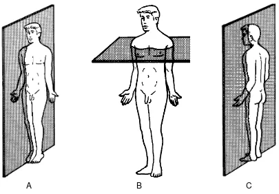
1-8. THE ANATOMICAL POSITION
The anatomical position is an artificial posture of the human body (see figure 1-2). This position is used as a standard reference throughout the medical profession. We always speak of the parts of the body as if the body were in the anatomical position. This is true regardless of what position the body is actually in. The anatomical position is described as follows:
a. The body stands erect, with heels together.
b. Upper members are along the sides, with the palms of the hands facing forward.
c. The head faces forward.
1-9. PLANES OF THE BODY
See figures 1-3A through 1-3C for the imaginary planes used to describe the body.
a. Sagittal planes are vertical planes that pass through the body from front to back. The median or midsagittal plane is the vertical plane that divides the body into right and left halves.
b. Horizontal (transverse) planes are parallel to the floor. They are perpendicular to both the sagittal and frontal planes.
A B C
Figure 1-3, A. The sagittal plane. B. The horizontal plane. C. The frontal plane.
1-10. DIRECTIONS
a. Superior, Inferior. Superior means above. Inferior means below.
b. Anterior, Posterior.
(1) Anterior (or ventral) refers to the front of the body.
(2) Posterior (or dorsal) refers to the back of the body.
c. Medial, Lateral. Medial means toward or nearer the midline of the body. Lateral means away from the midline or toward the side of the body.
d. Superficial, Deep. Superficial means closer to the surface of the body. Deep means toward the center of the body or body part.
1-11. NAMES
a. Names are chosen to describe the structure or process as much as possible. An international nomenclature was adopted for anatomy in Paris in 1955. It does not use the names of people for structures. (The single exception is the Achilles tendon at the back of the foot and ankle.)
b. Names are chosen to identify structures properly. Names identify structures according to shape, size, color, function, and/or location. Some examples are:
TRAPEZIUS MUSCLE
TRAPEZIUS = trapezoid (shape)
ADDUCTOR MAGNUS MUSCLE
AD = toward
DUCT = to carry (function)
MAGNUS = very large (size)
ERYTHROCYTE
ERYTHRO = red (color)
CYTE = cell
BICEPS BRACHII MUSCLE
BI = two
CEPS = head (shape)
BRACHII = of the arm (location)
Section III. CELLS
1-12. INTRODUCTION
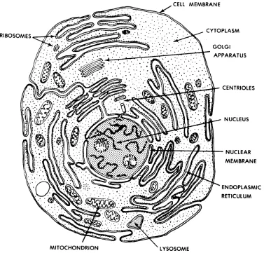
Figure 1-4. A "typical" animal cell (as seen in an electron microscope).
1-13. MAJOR COMPONENTS OF A "TYPICAL" ANIMAL CELL
a. Nucleus. The nucleus plays a central role in the cell. Information is stored in the nucleus and distributed to guide the life processes of the cell. This information is in a chemical form called nucleic acids. Two types of structures found in the nucleus are chromosomes and nucleoli. Chromosomes can be seen clearly only during cell
divisions. Chromosomes are composed of both nucleic acid and protein.
Chromosomes contain genes. Genes are the basic units of heredity which are passed from parents to their children. Genes guide the activities of each individual cell.
b. Cell Membrane. The cell membrane surrounds and separates the cell from its environment. The cell membrane allows certain materials to pass through it as they enter or leave the cell.
d. Mitochondria (Plural). Mitochondria are the "powerhouses" of the cell. The mitochondria provide the energy wherever it is needed for carrying on the cellular functions.
e. Endoplasmic Reticulum. The endoplasmic reticulum is a network of membranes, cavities, and canals. The endoplasmic reticulum helps in the transfer of materials from one part of the cell to the other.
f. Ribosomes. Ribosomes are "protein factories" in the cell. They are composed mainly of nucleic acids which help make proteins according to instructions provided by the genes.
g. Centrioles. Centrioles help in the process of cell division.
h. Lysosomes. Lysosomes are membrane bound spheres which contain enzymes that can digest intracellular structures or bacteria.
1-14. CELL MULTIPLICATION (MITOSIS)
Individual cells have fairly specific life spans. Some types of cells have longer life spans than others. During the processes of growth and repair, new cells are being formed. The usual process of cell multiplication is called mitosis. There are two important factors to consider:
a. From one cell, we get two new cells.
b. The genes of the new cells are identical (for all practical purposes) to the genes of the original cell.
1-15. HYPERTROPHY/HYPERPLASIA
Hypertrophy and hyperplasia are two ways by which the cell mass of the body increases.
a. With HYPERTROPHY, there is an increase in the size of the individual cells. No new cells are formed. An example is the enlargement of muscles due to exercise by the increased diameter of the individual striated muscle fibers.
b. With HYPERPLASIA, there is an increase in the total number of cells. An example of abnormal hyperplasia is cancer.
c. ATROPHY is seen when there is a loss of cellular mass.
EXERCISES, LESSON 1
REQUIREMENT. The following exercises are to be answered by completing the incomplete statement or by writing the answer in the space provided at the end of the question. After you have completed all the exercises, turn to "Solutions to Exercises," at the end of the lesson and check your answers.
1. What is anatomy?
2. What is the body type for each of the following individuals?
A broad individual: .
A slim individual: .
A person with average build: .
3. What kind of anatomical study is described by each of the items below?
Study of structures that cannot be seen with the unaided eye: .
Study of relationships between functions and structures: .
Study of the nervous system: .
Study of organ systems: .
4. What are the five levels or systems into which the body is organized, in ascending order?
5. What is a cell?
6. What is a tissue?
7. What is an organ?
8. What is an organ system?
9. What is the total organism?
10. What are the parts of the upper member? , , , , and .
11. What are the parts of the lower member? , , , , and .
12. What is one reason for studying terminology?
13. Describe the anatomical position.
a. The body stands with together.
b. The upper members are along the with palms facing .
14. Each plane in figure 1-5 is marked by a letter a, b, c, or d. Write the name of each plane in the appropriate space below.
a. plane.
b. plane.
c. plane.
d. plane.
15. In figure 1-6, three points are labeled a, b, and c, and two borders are labeled d and e. It is correct to say that a is to b and c, b is to a
and to c, and c is to a and b. We speak of d as the border. We speak of e as the border.
16. In figure 1-7, three portions of the arm are marked a, b, and c. The two ends of the arm are marked d and e. The portion marked a is the third. The portion marked c is the third. The end marked d is the end. The end marked e is the end.
Figure 1-7. Directions upon members (exercise 16).
18. In figure 1-8, parts of a "typical animal cell" are marked with the letters a through g. In the spaces below, provide the name of each structure.
a.
b.
c.
d.
e.
f.
g.
SOLUTIONS TO EXERCISES, LESSON 1
1. Anatomy is the study of the structure of the body. (para 1-1a)
2. A broad individual: endomorph. A slim individual: ectomorph.
A person with average build: mesomorph. (para 1-2)
3. Study of structures that cannot be seen with the unaided eye: microscopic anatomy.
Study of relationships between functions and structures: functional anatomy. Study of the nervous system: neuroanatomy.
Study of organ systems: gross anatomy by systems. (para 1-4)
4. The body is organized into cells, tissues, organs, organ systems, and the total organism. (para 1-5)
5. A cell is the smallest discrete living unit of the body construction. (para 1-5a)
6. A tissue is a grouping of like cells working together. (para 1-5b)
7. An organ is a structure composed of several different tissues performing a particular function. (para 1-5c)
8. An organ system is a group of organs performing an overall function together.
(para 1-5d)
9. The total organism is the individual human being. (para 1-5e)
10. The parts of the upper member are the shoulder, arm, forearm, wrist, and hand.
(para 1-6c(1))
11. The parts of the lower member are the hip, thigh, leg, ankle, and foot.
(para 1-6c(2))
12. One reason for studying terminology is to be successful in a medical field. Another reason is to be able to communicate well. (para 1-7a)
13. The anatomical position is described as follows:
a. The body stands erect, with heels together.
14. a. Midsagittal or median plane. b. Sagittal plane.
c. Horizontal or transverse plane. d. Frontal or coronal plane. (para 1-9)
15. It is correct to say that a is lateral to b and c, b is medial to a and lateral to c, and c is medial to a and b. We speak of d as the lateral border. We speak of e as the medial border. (para 1-10c)
16. The portion marked a is the distal third. The portion marked c is the proximal third. The end marked d is the distal end. The end marked e is the proximal end.
(para 1-10e)
17. A cell is the microscopic unit of body organization. (para 1-12)
18. a. Ribosomes. b. Mitochondrion.
c. Endoplasmic reticulum. d. Nucleus.
e. Centrioles. f. Cytoplasm.
g. Cell membrane. (fig 1-4)
LESSON ASSIGNMENT
LESSON 2 Tissues of the Body.
TEXT ASSIGNMENT Paragraphs 2-1 through 2-17.
LESSON OBJECTIVES After completing this lesson, you should be able to:
2-1. Define tissue.
2-2. Name four major types of tissues.
2-3. Define epithelial tissue, connective tissue, muscle tissue, and nervous tissue.
2-4. Given a description of epithelial tissue, matrix, fibrous connective tissue, cartilage connective tissue, bone connective tissue, fat connective tissue, smooth muscle tissue, striated muscle tissue, cardiac muscle tissue, nervous tissue, neuron, or glia, name it.
2-5. Name four major types of connective tissue (CT); name the characteristic cells of fibrous CT, cartilage CT, and bone CT; and describe the matrix of fibrous CT, cartilage CT, and fat CT.
LESSON 2
TISSUES OF THE BODY
Section I. GENERAL
2-1. DEFINITION
A tissue is a grouping of like cells working together.
2-2. TYPES OF TISSUES
There are several major types of tissues. The most common types are epithelial, connective, muscle, and nervous tissues. Later, this lesson will discuss each type.
2-3. TISSUES AND ORGANS
a. Tissues make up organs. An organ is a structure performing a particular function. An organ is composed of several different tissues. Examples of organs are the lungs and the heart.
b. In some cases, a term may be used to describe both a type of tissue and a kind of organ. For example, we speak of bone tissue and of bones. We speak of muscle tissue and of muscles.
Section II. EPITHELIAL TISSUES
2-4. DEFINITION
Epithelial tissue is tissue that covers surfaces and lines cavities. Here, it may protect, absorb, and/or secrete. Epithelial tissue covers the outer surface of the body. It lines the intestines, the lungs, and other hollow organs.
2-5. TYPES OF EPITHELIAL CELLS (BY SHAPE)
Figure 2-1. Epithelial cells.
2-6. TYPES OF EPITHELIAL TISSUES
a. Layers. In epithelial tissues, the cells are in single or multiple layers. If there is only one layer, the tissue is called a simple epithelium. If there is more than one layer, the tissue is called a stratified epithelium. See figure 2-2.
Figure 2-2. Types of epithelial tissues.
b. Naming. Epithelial tissues are named by the number of layers and the type of cell in its outermost layer. For example, if there are several layers and if the
outermost layer consists of squamous (flat) cells, then the tissue is called a stratified squamous epithelium.
c. Examples of Epithelial Tissues.
(1) A simple squamous epithelium called endothelium lines the heart and blood vessels.
(2) As serous membranes, simple squamous epithelial tissue lines the cavities of the abdomen (peritoneal lining) and the chest (pleural lining). Serous membranes are membranes which secrete a lubricating fluid.
d. Functions. According to its location, epithelial tissue has different functions. As the skin, epithelial tissue protects the tissues beneath. In the small intestines, the epithelial tissue absorbs. In the lungs, epithelial tissue is a membrane through which the gases pass easily. In the glands, epithelial tissue secretes.
Section III. CONNECTIVE TISSUES
2-7. DEFINITION
a. Connective tissue is tissue that supports other tissues, holds tissues together, or fills spaces.
b. Among and outside the cells of the connective tissues, there is a material called matrix. The matrix is manufactured by the connective tissue cells. Each type of connective tissue has its own particular type of matrix.
2-8. TYPES OF CONNECTIVE TISSUE
There are several major types of connective tissue (CT). These include fibrous CT (FCT), cartilage CT, bone CT, and fat CT. Blood is sometimes considered an additional type of CT.
2-9. FIBROUS CONNECTIVE TISSUE (FCT)
a. Fibroblasts. The characteristic cells of FCT are fibroblasts. Fibroblasts are able to form elongated fibers.
b. Matrix. These fibers make up the matrix of FCT.
c. Fibers. The fibers are either white or yellow.
(1) White fibers are made from a protein called collagen. White fibers tend to have a fixed length. White fibers are not very easily stretched.
(2) Yellow fibers are made from a protein called elastin. Yellow fibers are elastic. They can be stretched and then they can snap back (like a rubber band).
d. Types of FCT. The types of FCT are recognized by the arrangement of their fibers. These types include:
Loose areolar FCT is found widely throughout the body. An example is the superficial fascia (subcutaneous layer). The superficial fascia is the connective tissue which lies beneath the skin. Loose areolar FCT is the filling substance around most organs and tissues of the body.
(2) Dense FCT. The fibers of dense FCT are closely packed and parallel. There are no significant spaces between the fibers. Examples of dense FCT are ligaments and tendons. A ligament is a band of dense FCT that holds the bones together at a joint. A tendon attaches a muscle to a bone.
2-10. CARTILAGE CONNECTIVE TISSUE
a. Cartilage Cells. Cartilage cells are also called chondroblasts. Cartilage cells are clustered in microscopic pockets within the cartilage matrix. The cartilage cells produce the material of the matrix.
b. Matrix. The matrix produced by the cartilage cells appears homogeneous (the same throughout). The matrix also appears amorphous (shapeless).
c. Types of Cartilage CT.
(1) Hyaline cartilage CT. Hyaline cartilage CT appears homogeneous and clear.
HYALINE = clear
This type of cartilage helps to cover bone surfaces at joints. Hyaline cartilage is found as incomplete rings which keep the trachea (windpipe) open.
(2) Fibrous cartilage CT. Fibrous cartilage CT includes dense masses of fibers (of FCT). It is more rigid than hyaline cartilage. The auricle of the external ear is stiffened with fibrous cartilage.
(3) Calcified cartilage CT. Calcified cartilage CT is cartilage that has been stiffened by the addition of calcium salts. This is not the same as bone tissue. An example is the cartilages of the larynx (the voice box) which become calcified with age.
2-11. BONE CONNECTIVE TISSUE
b. Types of Bone Tissues. There are two major types of bone tissue. One is compact bone CT, which is dense. The other is cancellous bone CT, which is spongy. Compact bone CT forms the hard outer layers of bones as organs. Cancellous bone CT forms the inner, lighter portion of bones.
2-12. FAT CONNECTIVE TISSUE
a. Fat Cells. A large fraction of the volume of a fat cell is occupied by a droplet of fat. This droplet has its own membrane, in addition to the outer membrane of the cell. The remaining components of the fat cell, including the nucleus, are found in an outer layer of cytoplasm surrounding the droplet of fat.
b. Matrix. Fat connective tissue has a matrix of lipid (oil or fat). There may be yellow fat CT or brown fat CT.
c. Functions. Fat CT acts as a packing material among the organs, nerves, and vessels. Fat CT also helps to insulate the body from both heat and cold. Some fat CT serves as a high-energy storage area.
2-13. BLOOD "CONNECTIVE TISSUE"
Some experts consider blood to be a type of connective tissue. Blood will be discussed in lesson 9.
Section IV. MUSCLE TISSUES
2-14. DEFINITION
There are muscle tissues and there are organs called muscles. Muscles are made up of muscle tissues. Muscle tissues and the muscles they make up are
specialized to contract. Because of their ability to shorten (contract), muscles are able to produce motion.
2-15. TYPES OF MUSCLE TISSUES
See figure 2-3 for the three types of muscle tissue.
Figure 2-3. Types of muscle tissue.
b. Cardiac Muscle Tissue. The cells (muscle fibers) of cardiac muscle tissue are short, branched, contain one nucleus, and are striated. This tissue makes up the myocardium (wall) of the heart.
c. Smooth Muscle Tissue. The cells (muscle fibers) of smooth muscle tissue are spindle-shaped, contain one nucleus, and are not striated. Smooth muscle tissue is generally found in the walls of hollow organs such as the organs of the digestive and respiratory systems, the blood vessels, the ureters, urinary bladder, urethra, and reproductive ducts.
Section V. NERVOUS TISSUE
2-16. DEFINITION
Nervous tissue is a collection of cells that respond to stimuli and transmit information.
2-17. NERVOUS TISSUE CELLS
a. A neuron (figure 2-4), or nerve cell, is the cell of the nervous tissue that actually picks up and transmits a signal from one part of the body to another. A
synapse (figure 2-5) is the point at which a signal passes from one neuron to the next.
b. The neuroglia (also known as glia) is made up of the supporting cells of the nervous system (glial cells).
Figure 2-4. A neuron.
EXERCISES, LESSON 2
REQUIREMENT. The following exercises are to be answered by completing the incomplete statement or by writing the answer in the space provided at the end of the question.
After you have completed all the exercises, turn to "Solutions to Exercises," at the end of the lesson and check your answers.
1. What is a tissue?
2. What are the most common types of tissues?
a. .
b. .
c. .
d. .
3. What is epithelial tissue?
4. If an outer layer of epithelial tissue consists of flat cells and if there are several layers of cells in the tissue, then what is the type of epithelial tissue?
5. What is connective tissue?
6. What term is used for the material found among and outside the cells of connective tissue?
8. Characteristic cells of fibrous CT are . Cartilage cells are also called . Cells that make and repair bone are . Cells that tear down and remove bone are .
9. The matrix of fibrous CT consists of . The matrix produced by cartilage cells appears h and a . Fat CT has a matrix of .
10. Two major types of fibrous connective tissue (FCT) are FCT, which is a filling substance around most organs and tissues of the body, and FCT, which is found, for example, in ligaments and tendons.
11. What type of connective tissue has an amorphous, homogeneous matrix?
12. What type of connective tissue has a matrix of lipid (fat or oil)?
13. What are muscle tissues?
14. The cells of one type of muscle tissue are spindle-shaped, contain one nucleus, and are not striated. What is this tissue called?
15. Which type of muscle tissue has cells which have one nucleus and are short, branched, and striated?
16. Which type of muscle tissue has cells which have numerous nuclei and are long and cylindrical?
17. What is nervous tissue?
19. A nerve cell, which actually picks up and transmits a signal, is also known as a .
20. The supporting structure of the nervous system is known as the or the .
SOLUTIONS TO EXERCISES, LESSON 2
1. A tissue is a grouping of like cells working together. (para 2-1)
2. a. Epithelial. b. Connective. c. Muscle.
d. Nervous. (para 2-2)
3. Epithelial tissue is tissue that covers surfaces and lines cavities. (para 2-4)
4. If there are several layers and if the outer layer consists of flat cells, then the tissue is called a stratified squamous epithelium. (para 2-6b)
5. Connective tissue is tissue that supports other tissues, holds tissues together, or fills spaces. (para 2-7a)
6. The term used for material found among and outside the cells of connective tissue is matrix. (para 2-7b)
7. The four major types of connective tissue (CT) are fibrous CT, cartilage CT, bone CT, and fat CT. (para 2-8)
8. Characteristic cells of fibrous CT are fibroblasts. Cartilage cells are also called chondroblasts. Cells that make and repair bone are osteoblasts. Cells that tear down and remove bone are osteoclasts. (paras 2-9a, 2-10a, 2-11a)
9. The matrix of fibrous CT consists of fibers. The matrix produced by cartilage cells appears homogeneous and amorphous. Fat CT has a matrix of lipid.
(paras 2-9b, 2-10b, 2-12b)
10. Two major types of fibrous connective tissue (FCT) are loose areolar FCT, which is a filling substance around most organs and tissues of the body, and dense FCT, which is found, for example, in ligaments and tendons. (para 2-9d)
11. Cartilage CT. (para 2-10b)
12. Fat CT. (para 2-12b)
13. Muscle tissues are tissues whose contracting elements enable muscles to produce motion. (para 2-14)
14. Smooth muscle tissue. (para 2-15c)
16. Skeletal muscle tissue. (para 2-15a)
17. Nervous tissue is a collection of cells that respond to stimuli and transmit information. (para 2-16)
18. Nervous tissue. (para 2-16)
19. A nerve cell, which actually picks up and transmits a signal, is also known as a neuron. (para 2-17a)
20. The supporting structure of the nervous system is known as the glia, or the neuroglia. (para 2-17b)
LESSON ASSIGNMENT
LESSON 3 The Human Integumentary and Fascial Systems.
TEXT ASSIGNMENT Paragraphs 3-1 through 3-14.
LESSON OBJECTIVES After completing this lesson, you should be able to:
3-1. Define integumentary system, integument proper, integumentary derivatives, fascia, superficial fascia (subcutaneous layer), deep fasciae, and investing deep fascia.
3-2. Identify the three coverings, or envelopes, for the human body.
3-3. Name and describe the two layers of the skin.
3-4. Name and describe three types of
integumentary derivatives--hairs, glands, and nails.
3-5. Define serous cavities, describe a bursa, and give examples of serous cavities in the body.
LESSON 3
THE HUMAN INTEGUMENTARY AND FASCIAL SYSTEMS
Section I. GENERAL
3-1. DEFINITIONS
An organ system is a group of organs together performing an overall function. Portions of two organ systems, the integumentary and fascial systems, are represented in figure 3-1.
Figure 3-1. The integument and related structures.
a. Integumentary System. The integumentary system includes the integument proper and the integumentary derivatives. We know the integument proper as the skin. It is the outermost covering of the whole body. The integumentary derivatives include the hairs, nails, and various glands of the skin.
3-2. COVERINGS OF THE HUMAN BODY
The entire body is surrounded by three layers or envelopes, one inside the other. These coverings separate the body from the external environment. These envelopes include (from outside inward)--the skin (the integument proper), the subcutaneous layer, and the investing deep fascia.
Section II. THE HUMAN INTEGUMENTARY SYSTEM
3-3. THE INTEGUMENT PROPER
The integument proper is the outermost layer of the human body. It is usually known as the skin. The skin has two layers--the superficial or outer layer called the epidermis and the deeper or inner layer called the dermis.
a. The Epidermis. The epidermis is a stratified squamous epithelium. This means that it is made up of several layers of cells, the outermost being flat-type epithelial cells.
(1) The outer layers of the epidermis include cells which are transparent, flattened, dead, and without nuclei. These hardened cells of the outermost layers are completely filled with keratin and are known as cornified cells. These dead flat cells in the outermost layers resemble scales. Day by day, these cells are scraped away or just fall away from the body. They are replaced by cells from the intermediate layers.
(2) In the intermediate layers of the epidermis, the cells change their shapes. As the cells move towards the surface, they gain granules, begin to manufacture a hardening material called keratin, and lose their nuclei.
(3) The innermost layer of the epidermis is especially important because it is the source of all the other layers of the epidermis. It is known as the basal or germina-tive layer. The cells of this layer are capable of multiplication (mitosis). Its basic
structure is a single layer of columnar-type epithelial cells.
3-4. INTRODUCTION TO INTEGUMENTARY DERIVATIVES
The integumentary derivatives include the glands, hairs, and nails associated with the skin. All integumentary derivatives are formed from the tissues of the integument proper (dermis and epidermis). All are appended (attached) to the
integument proper and are often known as the appendages of the skin. See figure 3-2.
Figure 3-2. The integumentary derivatives (appendages).
3-5. HAIRS
a. A hair follicle is formed by the extension of the skin (dermis and epidermis) deeper into the surface of the body. Follicles may extend into the subcutaneous layer.
b. At the base of the hair follicle is the hair root. The hair shaft grows out from the root. The hair shaft is made of cells from the outermost layers of the epidermis.
3-6. GLANDS
The types of glands included are the sweat glands, the sebaceous (fat/ oil) glands, and the mammary glands (breasts). The ducts and secretory parts of these glands are made of epithelial tissues. Backup or supporting tissue is of FCT.
a. Sweat Glands. Sweat glands consist of a coiled secretory portion and a wavy duct which leads to the surface of the skin. The coiled secretory portion is located in the dermis or deeper. Sweat glands are found everywhere on the body in association with the skin.
b. Sebaceous Glands. Sebaceous glands produce an oily substance which lubricates the skin and hairs. The oil keeps the skin and hairs flexible. The sebaceous glands are usually found as a part of the walls of hair follicles and their oil flows into the follicle. In a few places without hairs, they open directly to the skin surface.
c. Mammary Glands. In the adult human female, the mammary gland lies in the subcutaneous layer anterior to the chest muscle (pectoralis major M.). Its function is to nourish the newborn. A nipple is located near the center of each breast. Around each nipple is a darkened area known as the areola. The tip of the nipple has many small openings to allow the passage of the milk from the milk ducts. These ducts are connected to lobes of glandular tissue located throughout the breast. Fat and fibrous CT fill in the spaces among the lobes.
3-7. NAILS
Nails are found on the ends of the digits (thumbs, fingers, and toes). Nails help to protect the ends of these digits. Each nail bed is attached to the top of the terminal phalanx (bone) of each digit. The nail itself is made up of cornified (hardened) outer cell layers of the epidermis. The nails grow continuously from their roots.
3-8. SKIN COLORATION
a. The skin includes red, black, and yellow pigments. The proportion of these pigments determine the skin color. This proportion is determined by genetics. The absence of all pigments is called albinism. In albinism, white light is reflected and a pink hue results from the color of the blood shining through the transparent skin.
Section III. THE FASCIAL SYSTEM OF THE HUMAN BODY
3-9. GENERAL
Most of the fibrous connective tissues (FCT) are fascial. These may occur as sheets or masses. NOT included in this definition are the tendons, ligaments, or aponeuroses (wide flat tendons). The different fasciae have varying proportions of white fibers, yellow fibers, fat, and tissue fluid. Some serve as membranes to inclose the body and its parts. Fasciae also help to support some organs and allow motions between other organs to be easier.
3-10. SUPERFICIAL FASCIA
a. The superficial fascia is the second envelope of the body. It is the layer between the skin (integument proper) and the investing deep fascial envelope. It is often called the subcutaneous layer, but it is technically not a part of the integumentary system as such.
b. The superficial fascia is made up primarily of loose areolar FCT with the spaces filled by fatty tissue and tissue fluid. It contains the superficial or cutaneous branches of nerves, arteries, veins, and lymphatics (NAVL) of the skin.
3-11. DEEP FASCIAE
a. The deep fasciae include various membranes made of consolidated or dense FCT. A deep fascia envelops the entire body as the third envelope. This third envelope is known as the investing deep fascia. It is beneath the skin and subcutaneous layer.
b. Deep fasciae also include the envelopes of the muscles and other organs. Around individual organs (for example, the kidney), it is called a capsule.
c. Another form of deep fascia is found in the collections of loose areolar FCT and fat that are found as filling among the organs. Similar deep fasciae attach organs to the body wall.
Section IV. SEROUS CAVITIES OF THE HUMAN BODY
3-12. GENERAL
3-13. BURSA
a. A bursa (figure 3-3) is the simplest of serous cavities. Each bursa is a small sac located between two moving structures, usually a muscle moving over a bony surface. The bursa reduces the friction between the two structures. For example, a bursa prevents excessive friction between the skin and patella (knee cap). This bursa, called the prepatellar bursa, allows the skin to move freely over the patella. (When injured, it produces excessive amounts of the serous fluid and is known as
"housemaid's knee.")
Figure 3-3. A bursa--the simplest serous cavity.
b. As a fibrous sac, each bursa has a central cavity which is lined with a serous membrane. This membrane is a simple squamous epithelium. The serous membrane secretes a serous fluid into the serous cavity. The serous fluid is the lubricant,
minimizing friction.
3-14. OTHER SEROUS CAVITIES OF THE BODY
b. Each serous cavity has an inner and an outer membrane. The inner membrane is intimately associated with the surface of the visceral organ. The outer membrane forms the outer wall of the cavity. The serous lining of the cavity secretes the serous fluid into the cavity to act as a lubricant between the membranes, allowing freer motion for the organs.
EXERCISES, LESSON 3
REQUIREMENT. The following exercises are to be answered by completing the incomplete statement or by writing the answer in the space provided at the end of the question.
After you have completed all the exercises, turn to "Solutions to Exercises," at the end of the lesson and check your answers.
1. What is included in the integumentary system?
2. What is another name for the integument proper?
3. What are three types of integumentary derivatives?
4. What is a fascia?
5. What is the subcutaneous layer (superficial fascia)?
6. Where are deep fasciae found in the body?
7. What is the investing deep fascia?
8. What is the outer layer of the skin?
9. What type of tissue makes up the epidermis?
What is the basic structure of the innermost layer of the epidermis?
What are characteristics of cells found in the outermost layers of the epidermis?
10. What type of tissue makes up the dermis?
What are papillae and what is their function?
What other structures are found in the dermis?
11. A hair follicle is formed by the . At the base of the hair follicle is the .
The hair shaft grows out from the . The hair shaft is made of cells from the .
12. Sweat glands consist of .
13. Sebaceous glands produce . Its function is to .
The sebaceous glands are usually found as a part of the walls of .
14. In mammary glands, milk ducts connect each nipple with .
What types of connective tissue fill in the spaces?
16. The term serous refers to a . Serous cavities are . These cavities serve as .
17. Each bursa is a small sac located between ,
usually a muscle moving over a . The bursa reduces the between two structures. The serous membrane lining the cavity within a bursa is a and it secretes a into the serous cavity.
18. Each lung is encased in a serous cavity called the . The heart lies in a serous cavity called the . The intestines move freely within the cavity.
SOLUTIONS TO EXERCISES, LESSON 3
1. The integumentary system includes the integument proper (skin) and the integumentary derivatives (hairs, nails, and glands of the skin). (para 3-1a)
2. Another name for the integument proper is the skin. (para 3-1a)
3. Three types of integumentary derivatives are the hair, nails, and various glands of the skin. (para 3-1a)
4. A fascia is a sheet or collection of fibrous connective tissue (FCT).
(para 3-1b)
5. The subcutaneous layer (superficial fascia) is the connective tissue which lies immediately beneath the skin. (para 3-1b)
6. Deep fasciae are found as envelopes for muscles and other organs and they fill spaces. (para 3-1b)
7. The investing deep fascia is the third envelope of the whole body beneath the skin and the subcutaneous layer. (para 3-1b)
8. The outer layer of the skin is the epidermis. The inner and deeper layer of skin is the dermis. (para 3-3)
9. The epidermis is a stratified squamous epithelium.
The basic structure of the innermost layer of the epidermis is a single layer of columnar-type epithelial cells.
The cells found in the outermost layers of the epidermis are transparent, flattened, dead, cornified, and without nuclei. (para 3-3a)
10. The dermis is dense FCT consisting of white and yellow fibers. Papillae are finger-like projections of the dermis that extend into the epidermis.
Papillae prevent the dermis and epidermis from sliding on each other. Other structures found in the dermis include blood vessels, lymph vessels, nerve endings, hair follicles, and glands. (para 3-3b)
11. A hair follicle is formed by the extension of the skin (dermis and epidermis) deeper into the surface of the body. At the base of the hair follicle is the hair root. The hair shaft grows out from the root. The hair shaft is made of cells from the outermost layers of the epidermis. (paras 3-5a, b)
13. Sebaceous glands produce an oily substance. Its function is to lubricate the skin and hairs and to keep them flexible. The sebaceous glands are usually found as a part of the walls of hair follicles. (para 3-6b)
14. In mammary glands, milk ducts connect each nipple with lobes of glandular tissue. Fat and fibrous CT fill in the spaces among the lobes. (para 3-6c)
15. Nails are found on the ends of the digits. Nails help to protect the ends of these digits. The nail itself is made up of cornified (hardened) outer cell layers of the epidermis. The nails grow continuously from their roots. (para 3-7)
16. The term serous refers to a watery-type fluid. Serous cavities are sacs lined with serous membranes. These cavities serve as lubricating devices.
(para 3-12)
17. Each bursa is a small sac located between two moving structures, usually a muscle moving over a bony surface. The bursa reduces the friction between the two structures. The serous membrane lining the cavity within a bursa is a simple squamous epithelium and it secretes a serous fluid into the serous cavity. (para 3-13)
18. Each lung is encased in a serous cavity called the pleural cavity. The heart lies in a serous cavity called the pericardial cavity, the intestines move freely within the peritoneal cavity. (para 3-14a)
LESSON ASSIGNMENT
LESSON 4 The Human Skeletal System.
TEXT ASSIGNMENT Paragraphs 4-1 through 4-14.
LESSON OBJECTIVES After completing this lesson, you should be able to:
4-1. Define skeleton.
4-2. Name four functions of the human skeleton.
4-3. Name the layers and describe the basic structure of an individual bone, name and describe the parts of an individual long bone, and describe the periosteum and the blood supply of an individual bone.
4-4. Describe the development of an individual bone.
4-5. Name four types of bones by shape.
4-6. Describe major categories used in classification of joints.
4-7. Name the major parts of a "typical" synovial joint.
4-8. Name and describe classifications of synovial joints according to the kind of motion and number of axes.
4-9. Name and define the two major subdivisions of the skeleton.
4-10. Describe a typical vertebra. Name the regions of the vertebral column and give the number of vertebrae in each region. Describe the
intervertebral discs and ligaments that hold vertebrae together.
4-11. Describe the thoracic cage.
4-13. Describe the general pattern of the bones of the upper and lower members.
LESSON 4
THE HUMAN SKELETAL SYSTEM
Section I. GENERAL
4-1. INTRODUCTION
The skeleton serves as a support or framework of the human body. It is a combination of bones joined together.
4-2. FUNCTIONS OF THE HUMAN SKELETON
The human skeleton serves the following functions:
a. Bodily Support. The skeletal system provides a framework for the human body.
b. Protection. The skeleton protects certain soft structures within the human body. An example is the skull, which surrounds the brain.
c. Motion. Muscles are attached to and move the bones. Bones provide leverage for motion.
d. Formation of Blood Cells (Hematopoiesis). Blood cells are manufactured in the red bone marrow, mainly found in flat bones.
4-3. PRIMARY STUDY AREAS
In this text, we study the skeletal system from four different viewpoints:
a. Bone As Tissues. This aspect of the human skeletal system was discussed in paragraph 2-11 and will not be further discussed here.
b. Bone As An Individual Organ. Section II of this lesson discusses bone as an individual organ.
c. Articulations (Joints)--Arthrology. Section III of this lesson introduces the study of joints, or arthrology.
Section II. BONE AS AN INDIVIDUAL ORGAN
4-4. BASIC STRUCTURE OF AN INDIVIDUAL BONE
a. Use of Bony Tissues to Form an Individual Bone.
(1) Cortex. The cortex is the outer layer of the individual bone. It is made up of compact (dense) bony tissue.
(2) Medulla. The medulla is the central portion of the individual bone. It generally consists of cancellous (spongy) bone tissue. In some bones, particularly long bones, the medulla may include a space without any bony tissue. This space is called the medullary or marrow cavity.
b. Marrow. Marrow serves as a filler of the inside of bones. There are two types of bone marrow--yellow bone marrow and red bone marrow. Yellow bone marrow is mostly yellow fat tissue. Red bone marrow is the only site in adults for the formation of red blood cells (hematopoiesis).
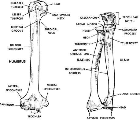
c. Named Parts of an Individual Long Bone.
(1) Shaft (diaphysis). The shaft is the central portion of a long bone. Here, the cortex is thickened as required by applied physical stresses.
(2) Ends (epiphyses). The ends of long bones are made up mainly of
cancellous (spongy) bone tissue. An articular cartilage covers each area where a bone contacts another bone(s). This articular cartilage is made up of hyaline cartilage tissue and provides a smooth surface for motions.
d. Periosteum. The periosteum is a covering of the bone surface area not covered by articular cartilage. It has two layers--the innermost layer and the fibrous layer.
(1) The innermost layer, which lies against the outer surface of the bone, consists of bone-forming cells (osteoblasts). It is the osteogenic (bone-forming) layer.
(2) The outermost layer is a FCT (fibrous connective tissue) layer.
(3) The periosteum is well supplied with blood vessels and sensory-type nervous tissue.
4-5. DEVELOPMENT OF AN INDIVIDUAL BONE
a. General. The human skeleton is "preformed" in the early fetus, but the early form is not of bony material. There are two types of bones according to their preformed basis: membranous bones and cartilage bones. These are in the location and have the general shape of the adult bones they will later become.
(1) Membranous bones. The outer skull bones are an example of
membranous bones. Osteoblasts invade a membrane to form a center of ossification (formation of bone). Bone-forming activity spreads out from this center until a full bone plate is formed.
(2) Cartilage bones. In the fetus, many bones, for example, long bones, exist first as models formed of cartilage.
b. Sesamoid Bones. Sesamoid bones are small masses of bone that develop in tendons at points where great forces are applied to the tendons. The most obvious and largest sesamoid bone is the patella, or kneecap.
c. Ossification Centers. An ossification center is a growing mass of actual bone within the preformed material, as noted above.
(1) Initial bone formation involves destruction of the preforming material and replacement with bony tissue.
(2) In the development of long bones, there are two types of ossification centers:
(a) Diaphyseal--in the shaft region.
(b) Epiphyseal--in the end(s).
(3) As a long bone grows in length, the preforming material grows faster than the ossification center can tear it down. Ultimately, with time, the preforming material is overcome and growth ceases.
d. Growth in Bone Width. A bone grows wider through the activity of the osteogenic layer of the periosteum. Remember, the periosteum covers most of the outer surface of the bone.
4-6. TYPES OF BONES
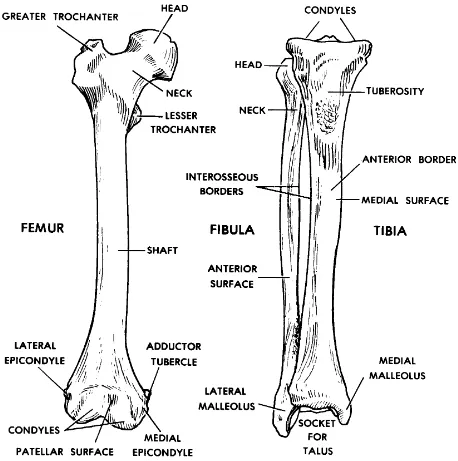
a. Long Bones. The basic structure of a long bone is illustrated in figure 4-1 and discussed in paragraph 4-4. Example: femur.
b. Short Bones. The short bones, such as those of the wrist and feet, have a thin layer of compact bone surrounding an inner mass of spongy bone.
Example: carpal bones.
c. Flat Bones. The flat bones are constructed with two plates of compact bone, which enclose between them a layer of spongy bone. The spongy bone is richly
supplied with blood vessels and red marrow. Example: the cranial frontal bone.
d. Irregular Bones. The irregular bones are those that do not fit into the three categories above. Example: a vertebra.
Section III. ARTHROLOGY--THE STUDY OF JOINTS (ARTICULATIONS)
4-7. DEFINITION
A joint, or articulation, is the location where two or more bones meet.
4-8. TYPES OF JOINTS
Joints are classified according to the kind of material holding the bones together and the relative freedom and kind of motion at the particular joint.
a. Fibrous Joints. Varying degrees of motion, from none to some, are possible in fibrous joints.
(1) Syndesmosis. When the bones are held together by FCT (fibrous connective tissue), the joint is referred to as a syndesmosis.
SYN = together
DESMOS = fiber (a tying material)
Example: The inferior tibio-fibular joint.
(2) Suture. When the bones are quite close together with a minimum of FCT, the joint is known as a suture. Example: the joints between the cranial bones.
b. Bony Joints. Should the bones be united by bony material, the joint is referred to as a synosteosis.
Example: The frontal bone. (The frontal bone of the skull is actually a bony fusion of two bones. Approximately 10 percent of the time, this fusion fails to take place; the original suture between the bones remains and is called a metopic suture.)
c. Cartilagenous Joints. These are also nonmovable joints.
(1) Synchondrosis. A cartilagenous joint in which the bones are held together by hyaline cartilage.
SYN = together
CHONDRO = cartilage
Example: Epiphyseal plate.
(2) Symphysis. A cartilagenous joint in which the bones are held together by a disc of fibrocartilage.
Example: Pubic symphysis.
d. Synovial Joints. In the synovial type of joints, the bones move on one another so as to allow various motions of the body parts. The "ovial" part of the name refers to the fact that the fluid substance seen in this type of joint appeared to the old anatomists to be like raw egg white (ovum = egg).
4-9. A "TYPICAL" SYNOVIAL JOINT
A "typical" synovial joint is one which has parts common to all of the synovial joints. In a sense, it is imaginary. It is not actually a specific synovial joint. It is a composite. It is illustrated in figure 4-2. The "typical" synovial joint has the following parts:
a. Bones. Bones are the levers of motion. They are the site of attachment for skeletal muscles.
b. Articular Cartilages. The "contact" points of the bones are usually covered with a layer of lubricated cartilage. Where these cartilages end, the synovial
Figure 4-2. A "typical synovial joint:--diagrammatic.
c. Synovial Membrane, Space, and Fluid.
(1) Synovial membrane. The synovial membrane lines the inner surface of the capsule. It secretes synovial fluid into the synovial space.
(2) Synovial space. Figure 4-2 exaggerates the amount of space between the bones. The space within the capsule allows movement.
d. Capsule. The "typical" synovial articulation is surrounded by a sleeve of dense FCT known as the capsule. The capsule encloses the articulation.
e. Ligaments. Primarily, ligaments hold bones together. Ligaments also may help restrain motion in certain directions and stabilize the articulation.
f. Muscles. Skeletal muscles apply the forces to produce a given motion.
NOTE: See table 4-1 for a summary of the structures in a "typical" synovial
articulation, the tissues composing each structure, and the actions attributed to each structure.
4-10. CLASSIFICATION OF SYNOVIAL JOINTS
Synovial joints are further classified according to the kind of motion and the number of axes of motions used.
a. Uni-Axial Synovial Joints.
(1) In uni-axial synovial joints, motion occurs in only one plane. The joints of the fingers (interphalangeal) flex and extend in the sagittal plane. These are commonly referred to as hinge joints.
(2) If a single rotatory (rotational) motion occurs around a post-like structure, the joint is a pivot joint. The atlas vertebra rotating around the dens (tooth like
projection) of the axis vertebra at the top of the neck (base of the skull) is a pivot joint.
b. Bi-Axial Synovial Joints. In bi-axial synovial joints, motion between the bones occurs in two planes. Here the surface in contact is curved or rounded in two directions.
(1) The proximal phalanx of a finger can flex and extend and move from side to side on the rounded head of the metacarpal bone. This is the MP or
metacarpophalangeal joint.
STRUCTURE TISSUE(S) FUNCTION(S)
1. BONE BONY (a) Serves as site of attachment for the
skeletal muscles.
Table 4-1. The tissues and functions of structures of a "typical" synovial articulation.
c. Multi-Axial Synovial Joints. In multi-axial joints, motion is possible in all three planes of space.
(1) The ball-and-socket-type synovial joint has the freest motion in all
(2) In the plane joint, the contact surfaces of the bones are essentially flat. These flat surfaces slide on one another (also called translatory motion). The
acromioclavicular joint of the shoulder region is an example of a plane joint.
4-11. THE ARTICULAR DISC
In three of the synovial joints of the human body, a special addition is seen. This addition is known as an articular disc. The joints with articular discs are the temporo-mandibular joint of the lower jaw, the sternoclavicular joint (at the sternum
(breastbone)), and the ulnocarpal joint of the distal end of the forearm.
a. An articular disc is a fibrocartilage plate. It is inserted between the articular surfaces of the bones of a synovial joint. In this way, it divides the synovial space into two spaces.
b. Joints having an articular disc are capable of having several different motions occurring at the same time. Mechanically, there are really two joints together here.
Section IV. THE HUMAN SKELETON
4-12. GENERAL
a. The human skeleton (figures 4-3A and 4-3B) is a collection of individual bones articulated (joined) together.
b. The major subdivisions of the skeleton are the axial skeleton and the appendicular skeleton.
4-13. THE AXIAL SKELETON
a. Vertebral Column (Spine). The vertebral column, or spine, is made up of a vertical series of bony blocks called vertebrae. These vertebrae are joined together in such a way as to form a semiflexible rod. The spine is the central support for the trunk, yet allows trunk movements.
(1) Anatomically and functionally, a typical vertebra (figure 4-4) is constructed of two major parts:
(a) The vertebral body is a drum-shaped cylindrical mass. Its superior and inferior surfaces are flat. Its function is primarily weight-bearing.
(b) The neural arch extends posteriorly, arching over and protecting the spinal cord of the central nervous system. From the neural arch are several processes. These processes serve as attachment areas for the trunk muscles. They also act as levers during various trunk motions.
Figure 4-4. A typical vertebra (superior and side views.
(2) The vertebral column has 32-33 vertebrae, one on top of the other. These vertebrae are arranged in regions. The vertebrae of each region have a characteristic shape. The regions are as follows:
(a) Cervical (neck) region, with seven cervical vertebrae.
(b) Thoracic (chest) region, with 12 thoracic vertebrae.
(d) The sacrum, which is a bony fusion of five sacral vertebrae.
(e) The coccyx (pronounced COCK-sicks, "tail"), with 3-4 coccygeal vertebrae together.
(3) The vertebrae are held together in two ways:
(a) The intervertebral disc holds the bodies of adjacent vertebrae together. The intervertebral disc is a fibrous ring with a soft center. This disc allows the vertebral bodies to move on one another. This joint between the vertebral bodies is a plane-type joint.
(b) The various parts of adjacent vertebrae are held together by ligaments. A ligament is a dense FCT structure which extends from bone to bone. These ligaments extend along the vertebral column from the base of the skull all the way down to the coccyx.
(4) The spine has four curvatures in the adult human. In the cervical (neck) region and the lumbar (low back) region, the spine curves forward. In the thoracic (chest) region and the sacro-coccygeal (pelvic- sacrum and coccyx) region, the spine curves backwards.
(5) When one examines the back of a person by sight and feel (palpation), certain landmarks are observed.
(a) At the upper shoulder region in the midline, a knob can be seen and felt. This is the tip of the spinous process of the seventh cervical vertebra. Since this is the first vertebra from the top that can be easily palpated, this bony landmark is called the vertebra prominens (the "prominent vertebra").
(b) From the vertebra prominens down to the beginning of the sacrum, one can feel the tip of the spinous process of each vertebra.
Figure 4-5. The human thorax with bones of the shoulder region.
(1) The sternum lies in the midline of the thorax anteriorly. It is made up of three parts: the manubrium at the top, the body as the main part, and the xiphoid process below. On the top of the manubrium is the jugular (sternal) notch, a common landmark. The junction between the manubrium and the body is a joint called the
sternal angle. This sternal angle is an important landmark clinically because the second rib attaches to the sternum at this junction. It is just a matter of simple counting after identifying the second rib to know where you are on the thoracic wall.
(2) The rib cage consists of the 12 thoracic vertebrae, 12 pairs of ribs, and the sternum. Each rib is curved laterally from back to front. All 12 pairs of ribs are attached posteriorly to the thoracic vertebrae. The upper six pairs of ribs are attached directly to the sternum by their costal cartilages. The seventh through tenth pairs of ribs are attached indirectly to the sternum through their costal cartilages (by attaching to the costal cartilage of the rib above). Rib pairs 11 and 12 do not attach to the sternum. Instead, they are embedded in the trunk wall muscles.
(1) The bones of the cranium form a spherical case around the brain. With age, the sutures between the cranial bones become more solid. The cranium has a base with several openings for the passage of blood vessels and nerves. The vault (or calvaria) is made up of flat bones arching over and covering the brain.
(2) The facial skeleton consists of bones which surround the nose and the mouth. These are mainly flat and irregular bones. Bones of the facial skeleton also form part of the orbit of each eye.
(3) Certain bones of the skull have air-filled spaces called the paranasal sinuses.
(4) The upper jaw (maxilla) and the lower jaw (mandible) are parts of the facial skeleton which surround the mouth.
(5) The hyoid bone is located at the junction between the head and the neck. It is not articulated directly with the other bones. It is held in place--and moved
around--by groups of muscles above and below. The root of the tongue is attached to its upper anterior surface. The larynx is suspended from its inferior surface. These three structures, together, form the hyoid complex. This complex is a functional unit for swallowing.
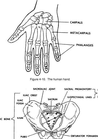
4-14. THE APPENDICULAR SKELETON
a. The appendicular skeleton is made up of the skeletal elements of the upper and lower members (often incorrectly referred to as the "extremities"). These members are appended (attached) to the axial skeleton.
b. The general pattern of construction of the upper and lower members is the same as follows:
(1) Girdle. The girdle is the actual attaching part. It attaches (appends) the limb (the member less the girdle) to the axial skeleton.
(2) Proximal limb segment. The proximal segment of the limb has a single long bone.
(3) Middle limb segment. The middle segment of the limb has two long bones parallel with each other.
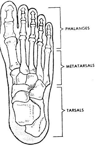
(4) Distal limb segment. The distal segment of the limb is made up of many long and short bones. These bones are arranged into a five-rayed pattern--the digits.
c. See table 4-2 for the main bones of the upper and lower members.