Correspondence:
Agung Budi Suiono, Department of Neurosurgery, Faculty of Medicine, Universitas Padjdajaran-Dr. Hasan Sadikin General Hospital
Jl. Pasteur No. 38 Bandung e-mail: agungbudis@gmail.com
of Internal Acousic Canal in the Vesibular Schwannoma
Abstract Objecive: To study the transformaion point of meningeal and periosteal dural at
the porus of internal acousic canal (IAC) in order to verify the diferent thickness of meningeal and periosteal dura in vesibular schwannomas (VS).
Methods: Three IAC cadaver specimens and ten samples of VS paients from porus were obtained and analyzed. Samples were stained by using Masson trichrome technique ater cuing in 6 micron of thickness. The samples were then observed under light microscopes to understand the meninges patern in the IAC.
Results: The meningeal dura is becoming thin at the porus and disappears at the meatal porion to form epineurium. However, the periosteal dura is lining coninuously to the fundus. In VS, the meningeal dura becomes thick and forms a pseudo-capsule in the middle of meatus, known as perineurium. The residual nerve ilament was compressed by the tumor parenchyma. Between the tumor and nerve interface, three or more perineureal layers are seen. The perineurium in the cisternal porion was consistently loose and forms the tumor and arachnoid nerve interface. Almost all the nerve ilaments are displaced to the tumor periphery near the pseudocapsule. In contrast, the periosteal dural of VS is becoming thin and disappear nearby the middle of meatal porion. This changing site establishes “meningo-periosteal ring” of VS because of the encircling nearby the porus.
Conclusions: In IAC, the meningeal dural becomes thin. The periosteal dura is lining coninuously to the fundus. In VS, the meningeal dura becomes thick, joins perineurium and forms pesudocapsule near the porus, but the periosteal dura disappeared. This changing point is called meningo-periosteal ring.
Keywords:Meningeal, periosteal, porus, vesibular schwannomas IJIHS. 2013;1(1):22–8
Agung Budi Suiono,1 M. Zafrullah Ariin,1 Ahmad Faried,1 Takayuki Ohira2
1Department of Neurosurgery, Faculty of Medicine, Universitas Padjadjaran-Dr. Hasan Sadikin General
Hospital
2Department of Neurosurgery, Keio University School of Medicine, Tokyo, Japan
Introducion
The concept from Yasargil et al.1,2 concerning
the origin of vesibular schwannomas (VS) came from the outside of subarachnoid space. It is stated the growth of VS pushes the arachnoid wall to the cerebelloponine cistern medially and duplicates the arachnoid layers. In addiion, the anatomical study in IAC revealed that the
dura mater and the arachnoid invaginates into the IAC from porus to fundus, creaing a lateral extension of cerebelloponine cistern, called acousicofacial cistern. VS originates from the vesibulocochleofacial complex which was contained in the acousicofacial cistern.3 A
Methods
Ethical approval was obtained from the Ethical Commitee of Keio University Hospital. Three formalin ixed adult cadaver head specimens were used and stained with Masson trichrome to observe the meninges structures in IAM (Fig. 1). Ten consecuive VS paients, operated by lateral suboccipital transmeatal approach, enrolled in this study ater providing informed consent. Two samples from meatal and cisternal porions near the porus were obtained from one paient and other eight samples near the porus entrance were obtained from the paients. The sample locaion and characterisics of VS paients are presented (Table 1). The surgical specimens were ixed in formaldehyde, cut perpendicularly to the tumor surface, then embedded in parain, and secioned at 4 micron of thickness. Stainings with hematoxylin eosin, Masson trichrome and the immunohistochemical myelin basic protein (MBP) were performed with Myelin basic protein staining performed using an enhanced, indirect immunoperoxidase technique, and the secions were lightly counterstained with hematoxylin. The primary anibody used was rabbit polyclonal ani–human MBP anibody.5
Results
Meninges patern in IAM
Axial histological secions of the right IAC were viewed and stained with Masson trichrome. The two layers of dura mater, with the meningeal dura facing the subdural space and periosteal
dura facing the bone, enter the IAM (Fig. 1) with the arachnoid invaginates into it. This leads to a creaion of a dural cul de sac muf3,6 and this
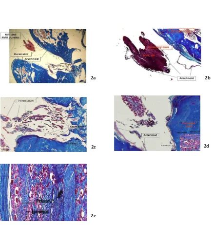
becomes thin in the fundus (Fig. 2a). However, the meningeal dura itself becomes thin and disappears at the middle of meatus (Fig. 2b) and the arachnoid transforms into the perineurium (Fig. 2c). Meanwhile, the periosteal dura drapes and follows the bone to the extracranial porion and becomes periosteum. The fasciovesibulo cochlear complex is wrapped with the arachnoid from porus and enters the subarachnoid space at the fundus level (Fig. 2a). The meningeal dura transforms into epineurium and the arachnoid transform into the perineurium. Meanwhile the endoneurium covers the nerve ilaments (Fig. 2d, e).
Meninges patern in the VS
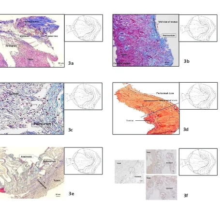
Histological secions of VS including the dura mater at the porus of IAC was stained with Masson trichrome. The thick meningeal dura follows the tumor and the thin periosteal dura lines on the bone surface and disappears ater passing the porus. It also joins the arachnoid and perineurium, which covers and directly interfaces with the tumor (Fig. 3a). The periosteal dura completely disappears near the porus inside the IAC wall with only meningeal dura let and it is connected to the perineurium (Fig. 3b). At the level of transformaion point from meningeal dura to the perineurium adjacent to the porus, the perineurium interfaces the tumor and also nerve en bloc (Fig. 3c). The histological secion which was stained with hematoxylin eosin shows a changing point of meningeal and periosteal
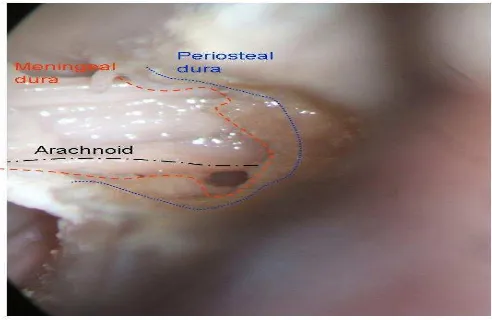
Fig. 1 The Right IAC from Cadaver Specimens. The posterior wall has been drilled out to expose the meatal porion. The meningeal and periosteal dural line the IAC, the arachnoid invaginates into the fundus and wraps the fasciocochleovesibular nerves complex
2a 2b
2c 2d
2e
The IAC Right Axial Secion. The VII and VIII nerve complexes course and enter the porus. The dura mater lining is found on the bone IAC wall and the arachnoid invaginates from cistern of cerebeloponine angele towards the fundus making dural cul de sac (Fig. 2b). Fig. 2a is an enlarged version at the cranial VII and VIII nerve complexes on which the meningeal dura becomes thin and periosteal dura coninues towards the fundus level. The arachnoid wraps the nerves (Fig. 2C). Fig. 2a is an enlarged version at the fundus level, towards the facial canal. The arachnoid wraps the nerves and transforms into epineurium (Fig. 2d). Fig. 2a is an enlarged version at the fundus level towards the vesibular nerve. The periosteal dura becomes thin, arachnoid wraps the nerves and transforms into perineurium. Enlarging Fig. 2d on the right below, the endoneurium is seen at the outer layer of myelin sheat (Fig. 2e). Fig. 2c is the enlarged version. Periosteal dura, meningeal dura, arachnoid, epineurium, perineurium straiicaion are seen from outside to the inside nerve ilament bundle
Agung Budi Suiono
dura at the level of porus from the cisternal to the meatal porion. Periosteal dura becomes thin, meanwhile the meningeal dura is thicker than periosteal dura and transforms into perineurium (Fig. 3d). The Masson trichrome staining in the cisternal porion close to the porus shows that the perineurium forms pseudocapsule and is located between the arachnoid and the tumor, creaing an arachnoid-tumor interface (Fig. 3e). The immunohistochemical myeline basic protein reveals the nerve ilaments located on the pseudocapsule near the arachnoid due to the compression of the tumor parenchyma (Fig. 3f).
Discussion
The fate of meningeal-periosteal dura patern in VS has not given considerable discussions in the past. This study shows that the patern of meninges has diferent dura mater characterisics in the porus IAC with VS compared to the normal anatomy. The meningeal dura joins perineurium and intermingles gradually, ightly adheres with the tumor (Fig. 3a, b, c). It was considered that the pseudocapsule formaion starts near the porus of IAC wall but it is less prominent. Periosteal dura becomes thin and disappears ater passing through the porus in the meatal porion (Fig. 3d). Tumor beyond the meatus is covered by perineurium and arachnoid leading to pseudocapsule development (Fig. 3e). The applicaion of immunohistochemical myelin
basic protein staining shows that the residual vesibular nerve ilament is compressed by the Schwann cell tumor. The residual nerve ilaments are displaced near the tumor capsule (Fig. 3f).
Concurrently, the cadaver specimen staining shows normal strucutures of IAC axial secion (Fig. 1). The dura mater consists of periosteal and meningeal dura which cover the bonny wall of IAM at the porus level. The periosteal dura gradually becomes thin towards the fundus, but the meningeal dura disappears at the middle part of IAC (Fig. 2a, b). The periosteal dura remains and coninues lining into the fundus level to the extracranial porion to become periosteum (2c, d). The meningeal dura joins perineurium that is transformed from arachnoid (Fig. 2e). dura disappears. Conversely, in the VS specimen the periosteal dura disappears ater passing the porus. However, the meningeal dura transforms into epineurium and the arachnoid becomes perineurium. Both meninges patern forms a meningoperiosteal dura circle that lines the meatal wall to create a “meningoperiosteal ring” (Fig. 4a, b). The meningoperiosteal ring is an overlying two types of dura mater. It is located between meningeal dura and periosteal dura in which the periosteal dura faces the bone and disappears. The meningeal dura connects to the
Table 1 Characterisic Paients with Vesibular Schwannoma
No. Gender/Age (years) Size (cm) Symptoms Sample locaion
1 F, 45 3.7 Tinnitus and hearing disturbance Inside and outside meatus near porus separately
2 M, 30 1.4 Tinnitus and hearing disturbance Porus inside meatus
3 M, 58 2.4 Hearing disturbance Inferior vesibular nerve en bloc near porus inside meatus
4 F, 31 1.2 Hearing loss Inside meatus near porus including duramater
5 F, 62 3.2 Hearing loss and dizziness Inside meatus near porus including arachnoid and duramater
6 F, 62 2.3 Hearing loss Outside meatus near porus
7 M, 69 4.8 Hearing disturbance Cysic outside meatus near porus
8 M, 54 1.7 Tinnitus Porus entrance including duramater
9 F, 41 2.6 Tinnitus Porus entrance including duramater
3a 3b
3c 3d
3e 3f
VS Sample Stained with Masson Trichrome was Taken Near Porus as Shown on the Scheme Figure on the Right Upper. The meningeal dura and periosteal dura is folding due to the staining process. The meningeal dura is thicker than periosteal dura, then periosteal dura disappears. The arachnoid transforms into perineurium, faces the tumor and connects to the meningeal dura (Fig. 3b). The sample is taken from meatus, nearby the fundus, from the meatal wall. The epineurium is interconnecing with meningeal dura. It might be the beginning of pseudocapsule but less prominent. Note that the periosteal dura is not observed on this secion, although the sample is taken from the dura which ataches on the meatal bone (Fig. 3c). Sample was taken from meatus nearby porus. The meningeal dura connects to the perineurium. The perineurium borders the nerve and tumor interface (Fig. 3d).Sample stained with Hematoxylin eosin was taken from cistern to the meatus porion, passing through the porus. The periosteal dura which faces the bone becomes thin and disappears ater passing the porus. The thick meningeal dura remains and transforms into perineurium in the meatal porion. The tumor interfaces the perineurium forming a pseudocapsule in the cisternal porion (Fig. 3e). Sample was taken from meatal porion. The perineurium interfaces the tumor and arachnoid, creaing a pseudocapsule (Fig. 3f). Sample from the meatal porion was stained with myelin basic protein to observe nerve ilament. The residual nerve ilaments exist on the tumor surface near the arachnoid
Agung Budi Suiono
perineurium to form the pseudocapsule. The meningoperiosteal ring can be found on the meatal, porus, or cisternal porions due to the widening and compression of the tumor (Table 2). However, it needs further invesigaions.
In the process of the tumor enlargement, the tumor compresses and displaces the perineurium from the origin of tumor nerve ilament towards the meatal wall to develop a perineural patern with diferent thickness. The perineurium thickness variaions before
it transforms into meninges could be related to the topographical anastomoses between facial and vesibular nerves. The anastomoses between facial and vesibular nerves are found in 83%, of which 67% of them are near porus and 33% are between intermedial and lateral porions of IAM. The other variaion is also found regarding the posiion of facial nerve towards the vesibulochoclear nerve, of which the anterioinferior to the vesibulochoclear nerve is more dominant than the anterosuperior.7-11
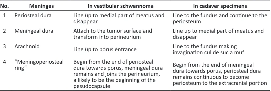
Table 2 Comparison of Meningeal and Periosteal Dural Patern of in VS and Cadaver Specimens
No. Meninges In vesibular schwannoma In cadaver specimens 1 Periosteal dura Line up to medial part of meatus and
disappear Line to the fundus and coninue to the periosteum 2 Meningeal dura Atach to the tumor surface and
transform into perineurium Line up to medial part of meatus and disappear 3 Arachnoid Line up to porus entrance Line to the fundus making
invaginaion cul de suc a muf 4 “Meningoperiosteal
ring” Begin from the end of periosteal dura towards porus, meningeal dura remains and joins the perineurium, a likely to be the beginning of the pesudocapsule
Begin from the end of meningeal dura towards porus, periosteal dura remains coninuous to become periosteum to the extracranial porion
4a 4b
Conceptual Scheme of the Meningeal Dura and Periosteal Dura. In the normal structure of IAC, the periosteal dura lines the bone of the meatal wall from porus toward fundus coninuously, become periosteum to the extracranial part. The meningeal dura which faces the meatal cistern becomes thin near the middle of meatus and disappears. From this point a “meningoperiosteal ring” begins towards the porus (Fig. 4b). The scheme of VS shows that the periosteal dura that lines the bone becomes thin and disappears near the middle of the meatal porion, and this point is the beginning of the “meningoperiosteal ring” towards the porus. The meningeal dura remains and joins with the perineurium, interfacing the tumor and likely to be the beginning of pseudocapsule
References
1. Yasargil M, Smith R, Gasser J. Microsurgical approach to acousic neurinomas. In: Krayenbuhl H, editor. Advanced and technical standards in neurosurgery. Vol. 4. Vienna, Austria: Springer-Verlag; 1977. p. 93–129
2. Yasargil MG, Kasdaglis K, Jain KK, Weber HP. Anatomical observaions of the subarachnoid cisterns of the brain during surgery. J Neurosurg. 1976;44:298–302.
3. Lescanne E, Velut S, Lefranco T, Destrieux C. The internal acousic meatus and its meningeal layers a microanatomical study. J Neursurg. 2002;97:1191–97.
4. Sasaki T, Shono T, Hashiguchi K, Yoshida F, Suzuki SO. Histological consideraions of the cleavage plane for preservaion of facial and cochlear nerve funcions on vesibular schwannoma surgery. J Neurosurg. 2009 Apr;110(4):648–55.
5. Keita M, Magy L, Richard L, Piaser M, Vallat J. LR white post-embedding colloidal gold method to immunostain MBP, PO, NF and S100 in glutaraldehyde ixed peripheral nerve issue. J Peripheral Nerv Syst. 2002;7:128–33.
6. Lescanne E, Francois P, Velut S. Cerebelloponice cistern: microanatomy applied to the vesibular schwannomas. Prog Neurol Surg. 2008;21:43–53. 7. Tian G, Da chuan Xu, Huang D, Liao H, Huang M.
The topographical relaionship and anastomoses of the nerves in the human internal auditory meatus. Surg Radiol Ant. 2008;30:234–47. 8. Kuo TC, Jackler RK, Wong K, Blevins NH, Pits LH.
Are acousic neuromas encapsulated tumors? Otolaryngol Head Neck Surg. 1997;117:606–9. 9. Ohata K, Tsuyuguchi N, Morino M, Takami T, Goto
T, Hakuba A, et al. A hypothesis of epiarachnoidal growth of vesibular schwannoma at the cerebello-ponine angle: surgical importance. J Postgrad Med. 2002;48:253–9.
10. Thomas PK. Change in the endoneural sheaths of peripheral myelinated nerve ibers during wallerian degeneraion. J Anat. 1964;98(2):175– 82.
11. Lihua C, Ling C, LiXu L, Feng L, Xianrui Y, Jiasheng F, et al. Vesibular schwannoma microsurgery with special reference to facial nerve preservaion. Clin Neurol Neurosurg. 2009;111:47–53.
In conclusion, in the IAC, the meningeal dural becomes thin and disappears. The periosteal dural lines coninuously and follows the bone to the fundus to form the periosteum. Conversely, in VS, the meningeal dura becomes thick and transforms into perineurium to form pesudocapsule in the middle part of meatus towards porus entrance; but in this part, the periosteal dura disappears. This changing point
is called the “meningoperiosteal ring”. Acknowledgement