Juan José LÓPEZ DÍEZ
REPRODUCTIVE STRATEGY OF THE
LEAFHOPPER
Orientus ishidae
Matsumura
(Hemiptera: Cicadellidae)
M. SC. THESIS
Juan José LÓPEZ DÍEZ
REPRODUCTIVE STRATEGY OF THE LEAFHOPPER
Orientus ishidae
Matsumura (Hemiptera: Cicadellidae)
M. SC THESIS
Master Study Programmes
PARITVENA STRATEGIJA JAPONSKEGA ŠKRŽATKA
Orientus ishidae
Matsumura (Hemiptera: Cicadellidae)
MAGISTRSKO DELO
Magistrski študij –
2. stopnja
The M. Sc. thesis is a completion of the Master Study Programme in Horticulture at Biotechnical Faculty, University of Ljubljana. The experimental part was carried out in the laboratories of the Department of Organisms and Ecosystems Research at the National Institute of Biology, Slovenia.
Magistrsko delo je zaključek Magistrskega študija hortikulture na Katedri za sadjarstvo, vinogradništvo in vrtnarstvo Oddelka za agronomijo Biotehniške fakultete Univerze v Ljubljani. Eksperimentalni del je bil opravljen v laboratoriju Oddelka za raziskave organizmov in ekosistemov na Nacionalnem inštitutu za biologijo, Slovenija.
The Council of the 1. and 2. study cycle (agronomy department) appointed Professor Denis Rusjan, PhD, as a supervisor, and Professor Meta Virant-Doberlet, PhD, as a co-supervisor. Študijska komisija Oddelka za agronomijo je za mentorja imenovala prof. dr. Denisa Rusjana in za somentorico prof. dr. Meto Virant-Doberlet.
Commission for assessment and defence / Komisija za oceno in zagovor: President/Predsednica: prof. dr. Zlata Luthar
University of Ljubljana, Biotechnical faculty,
Department of Agronomy Member/Član: prof. dr. Denis RUSJAN
University of Ljubljana, Biotechnical faculty, Department of Agronomy
Member/Članica: prof. dr. Meta VIRAN-DOBERLET
National Institute of Biology, Department of Organisms and Ecosystems Research
Member/Član: prof. dr. Žiga LAZNIK
University of Ljubljana, Biotechnical faculty, Department of Agronomy
KEY WORDS DOCUMENTATION (KWD)
DN Du2
DC UDC 634.8:632.7(043.2)
CX mating behaviour, Orientus ishidae, leafhopper, phytoplasma, Flavescence dorée, mating disruption, vibrational communication
AU LÓPEZ DÍEZ, Juan José
AA RUSJAN, Denis (supervisor), VIRANT-DOBERLET Meta (co-advisor) PP SI-1000 Ljubljana, Jamnikarjeva 101
PB University of Ljubljana, Biotechnical Faculty, Department of Agronomy, Master Study Programme in Horticulture
PY 2019
TI REPRODUCTIVE STRATEGY OF THE LEAFHOPPER Orientus ishidae
Matsumura (Hemiptera: Cicadellidae) DT M. Sc. Thesis (Master Study Programmes) NO XI, 64 p.,7 tab., 37 fig., 90 ref.
LA en AL en/sl
AB Orientus ishidae (Matsumura) (Hemiptera, Cicadellidae) is an invasive leafhopper from eastern Asia. Since it has a capacity to transmit Flavescence Dorée phytoplasma, this insect is a potential pest in vineyards. We studied vibrational communication in O. ishidae in order to understand the reproductive behaviour of this species and obtain necessary information needed to design control measures that can be applied to an Integrated Pest Management strategy and ecological viticulture. Vibrational repertoar of O. ishidae was rich and complex. Mating behaviour sequence can be divided into three phases, (1) recognition duet, (2) courtship phase that includes early and late courtship and (3) wing phase. Each phase was characterized by different types of male and female vibrational signals and duet structure. Each phase consisted of several cycles in which signals and phrases were repeated in a stereotyped pattern and progressively changed throughout the phase. Males had to complete all stages in order to obtain copulation. Male competitive behaviour was expressed in emission of high-amplitude rivarly signals emitted in the female reply window witin the duet with another male, as well as in satellite behaviour.
KLJUČNA DOKUMENTACIJSKA INFORMACIJA (KDI) DN Du2
DC UDK 634.8:632.7(043.2)
CX paritveno vedenje, Orientus ishidae, škržatek, fitoplazma, zlata trsna rumenica, prekinitev parjenja, vibracijska komunikacija
AU LÓPEZ DÍEZ, Juan José
AA RUSJAN, Denis (mentor), VIRANT-DOBERLET Meta (somentor) PP SI-1000 Ljubljana, Jamnikarjeva 101
PB Univerza v Ljubljani, Biotehniška fakulteta, Oddelek za agronomijo, Magistrski študijski program druge stopnje Hortikultura
PY 2019
TI PARITVENA STRATEGIJA JAPONSKEGA ŠKRŽATKA (Orientus ishidae
Matsumura; Hemiptera: Cicadellidae)
DT Magistrsko delo (Magistrski študij Hortikultura – 2. stopnja) NO XI, 64 str., 7 pregl., 37 sl., 90 vir.
LA en AL en/sl
AB Japonski škržatek (Orientus ishidae Matsumura; Hemiptera, Cicadellidae) je invazivna vrsta škržatka, ki izvira iz Vzhodne Azije. Zaradi zmožnosti prenašanja sevov fitoplazem Flavescence Dorée je lahko v vinogradih potencialni škodljivec. Preučevali smo vibracijsko komunikacijo japonskega škržatka, kar bi omogočilo razumevanje paritvenega vedenja vrste in odprlo možnost razvoja metod za kontrolo škodljivca, ki bi jih lahko uporabili v strategiji integriranega in ekološkega varstva rastlin. Vibracijski repertoar japonskega škržatka je bogat in kompleksen. Zaporedje paritvenega vedenja lahko razdelimo v tri faze, (1) duet prepoznavanja, (2) faza dvorjenja, ki vključuje zgodnje in pozno dvorjenje, in (3) faza kril. Vsako od faz so zaznamovali različni tipi vibracijskih signalov samcev in samic ter sestava dueta. Faze je sestavljalo po več ciklov, znotraj katerih so se ponavljale fraze v stereotipnem zaporedju in se postopoma spreminjale proti koncu faze. Samci so morali zaključiti vse faze, da so prišli do parjenja. Tekmovalno vedenje samcev se je izrazilo v obliki oddajanja rivalnih signalov z visoko amplitudo, oddanih v delu dueta, kjer bi moral priti samičin odgovor na signale drugega samca, kot tudi v obliki satelitskega vedenja.
TABLE OF CONTENTS
KEY WORDS DOCUMENTATION (KWD) ... III KLJUČNA DOKUMENTACIJSKA INFORMACIJA (KDI) ... IV TABLE OF CONTENTS ... V INDEX OF TABLES... VII INDEX OF FIGURES ... VIII ABBREVIATIONS AND SYMBOLS ... XI
1 INTRODUCTION ... 1
1.1 RESEARCH BACKGROUND ... 1
1.2 WORKING HYPOTHESIS ... 1
1.3 OBJECTIVE OF THE RESEARCH ... 1
2 LITERATURE REVIEW ... 2
2.1 PHYTOPLASMAS ... 2
2.1.1 Symptoms in plants ... 3
2.1.2 Flavescence dorée ... 5
2.2 BIOLOGY OF Orientusishidae ... 7
2.3 VIBRATIONAL COMMUNICATION ... 12
2.3.1 Leafhopper vibrational communication ... 12
2.3.2 Exploiting vibrational signals for pest management ... 13
3 MATERIALS AND METHODS ... 15
3.1 MATERIALS ... 15
3.1.1 Insect collection and rearing ... 15
3.1.2 Experimental set-up ... 15
3.2 METHODS ... 16
3.2.1 Playback experiments ... 16
3.2.1.1 Short stimulation protocol ... 17
3.2.1.2 Stimulation protocol ... 18 3.2.1.3 Rival protocol ... 18 3.2.1.4 “Wings” protocol ... 19 3.2.2 Behavioural experiments ... 20 3.2.2.1 Individual behaviour ... 20 3.2.2.2 Mating behaviour ... 21
3.2.2.3 Rivalry interaction ... 21
3.3 TERMINOLOGY AND SIGNAL CHARACTERISATION ... 21
4 RESULTS ... 23
4.1 INDIVIDUAL BEHAVIOUR ... 23
4.1.1 Male calling phrase ... 23
4.1.2 Female pulses ... 25
4.2 MATING BEHAVIOUR ... 28
4.2.1 Phase 1: Recognition duet... 28
4.2.1.1 Female quiet pulse ... 29
4.2.1.2 Click trains ... 30
4.2.1.3 End of cycle phrase ... 31
4.2.1.4 Crackle signal ... 32
4.2.2 Courtship phase... 33
4.2.2.1 Early courtship: overview ... 34
4.2.2.2 Late courtship overview ... 36
4.2.2.3 Rattle signal ... 38
4.2.2.4 Excitement signal... 41
4.2.2.5 Overlapping female ... 41
4.2.3 “Wings” phase ... 42
4.3 RIVALRY INTERACTION ... 44
5 DISCUSSION AND CONCLUSIONS ... 47
5.1 DISCUSSION ... 47 5.2 CONCLUSIONS ... 49 6 SUMMARY (POVZETEK) ... 50 6.1 SUMMARY ... 50 6.2 POVZETEK ... 52 7 REFERENCES ... 58 AKNOWLEDGMENTS
INDEX OF TABLES
Table 1: Plant species were O. ishidae nymphs and adults were captured (Felt and Bromley, 1941; Rosenberg and Jones, 1977; Valley and Wheeler, 1985; Johnson and Freytag, 2001; Günthart and Mühlethaler, 2002; Seljak, 2004; Guglielmino, 2005; Mazzoni, 2005; Nickel, 2010; Koczor et al., 2013; Lessio et al., 2016; Klejdysz et al., 2017; Parise, 2017)... 8 Table 2: Expansion of O. ishidae in Europe since its appearance in 1998 (modified according to EPPO Global Database, 2015) ... 11 Table 3: List of protocols used in playback experiments to study O. ishidae behaviour. ... 17 Table 4: Number of female signals recorded in each of the ten mating experiments ... 30 Table 5: Number of cycles and their duration (s) in early and late courtship. Mean duration of a cycle is given in relation to the total duration of the sub phases. Couples number 5 and 7 did not exhibit the early courtship subphase. ... 34 Table 6: Number of rattle signals with a crackle (RC) or alone (R) during the whole courtship phase of one of our experiments. RC/R duration refer to the total time emitting the signals within a section. Mean is calculated by division of RC/R duration between the number of signals per section. ... 40 Table 7: Copulation time (s) of every couple of O. ishidae used in the mating behaviour experiments. ... 44
INDEX OF FIGURES
Figure 1: Reddening of pear leaves infected by pear decline phytoplasma (EPPO, 2006)... 4 Figure 2: Phyllody in Callistephus chinensis (L.) Nees infected with phytoplasma (left). Non-infected plants are shown in the right (Amityadav8, 2016). ... 4 Figure 3: “Witches’ broom” in Hibiscus rosa-sinensis (L.)infected with phytoplasma (left). On the right a healty branch is shown. (Montano et al., 2001) ... 5 Figure 4: Damage of FD to grape bunches (up left), leaves (up right) and the whole plant (bottom) in red grape varieties in Catalonia, Spain (Rahola et al., 1997) ... 6 Figure 5: Yellowing of leaves of grapevine 'Chardonnay' variety infected with FD (EPPO, 2007). ... 6 Figure 6: Winged adults of O. ishidae and 5th instar nymphs with their abdomen pointing up (photo: Seljak, 2009-2010) ... 10 Figure 7: Schematic representation of the 5th nymph instar and eggs of O. ishidae (modified
according to Valley and Wheeler, 1985). ... 10 Figure 8: Experimental set-up for recording vibrational signals and behaviour of O. ishidae.
16
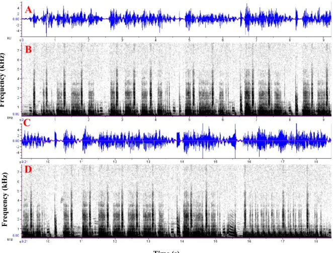
Figure 9: Short stimulation protocol to induce signalling in O. ishidae adults. A - oscillogram; B - spectogram ... 17 Figure 10: Schematic presentation of the “Stimulation protocol” used for females. RT: Repetition time. P: Inter-signal pause. PT: Pulse train pause. ... 18 Figure 11: Rival protocol. (A, C – oscillogram; B, D - spectrogram). ... 19 Figure 12: Wings protocol. The high amplitude vibrations are caused by the wing movements. ♂ represents the male pulse, arrows mark the crackles and the horizontal bars show the drum pulses. Above: oscillogram; below: spectrogram. ... 20 Figure 13: Male calling phrase of O. ishidae composed of the tapping sequence (A) and three male pulses (B). Above: oscillogram; below: spectrogram.. ... 23 Figure 14: Male calling phrase emitted after short stimulation. Note the high number of taps in the tapping sequence. Above: oscillogram; below: spectrogram ... 24 Figure 15: Decreasing frequency during the course of the male pulse in the calling phrase. Order refers to each of the 18 intervals in which we divided the pulse, from the beginning to the end. ... 25 Figure 16: Female pulses spontaneously emitted by an O. ishidae female. Above: oscillogram; below: spectrogram. ... 26 Figure 17: Female replies (red arrows) during the last section of the stimulation protocol. Above: oscillogram; below: spectrogram. ... 26 Figure 18:Female pulses. Above: oscillogram; below: spectrogram. The red arrows mark the dominant frequency around 600 Hz in A and 300 Hz in B ... 27
Figure 19: Decreasing frequency during the course of the female pulse. Order refers to each of the 18 divisions to which we divided the pulse, from the beginning to the end. .28
Figure 20: Male calling phrase followed by the first cycle of a recognition duet. Male and female signals are marked with their respective simbols. Above: oscillogram; below: spectrogram. ... 29 Figure 21: Female pulse and female quiet pulse (red arrows) in response to male pulses. Above: oscillogram; below: spectrogram. ... 30 Figure 22: Click trains in a cycle of the recognition duet (red round brackets) ... 31 Figure 23: End of cycle phrase, composed by four drum pulses, male pulse, whistle signal and two abdomen hits ... 31 Figure 24: Frequency changes during the course of whistle signal. Order refers to each of the 18 intervals to which we divided the pulse, from the beginning to the end. . 32 Figure 25: “Crackles” in the recognition duet (red arrows). Clicks are marked with black arrows. ... 32 Figure 26:Structure of a cycle in the early courtship phase. The phrase usually started with a series of crackle signals (red arrows), to which the rattle signal is added. Near the “end of cycle” phrase is were the male would emit his pulses, preceded by 1-4 drum pulses. Between 1-3 male pulses (not counting the one in the end of cycle sequence) would be emitted per cycle. Males tended to increase the duration of their rattle signals as the cycle progresses, as shown in the image. With dark arrows the rattle signals emitted without crackles are marked, normally before or after the male pulses. During the end of cycle phrase, no rattle signals were emitted and click trains were also rare. Above: oscillogram; below: spectrogram. ... 35 Figure 27: Structure of a cycle in the late courtship phase. ♂, ♀ and ♀* represent the male, female and quiet female pulses, respectively. Black arrows are marking rattle signals alone. Red bars mark rattle signals with a crackle. The black bar shows the modified “end of cycle” with the excitement signal. Above: oscillogram; below: spectrogram. ... 37 Figure 28: Rattle signal of 1 second composed by 54 drum pulses. Two images of the same signal in different time scales are presented to acknowledge the drum pulses. Above: oscillogram; below: spectrogram. ... 38 Figure 29: Rattle signals, alone (black arrows) or followed by crackle (red arrows). Spectrogram is shown below the corresponding oscillogram. ... 39 Figure 30: Relation between mean duration of rattle signal with a crackle (A) or alone (B) and number of signals during a complete courtship phase. RC: Rattle and crackle, R: Rattle.The arrow indicates the start of late courtship. ... 40 Figure 31: End of cycle with excitement signal. The crackles are marked with red and the clicks with black arrows. Above: oscillogram; below: spectrogram. ... 41 Figure 32: Female pulse overlapping the whistle (red arrow) and appearing before the excitement signal (black arrow) at the end of cycle phrase in the late courtship. Above: oscillogram; below: spectrogram ... 42
Figure 33: Double female pulses in the late courtship. Above: oscillogram; below: spectrogram. ... 42 Figure 34: Structure of the wings phase. The “end of the cycle” phrase belongs to the last cycle of the precedinglate courtship. The high-frequency oscillations in the spectrogram where caused by wing movements, and between them we can see several male pulses (marked with ♂ symbols). Red arrows mark the crackles. Above: oscillogram; below: spectrogram. ... 43 Figure 35: Male rival pulses (red arrows). Red square brackets mark the rival playback. Above: oscillogram; below: spectrogram. ... 45 Figure 36: Rival pulses (red arrows) emitted between wing movements during wings protocol. Above: oscillogram; below: spectrogram. ... 45 Figure 37: Male rival signals during early courtship (red arrows). Above: oscillogram; below: spectrogram. ... 46
ABBREVIATIONS AND SYMBOLS AYP Aster yellow phytoplasma
DNA Deoxyribonucleic acid
EPPO European and Mediterranean Plant Protection Organization
FD Flavescence dorée
Hz Hertz
IRPCM International Research Program for Comparative Mycoplasmology
ms Milliseconds
NIB National institute of Biology, Ljubljana PDP Pear decline phytoplasma
rRNA Ribosomal RNA
RT Repetition time
1 INTRODUCTION
1.1 RESEARCH BACKGROUND
Plant diseases associated to phytoplasmas are one of the major threats to worldwide agriculture, especially in the viticulture sector, where Flavescence dorée (FD) causes severe yield losses (up to 90% of the vineyard) due the lack of prophylaxis and rapid spread through vectors (Scattini et al., 2000). Only in Italy, the Italian government and the European Community spend a total of 34 million € to refund yield losses in 2005 (Belli et al., 2010). In the recent years, many alien insect species from the hemipteran suborder Auchenorrhyncha, which are known to be the vectors of phytoplasmas, have rapidly spread through Europe. To improve the efficiency of the pest management methods for these vectors, more research is needed to understand their life cycle and biology. Orientus ishidae
(Matsumura) (Hemiptera, Cicadellidae) is an invasive leafhopper from eastern Asia. It was first detected in Slovenia in 2002 and has since then spread all over the country (Seljak, 2004). Its vector capacity for FD was confirmed in different studies (Mehle et al., 2010) and even though vines are not its main host, its presence has been confirmed inside the vineyards. Improved knowledge about this pest is necessary for setting up efficient Integrated Pest Management and ecological management strategies.
1.2 WORKING HYPOTHESIS
As revealed in studies of other members of the Cicadellidae family, vibrational communication during the mating sequence follows the same general pattern, even across different species. The pair formation begins with emission of male vibrational signals, sexually receptive female replies and after establishing a vibrational contact, the exchange of signals continues as the male searches for a stationary replying female. We expect that also O. ishidae will follow this pattern.
1.3 OBJECTIVE OF THE RESEARCH
The main objective of this research is to characterize the mating behaviour of O. ishidae. As it has been described in several other leafhopper species, we may find in O. ishidae rivalry behaviour involving disruptive vibrational signals that can potentially be applied for the management of this pest.
2 LITERATURE REVIEW 2.1 PHYTOPLASMAS
Phytoplasmas are gram-positive prokaryotes, unicellular organisms that do not contain membrane-bound organelles such as nucleus or mitochondria, and obligate intracellular parasites of plants and insect vectors. They are pathogenic agents that cause mostly lethal diseases to hundreds of plant species. In plants they infect the sieve cells present in the phloem’s sieve tubes, while in insects they can infect different organs and tissues, including haemolymph, intestine, genitals and salivary glands. With a size around 1 μm, they can cross through the pores of the phloem sieve cells. The phytoplasma cell is surrounded by an approximately 10 nm thick trilaminar plasma membrane composed by proteins and lipids. Its cytoplasm contains ribosomes for the synthesis of proteins and a double circular DNA molecule. The presence of extrachromosomal DNA has also been detected (Nishigawa et al., 2001).
Phytoplasmas are transmitted through phloem-feeding insect vectors belonging to the order Hemiptera, best known examples are from the families Cicadellidae, Psyllidae, Cixiidae, Cercopidae, Delphacidae, Derbidae, Flatidae and Fulgoridae, and they multiply inside the insect and persist in it until its death. Nymphs and adults can acquire the phytoplasma when they feed on infected plant and then can transmit it to healthy ones. Although phytoplasmas are not commonly transmitted to offspring, transmission of phytoplasmas from adults to eggs and nymphs has been demonstrated in few species, like in Scaphoideus titanus Ball (Alma et al., 1997) and Matsumuratettix hiroglyphicus Matsumura (Hanboonsong et al., 2002), vectors of Flavescence dorée and Sugarcane white leaf phytoplasmas respectively.
Phytoplasmas can be transmitted by one or several insect vectors, depending on the degree of specificity in the phytoplasma-insect interaction. There are phytoplasmas associated with one main vector, and less specific ones, able to be transmitted by different species, whereas vectors can be infected by more than one species of phytoplasma (Weintraub and Beanland, 2006). The range of host plants for each phytoplasma depends on the feeding behaviour of the vector. Oligophagous vectors can disseminate the phytoplasma among one or a few plant species, such as Psylla piri L.(formerly Cacopsylla pyri L.) and pear decline phytoplasma (PDP) (Carraro et al., 2001). Contrary, if the insect feeds on different plant species, the phytoplasma will affect a greater range of plants, as is the case of Macrosteles quadrilineatus
Forbes, related to Aster yellow phytoplasma (AYP) (Maramorosch, 1956). Phytoplasmas can have a beneficial, adverse or no impact on their insect vectors. As an example, life span and fecundity was increased in females of M. quadrilineatus associated with AYP (Beanland et al., 2000), while it had the opposite impact on the S.titanus adults containing FD (Bressan et al., 2005). While some phytoplasmas can have a negative impact on the vectors, they rarely kill the insects, which would prevent their spread (Weintraub and Beanland, 2006).
Regarding taxonomy, the analysis of the 16 S rRNA gene sequence is probably the most widely used method for the determination of phylogenetic relationships and molecular classification of microorganisms (Fox et al., 1992). After using this molecular technique, all phytoplasmas were grouped in a new designed taxon named “Candidatus phytoplasma” (Ca
phytoplasma) (IRPCM, 2004). This approach has been widely used for the classification of microorganisms at genus or higher taxonomic levels. However, the 16S rRNA gene sequence is not as good for species level differentiation (Stackebrant and Goebel, 1994). Therefore, biochemical (antibody specificity, genes) or biological criteria (host range, vector transmission) are required for an adequate determination of different phytoplasma species. For example, Apple proliferation, Pear decline and European stone fruits yellows are three strains of Ca Phytoplasma spp. that show a 99% homology in their 16 rDNA sequences, but differ in their insect vectors (Seemüller and Schneider, 2004)
2.1.1 Symptoms on plants
Generally, plant diseases associated with the presence of phytoplasmas are recognized by a set of symptoms that suggest profound alterations in the plant’s biochemical molecules, photosynthesis or the reserve substances (Bertaccini and Duduk, 2010; Rusjan et al., 2012). The most common symptoms at the infected plants are:
• yellowing or chlorosis,
• reddening of leaves (Figure 1),
• virescence of flowers, where they lose their common colour and petals appear green,
• sterility of flowers,
• dwarfism,
• vegetative disorders, such as development of large clusters of leaves or undeveloped flowers,
• leaves curling down,
• general plant decline,
• phyllody, or transformation of floral organs into foliar structures, showing a green colour without the characteristic flower colour (Figure 2) and
• proliferation of adventitious buds that develop into many branches in a single site, creating what is known as the “witches’ broom” (Figure 3).
Figure 1: Reddening of pear leaves infected by pear decline phytoplasma (EPPO, 2006)
Slika 1: Rdečenje listov hruške okužene s fitoplazmo Pear decline (EPPO, 2006)
Figure 2: Phyllody in Callistephus chinensis (L.) Neesinfected with phytoplasma (left). Non-infected plants are shown in the right (Amityadav8, 2016)
Slika 2: Filodij pri vrtni astri (Callistephus chinensis (L.) Nees), okuženi s fitoplazmo (levo).
Figure 3: “Witches’ broom” in Hibiscus rosa-sinensis (L.)infected with phytoplasma (left). On the right a healty branch is shown. (Montano et al., 2001)
Slika 3: Metlasta rast osleza (Hibiscus rosa-sinensisL.) okuženega s fitoplazmo (levo). Na desni prikaz zdrave vejice (Montano in sod., 2001)
2.1.2 Flavescence dorée
Flavescence dorée (FD) is, economically, one of the most desctructive diseases of grapevines caused by a phytoplasma, affecting many cultivars of Vitis vinifera L. among several European countries. Its main vector is S. titanus (Alma et al., 1997), a specialist that feeds of several species of the Vitaceae family, considered an invasive and quarantine pathogen in Europe. In Slovenia, monitoring of S. titanus with yellow sticky traps, pesticide control in case of its ocurrence and yearly inspection for FD symptoms are mandatory in each vineyard (Pravilnik o ukrepih …, 2014). It was introduced in Europe at the beginning of 20th century from North America, the species original area (Bertin et al., 2007) probably in egg form layed on imported grapevine canes. If adults or nymphs feed on infected plants, the phytoplasma passes to the intestine of the insect, where it reproduces, and later to the hemolymph, reaching the salivary glands 4 to 5 weeks after ingestion. From this moment and until its death, the animal act as phytoplasma vector (Chuche and Thiéry, 2014). Affected strains of grapevine show the following symptoms:
• delay in sprouting,
• total or partial mortality of strains,
• lack of production: bunches get dried and do not mature,
• curling of leaves towards the underside,
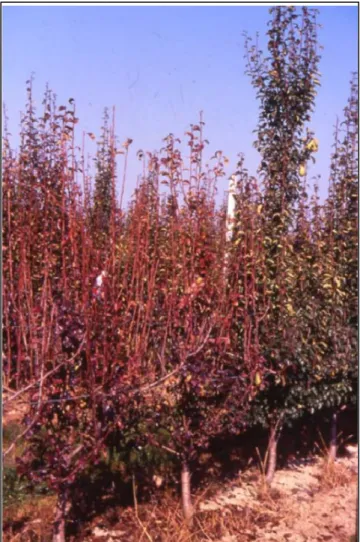
• general decline (Figure 4),
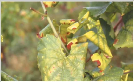
• red or yellow colouring of the leaves, depending on whether they are red or white varieties (Figure 5) and
Figure 4: Damage of FD to grape bunches (up left), leaves (up right) and the whole plant (bottom) in red grape varieties in Catalonia, Spain (Rahola et al., 1997)
Slika 4: Poškodba zaradi FD na grozdu (zgoraj levo), listih (zgoraj desno) in celi rastlini (spodaj) pri rdečih sortah iz Katalonije, Španija (Rahola in sod., 1997).
Figure 5: Yellowing of leaves of grapevine 'Chardonnay' variety infected with FD (EPPO, 2007) Slika 5: Porumenitev listov žlahtne vinske trte sorte ‘Chardonnay’, okužene s FD (EPPO, 2007)
There is no direct control method for FD once the plant has been infected. Only some antibiotics (tetracyclines) could have some efficacy (Caudwell, 1990), but its use is totally unfeasible since its application to the plant requires an extremely complicated technique, and it is prohibited by European law.
Prevention methods include (Pravilnik o ukrepih … , 2014; Laznik and Trdan, 2015):
• use of healthy plant material,
• vector control (S. titanus) and
• destruction of contaminated and/or abandoned vineyards.
Plant material can be cleaned of phytoplasmas by thermoterapy. This measure consists of immersing the plant material in the hot water thermostated on 50 °C for 45 min (EPPO, 2012). This practice can ensure the health of treated material, being also effective against bacteria (Agrobacterium tumefaciens Smith and Townsend, Xylella fastidiosa Wells et al.), fungi (Phytophthora cinnamoni Rands) and eggs of Cicadellidae (Grondeau and Samson, 1994). In Europe, control of S. titanus is done by monitoring and mandatory sprays of organophosphate pesticides (Chuche and Thiéry, 2014). In Slovenia the use of Thiamethoxam, a pesticide of the neonicotinoids class, showed the highest efficacy in the control of S. titanus (Žežlina et al., 2013). Other insecticides that are effective against S. titanus are neonicotinoids, organophosphates and those from the phyretrins group (Peterlin, 2015). However, against S. titanus three insecticide interventions are recommended (Rahola et al., 1997):
• First treatment. Taking into account that the insects are not infectious until about 4 weeks after adquiring the phytoplasma of a diseased plant, it will be between three weeks and a month after the first nymphs are observed. This first treatment is essential.
• Second treatment. It is carried out at the end of the hatching period, approximately 15 days after the first treatment.
• Third treatment. The first two treatments must cover the entire S. titanus egg hatching period. The third treatment will allow to destroy them in their adult state, which is when there is a greater displacement of the animals and therefore they can spread the disease to a greater distance. It should be done about 30 days after the second treatment.
Other alternavite methods of pest management involving the rival behaviour of Cicadellidae via vibrational communication are being developed to control S. titanus (explained in section 2.3.2).
2.2 BIOLOGY OF Orientus ishidae
Orientus ishidae (Matsumura, 1902), previously known as Phlepsius tinctorius (Sanders and DeLong, 1919)is a hemipteran insect, member of the Cicadellidae family, commonly known as mosaic leafhopper or Japanese leafhopper. The common name its given after the distinctive mosaic-like coloured pattern that adults of this insect present in their wings. It is
hemimetabolous (incomplete metamorphosis with 5 nymph stages) and has an univoltine life cycle (produces one generation per year). Adults can be observed from early July to October, then they lay eggs overwinter, then the next year nymphs will hatch around mid May and can be found until early August (Lessio et al., 2016). Adults and nymphs are polyphagous and feed on a wide range of plants (Table 1) by sucking the sap of phloem after piercing the plant vascular tissue, as other members of the Cicadellidae family. Due to its wide range of host plants, O. ishidae has been found in many different ecosystems, like forests, urban areas, ornamental landscape trees, orchards and vineyards.
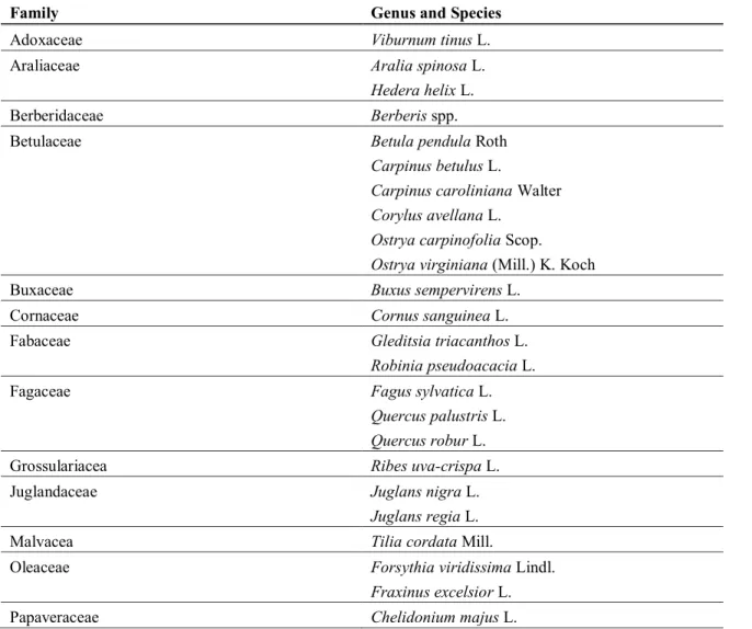
Table 1: Plant species were O. ishidae nymphs and adults were captured (Felt and Bromley, 1941; Rosenberg and Jones, 1977; Valley and Wheeler, 1985; Johnson and Freytag, 2001; Günthart and Mühlethaler, 2002; Seljak, 2004; Guglielmino, 2005; Mazzoni, 2005; Nickel, 2010; Koczor et al., 2013; Lessio et al., 2016; Klejdysz et al., 2017; Parise, 2017).
Preglednica 1: Rastlinske vrste, na katerih so našli nimfe in odrasle osebke japonskega škržatka (Felt in Bromley, 1941; Rosenberg in Jones, 1977; Valley in Wheeler, 1985; Johnson in Freytag, 2001; Günthart in Mühlethaler, 2002; Seljak, 2004; Guglielmino, 2005; Mazzoni, 2005; Nickel, 2010; Koczor in sod., 2013; Lessio in sod., 2016; Klejdysz in sod., 2017; Parise, 2017)
Family Genus and Species
Adoxaceae Viburnum tinus L. Araliaceae Aralia spinosa L.
Hedera helix L. Berberidaceae Berberis spp. Betulaceae Betula pendula Roth
Carpinus betulus L.
Carpinus caroliniana Walter
Corylus avellana L.
Ostrya carpinofolia Scop.
Ostrya virginiana (Mill.) K. Koch Buxaceae Buxus sempervirens L.
Cornaceae Cornus sanguinea L. Fabaceae Gleditsia triacanthos L.
Robinia pseudoacacia L. Fagaceae Fagus sylvatica L.
Quercus palustris L.
Quercus robur L. Grossulariacea Ribes uva-crispa L. Juglandaceae Juglans nigra L.
Juglans regia L. Malvacea Tilia cordata Mill.
Oleaceae Forsythia viridissima Lindl.
Fraxinus excelsior L. Papaveraceae Chelidonium majus L.
continuation of Table 1
Papaveraceae Chaenomeles speciosa L.
Rosaceae Amelanchier spicata (Lam.) K.Koch
Craegus oyacantha L.
Crataegus rhipidophylla Gand.
Cydonia oblonga (Sweet) Nakai
Malus domestica Borkhausen
Malus sylvestris Mill.
Prunus avium Mill.
Prunus domestica L.
Prunus laurocerasus L.
Prunus virginiana L.
Rosa canina L.
Rubus fruticosus L Salicaceae Populus alba L.
Populus nigra L.
Salix alba L. Salicaceae Salix babylonica L.
Salix caprea L.
Salix cinerea L.
Salix purpurea L.
Salix x rubens Schrank Sapindaceae Acer campestre L. Ulmaceae Ulmus spp. Urticaceae Urtica dioica L. Vitaceae Vitis vinifera L.
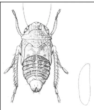
Adults of O. ishidaehave a length of 4.5-6.5 mm (personal observations), and present sexual dimorphism, females being bigger than males. The length of nymphs is 3.6-5.0 mm, the later nymph instars being bigger, and having a strong coloration with variable patterns. They are noted for pointing up their abdomen tips in presence of danger. Eggs of O. ishidae are 1.10-1.22 mm long and 0.28-0.34 mm wide, nearly S-shaped and with the posterior part rounded (Valley and Wheeler, 1985). Pictures and schemes from O. ishidae adults, nymphs and eggs can be seen in Figures 6 and 7.
Figure 6: Winged adults of O. ishidae and 5th instar nymphs with their abdomen pointing up (photo: Seljak, 2009-2010)
Slika 6: Krilati odrasli japonski škržatki in nimfe v petem stadiju s kvišku obrnjenimi zadki (foto: Seljak, 2009-2010)
Figure 7: Schematic representation of the 5th nymph instar and eggs of O. ishidae (modified according
to Valley and Wheeler, 1985)
Slika 7: Shematičen prikaz nimfe v 5. stadiju in jajčeca japonskega škržatka (O. Ishidae) (prirejeno po Valley in Wheeler, 1985)
O. ishidae originates from Asia and was first described by Shōnen Matsumura in Japan in 1902, but it has been introduced in numerous countries around the world. In the USA, it was found in 1941, probably introduced with ornamental plants from Asia (Felt and Bromley, 1941), and its expansion through different states, together with tree species were it was found, was reported by several authors (Johnson and Freytag, 2001; Rosenberg and Jones, 1977; Sanders and DeLong, 1919; Valley and Wheeler, 1985). In Europe it was discovered for the first time in 1998 in Northern Italy (Guglielmino, 2005) and 2000 in Switzerland (Günthart and Mühlethaler, 2002), and since then its expansion has been reported by different authors, together with anonymous reports on various internet forums (Table 2).
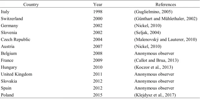
Table 2: Expansion of O. ishidae in Europe since its appearance in 1998 (modified according to EPPO Global Database, 2015)
Preglednica 2: Razširjanje japonskega škržatka v Evropi od njegove prve pojavitve leta 1998 (prirejeno po EPPO Global Database, 2015)
Country Year References Italy 1998 (Guglielmino, 2005)
Switzerland 2000 (Günthart and Mühlethaler, 2002) Germany 2002 (Nickel, 2010)
Slovenia 2002 (Seljak, 2004)
Czech Republic 2004 (Malenovský and Lauterer, 2010) Austria 2007 (Nickel, 2010)
Belgium 2008 Anonymous observer France 2009 (Callot and Brua, 2013) Hungary 2010 (Koczor et al., 2013) United Kingdom 2011 Anonymous observer Slovakia 2012 Anonymous observer Spain 2012 Anonymous observer Poland 2015 (Klejdysz et al., 2017)
In 2009, O. ishidae was found to host phytoplasma from the 16SrV group, related to Flavescence dorée phytoplasma strains in Slovenia (Mehle et al., 2010). Although it is rarely found feeding on vines, its presence in Northern Italian vineyards and their surroundings have been confirmed by different authors (Gaffuri et al., 2011; Lessio et al., 2016), meaning a potential danger for the plants and a new vector for the disease. Furthermore, laboratory experiments by Lessio et al. (2016) found that O. ishidae can lay eggs in grapevine, after obtaining up to 40 nymphs from pruned grapevine wood. Nevertheless, O. ishidae presence in vineyards is small as compared with the main vector of FD, S. titanus, a grapevine specialist. The little presence of O. ishidae may be explained by the mandatory use of insecticide treatments to stop the spreading of S. titanus in Europe (Žežlina et al., 2013).
As a possible threat for vineyards, more research regarding possible control of this insects should be done, especially focused on an Integrated and Ecological Pest Management approach, to reduce the use of pesticides. Experiments performed by Parise (2017) revealed some possible natural enemies of O. ishidae, including parasitic activity from larvae of the mites Charletonia cardinalis (C.L. Koch, 1837) and Erythrae jowitae (Haitlinger, 1987) (Acari, Erythraeoidae) and parasitic attacks from Anteon fulviventre (Haliday, 1828) (Hymenoptera, Dryinidae), but these are general parasitoids that feed on a wide range of insects and no record was found of O. ishidae specialist predators/parasitoids. New insights in pest control of Cicadellidae include the disruption of their mating behaviour via vibrational playback (Polajnar et al., 2016a), a strategy that potentially could be applied to
O. ishidae, but more knowledge about their mating behaviour is required. The use of vibrations by members of Cicadellidae family and possible disrupting method is explained below.
2.3 VIBRATIONAL COMMUNICATION
Exchange of information via mechanical signals transmitted through the substrate on which the animals are standing is one of the oldest and most widespread forms of animal communication (Virant-Doberlet and Čockl, 2004; Cocroft and Rodríguez, 2005; Hill, 2008; Cocroft et al., 2014). Vibrational communication is particularly widespread among arthropods, where it is estimated that is used by more than 220.000 species (Cocroft and Rodríguez, 2005). Insects use vibrational signals in many behavioural contexts, for example for finding a partner (Čokl and Virant-Doberlet, 2003; de Groot et al., 2012; Nieri et al., 2017; Derlink et al. 2018), in social interactions (Cocroft and Hamel, 2010), for defence (Cocroft, 1999) and alarm (Hager and Kirchner, 2014).
Vibrational signals are usually species- and sex specific (Cocroft et al., 2010; Derlink et al., 2018;) and insects produce vibrational signals by several different manners (Virant-Doberlet and Čokl, 2004). The most common and widespread way of producing substrate-borne vibrations is drumming, where animals strike the substrate with different body parts (Stewart and Sandberg, 2006). Signal production mechanism named tremulation involves oscillations of the body or body parts without touching the substrate (Čokl, 2008). Stridulation is a signal production mechanism where animals rub different body parts against each other (Čokl and Virant-Doberlet, 2003), while tymbal mechanism involves buckling of a membrane plate to which strong muscles are attached (Wessel et al., 2014).
Insects also have several receptors for detecting vibrational signals, most of them located in legs (Yack, 2004). The most investigated vibroreceptor is a subgenual organ located in the tibia, which is also the most sensitive one and responds to displacements of the substrate smaller than 1 nm (Stritih Peljhan and Strauß, 2018). The morphology and sensitivity and frequency response characteristics of vibroreceptors differ among insect groups and species (Virant-Doberlet and Čokl, 2004).
2.3.1 Leafhopper vibrational communication
In leafhoppers, mating sequence is usually stereotyped, and partners communicate exclusively via species- and sex specific vibrational signals (Čokl and Virant-Doberlet, 2003). So far, mating behaviour and associated vibrational signals have been studied in a number of species (de Groot et al., 2012; Nieri and Mazzoni, 2017; Nieri et al., 2017; Abt et al., 2018; Derlink et al., 2018). The common pattern of mating and signalling behaviour revealed in these studies could be summarized as following: (a) vibrational communication between a male and a female begins with spontaneously emitted male advertisement call; (b) sexually receptive female replies and partners establish a coordinated duet with species-specific structure; (c) male searches for a replying, stationary female and vibrational exchange continues throughout the searching phase.
While the general pattern of leafhopper mating sequence appears to be similar in all species, the repertoire and complexity of vibrational signals can differ greatly among species (de Groot et al., 2012; Derlink et al., 2014; Nieri and Mazzoni, 2017; Nieri et al., 2017; Derlink et al., 2018). Here, females usually produce only one type of signal as a reply, whereas male vibrational signals are often composed of different elements and can also differ in different stages of mating behaviour (de Groot et al., 2012). In leafhoppers, the main production mechanism of vibrational signals in males and females is supposed to be tymbal (Ossiannilsson, 1949; Wessel et al., 2014), however, during the close-range courtship male wing flapping has been observed in several species (Derlink et al., 2018).
Leafhoppers also have well developed rivalry behaviour that includes emission of rivalry signals and satellite behaviour. (Mazzoni et al., 2009; Kuhelj and Virant-Doberlet, 2017; Nieri and Mazzoni, 2017; Derlink et al., 2018). Masking signals that overlap female reply and disrupt the ongoing duet are the most common form of rivalry signals.
2.3.2 Exploiting vibrational signals for pest management
Recent advances in understanding vibrational communication have prompted research into use of this communication modality for pest control, analogous to disruption of mating and mass trapping of insects using synthetic pheromones, which are two of the most widespread modern alternatives to pesticide use (Witzgall et al., 2010). Behavioural manipulation using pheromones is, unfortunately, not applicable to Auchenorrhyncha and several other insect groups in which chemical communication is largely absent, but they still comprise important agricultural pests (Polajnar et al., 2016b).
Vibrations open promising new avenues for developing alternative pest management practices. One of them is improving early detection of pest insects, exploiting either signals or incidental vibrational emissions (result of feeding, moving etc.), but knowledge of temporal and spectral structure of these emissions is crucial for reliable identification (Čokl and Millar, 2009; Mankin et al., 2011; Laumann et al., 2017). This approach is most actively researched in connection with concealed pests of wood and stored products (Mankin et al., 2010; Zorović and Čokl, 2015). Other options involve behavioural manipulation using artificial playback – here, vibrations may serve for attracting pests to an area where they can be conveniently eliminated, repelling them from protected resources (“push-pull” techniques) or interfering with key behaviours such as locating hosts or attracting mates (Polajnar et al., 2015)
The idea of using rival vibrational emissions for mating disruption arose from early observations of mating behaviour of the leafhopper S. titanus which revealed that rival vibrational emissions used in antagonistic interactions among males are efficient in masking vibrational signalling between a courting male and female (Mazzoni et al., 2008). The underlying idea of using artificial noise in the vibrational channel for mating disruption is
several decades older (Saxena and Kumar, 1980), but was ignored for a long time for various reasons. Because leafhoppers rely exclusively on vibrations in long-range sexual communication, sufficiently intense vibrational noise should, in theory, block the information transfer entirely and thus prevent potential partners from recognizing and locating each other. On this basis, a system of electromagnetic shakers was recently set up in an experimental vineyard, artificially reproducing male rival emissions throughout the mating season and reducing reproductive success in semi-field conditions (Polajnar et al. 2016a). While S. titanus has several advantageous traits that facilitate this approach (monophagy, low dispersal capability, univoltine life cycle; Chuche and Thiéry, 2014), the success of early trials nevertheless demonstrates the value of studying basic reproductive biology of a pest species even if practical use is not yet apparent.
3 MATERIALS AND METHODS 3.1 MATERIALS
3.1.1 Insect collection and rearing
O. ishidae adults and nymphs were catched up on different species of Salix genus in urban areas of Nova Gorica (45°57'26.6"N 13°39'06.4"E) and the vicinity of the National Institute of Biology (NIB) in Ljubljana (46°03'09.4"N 14°28'08.8"E) at the end of June of 2017. The two main willow species from which leafhoppers were collected were Salix cinerea L. (grey willow) and Salix alba L. (white willow). Catching was conducted with a sweep net and aspirator and animals were transported in net cages (36 x 22.5 x 25 cm) containing fresh willow branches. In Ljubljana, they were reared at Department of Organisms and Ecosystems Research at NIB at 24±1 °C, 16:8 (L:D) photoperiod and 70±10 % relative humidity. Once in the laboratory, the sex of captured adult males and females was determined by observing genital structure under the dissecting microscope according to description in Guglielmino (2005), and individuals were kept separated in transparent plastic containers (8 x 8 x 13.5 cm) containing a small glass vial with water to keep the willow leaves fresh. Nymphs were kept together in net cages (36 x 22.5 x 25 cm) that were checked daily freshly new moulted adults. Containers were cleaned and provided with fresh willow cuttings twice a week. In experiments, only virgin adults were used (48 and 36 adult males and females, respectively). We considered males and females sexually mature and ready to use for mating experiments if they emitted vibrational signals spontaneously (in case of males) or if answered to our stimulation protocols (see below). All the animals were pre-tested to establish their sexual maturity prior the experiments by recording their vibrational activity in our set-up for 5 min without any stimulus, and then stimulated with one of the playback protocols, if needed.
3.1.2 Experimental set-up
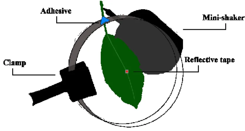
All recordings of vibrational signals were performed at NIB in July 2017 between 9:00-15:00, at temperatures of 24±1 °C and with a relative humidity of 70±10 %. The set-up for the recordings consisted of a S. cinerea leaf (size approximately 4 x 8 cm) placed inside a covered petri dish (100x15mm) with a small hole through which the petiole was pulled out and fixed with adhesive putty (BlueTack) (Figure 8). The petiole was attached to an electromagnetic mini-shaker (Type 4810, Brüel and Kjær Sound and Vibration A/S, Nærum, Denmark), that was used to deliver stimulatory signals to the leaf in playback experiments (see below). The minishaker was driven from the computer via X-fi surround 5.1 Pro sound card (Creative Labs Inc., Ireland) by Cool Edit Pro 2 (Syntrilium Software, Phoenix, USA). Each day, after finishing the experiments, the petiole was kept in water in Eppendorf PCR tubes tightened with parafilm to keep it hydrated. In case of wilting, a leaf from the same willow tree and of similar dimensions was used as replacement. During the experimental
season, we had to replace the leaf three times. In the centre of the leaf, near or across the midvein, we placed a small piece of reflective tape (~0.3 cm2), on which we focused the
laser beam (Figure 8). The petri dish was suspended vertically with a utility clamp for better video recording and the whole set-up was placed on a custom-made vibration isolated table to minimize the background vibrational noise.
Vibrational signals were registered on the leaf lamina by using a laser vibrometer (PDV 100, Polytec GmbH, Waldbronn, Germany). Signals were digitized with 44100 Hz sample rate and 32-bit resolution and stored directly onto a hard drive of a computer using X-fi surround 5.1 Pro sound card (Creative Labs Inc., Ireland) and Audacity 2.2.2 software (Audacity Team, 2007). Recorded vibrational signals were analysed using the computer program Raven Pro 1.5 (The Cornell Lab of Ornithology, 151 Ithaca, NY, USA. In addition, during our behavioural experiments, the behaviour of O. ishidae together with recorded vibrations was filmed with a Panasonic HC-VXF990 video camera (Panasonic corporation, Japan), and the video was transferred to a computer using Windows Movie Maker 2.0. Videos helped us to identify which animal was emitting the signals and to associate some of the signals with specific movements. In our work, visual representation of sound will be done by oscillogram and spectrogram pictures.
Figure 8: Experimental set-up for recording vibrational signals and behaviour of O. ishidae
Slika 8: Eksperimentalna postavitev za snemanje vibracijskih signalov in vedenja japonskega škržatka (O. Ishidae)
3.2 METHODS
3.2.1 Playback experiments
To study signalling and rivalry behaviour we performed playback experiments. Female leafhoppers rarely emit vibrational signals spontaneously (de Groote et al., 2011; Nieri et al., 2017) and we induced their signalling by playback of vibrational signals emitted by single males. Four protocols were designed for our different experiments (Table 3). The amplitude
of our playback signals was adjusted to the amplitude range of signals emitted by live leafhoppers to maintain the frequency characteristics. A more detailed description of signals used in our protocols is provided in the results section.
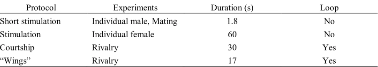
Table 3: List of protocols used in playback experiments to study O. ishidae behaviour
Preglednica 3: Seznam protokolov, uporabljenih v poskusih za preučevanje vedenja japonskega škržatka (O. Ishidae)
Protocol Experiments Duration (s) Loop Short stimulation Individual male, Mating 1.8 No Stimulation Individual female 60 No
Courtship Rivalry 30 Yes
“Wings” Rivalry 17 Yes
3.2.1.1 Short stimulation protocol
The short stimulation protocol was designed to induce signalling in individual males (see section 3.2.2.1), as well as in mating experiments (see section 3.2.2.2), when individuals did not emit any signals three minutes after the start of the experiment. From our own recordings we used a male calling phrase that was composed by a tapping sequence of five taps, separated by a 30 ms interval from the first two male pulses of 80 ms length, and interval 65 ms between them (Figure 9). The dominant frequency of the male pulses was 560 Hz.
Figure 9: Short stimulation protocol to induce signalling in O. ishidae adults. A - oscillogram; B - spectogram Slika 9: Protokol “Short stimulation” za sprožitev oglašanja pri odraslih japonskih škržatkihO. ishidae A - oscilogram; B - spektogram
A
B
Time (s) F re q u en cy (k Hz )3.2.1.2 Stimulation protocol
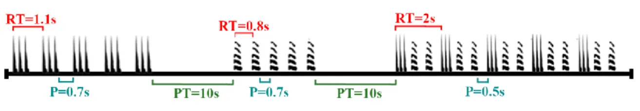
The stimulation protocol was created using elements of the recorded calling phrase used in short stimulation protocol. It was composed by three sections, the first one was a tapping sequence (composed by five taps), the second with individual male pulses, and the third one with a complete male calling phrase. Every element in the sections was repeated ten times, since we also wished to check whether a single element of the phrase was sufficient to trigger the female reply. The repetition time (RT, time between the beginning of an element and the start of the next one) and inter-signal pause (P) between the elements of the phrase are explained in the Fig. 10. Among the different sections we inserted a pulse train pause (PT) of ten seconds (Figure 10).
Figure 10: Schematic presentation of the “Stimulation protocol” used for females. RT: Repetition time. P:
Inter-signal pause. PT: Pulse train pause
Slika 10: Shema protokola “Stimulation”za samice. RT: Ponavljalni čas. P: Premor med signali. PT: Premor
med pulzi
3.2.1.3 Rival protocol
The vibrational sequence used in this protocol consisted of a complete cycle of the “late courtship” phrase (Figure 11) emitted by a male in our behavioural experiments. Briefly, the courtship phase is characterised by repeating the male courtship phrase, and this fact allowed us to loop the playback so that the male would perceive it as an ongoing courtship. The playback contained eight male and five female pulses with dominant frequency of around 600 and 690 Hz, respectively. The female in this recording answered to the last five male pulses after a small gap of around 100 ms. The rest of the signals were “crackles”, “rattle”, drum pulses preceding the male pulse, “end of cycle” and “excitement”, explained in detail in the results section.
Figure 11: Rival protocol. (A, C – oscillogram; B, D - spectrogram) Slika 11: Protokol “Rival”. (A, C – oscilogram; B, D - spektrogram)
3.2.1.4 “Wings” protocol
The “wings” protocol, given in Figure 12, was designed after our experiments with the courtship protocol to study if we could observe rival behaviour in the last section of the mating behaviour, the “wings” phase. In it the males perform fast wings movements alternated with one or two male pulses. The wing movements and pulses had a mean RT of 1.165 seconds. The male pulses had a length of around 60 ms and a mean peak frequency of 646 Hz. In the cases where two male pulses were present between the wing movements, there was an interval of 60 ms between them. Single male pulses were preceded by three drum pulses. Preceding all the wing movements there was a crackle signal.
A
B
C
D
F re q u en cy (k Hz ) F re q u en cy (k Hz ) Time (s)Figure 12: Wings protocol. The high amplitude vibrations are caused by the wing movements. ♂ represents the male pulse, arrows mark the crackles and the horizontal bars show the drum pulses. Above: oscillogram; below: spectrogram.
Slika 12: Protokol "Wings". Visoka amplituda vibracij je posledica gibanja kril. ♂predstavlja samčev pulz, puščice označujejo prasketanje, vodoravne črte pa prikazujejo bobnajoče pulze. Zgoraj: oscilogram; spodaj: spektrogram.
3.2.2 Behavioural experiments 3.2.2.1 Individual behaviour
Males (N=15) and females (N=15) were placed individually in our set-up and recorded for 10 minutes or until vibrational signals were emitted. In case no signals were emitted after five minutes, “Short stimulation protocol” was applied to the petiole.
To describe the structure of the male calling phrase we measured the following parameters: (i) time from the beginning of the trial to the spontaneous emission of the first male signal, (ii) duration of the signals,
(iii) repetition time,
(iv) dominant frequency of the male pulse and (v) number of taps in the taping sequence.
We also noted potential interaction with short stimulation playback. To describe the frequency modulation in the male pulse we took 30 random male pulses from six different males and divided each pulse into 18 intervals (5 ms) from the beginning until the end of the pulse.
In individual behaviour experiments with females we only addressed how females interacted with the stimulation protocol and the duration. In those cases when females emitted signals spontaneously, we also noted the time of the emission. We also reproduced the stimulation protocol to mated females in order to check whether they retain their receptivity.
Time (s) F re q u en cy (k Hz ) ♂♂ ♂♂ ♂ ♂ ♂
3.2.2.2 Mating behaviour
20 pairs of virgin males and females were used to describe mating behaviour. The leafhoppers were placed randomly on the surface of the leaf and females were always placed first. If leafhoppers did not emit vibrational signals for 3 minutes, we played them the “Short stimulation protocol”, in order to induce signalling. We recorded the emitted vibrational signals and videotaped the behaviour throughout the whole courtship, copula and five minutes after the copula to observe their post-copulation behaviour. If animals did not emit vibrational signals for 10 min, we stopped the trial. For detailed analyses we randomly chose 10 of these experiments that ended with a copula.
Because in these experiments we recorded the highest number of female signals (N=720), we analysed the temporal and frequency parameters with this data pool. To describe the structure of the female pulse we measured its duration, dominant frequency, as well frequency modulation. As female pulses vary in duration, only pulses of 200 ms were used for the frequency modulation analysis, with 18 intervals of 11 ms length. As with the description of the modulated frequency of individual males (see above), we used 30 random female pulses from 6 different females.
To describe the frequency modulation in the “whistle” signal, we randomly chose five of these signals from each male used in courtship (N=10) and divided each signal into 18 intervals (12 ms) from the beginning to the end of the signal. One male was not used in our analysis due poor signal quality.
3.2.2.3 Rivalry interaction
To investigate male rivalry behaviour, we performed two types of experiments. In the first experiment, we played single males (n=15) the “Rival protocol” (see above). In the second type of experiment, ten trios composed of two live males and one live female were placed in the set-up and their emitted signals and behaviour were recorded until one male and a female copulated, or the trial was stopped earlier if leafhoppers did not emit vibrational signals for 10 minutes.
“Short stimulation protocol” was used to induce signalling if after placement to the leaf animals did not emit any vibrational signal for three minutes. As with the reproductive behaviour experiments, males that mated in previous experiments were not used in other trials and different males were used in every trio.
3.3 TERMINOLOGY AND SIGNAL CHARACTERISATION
Vibrational signals and phrase elements were assigned unique labels to enable better characterisation of the animal behaviour. Signal characterisation has been performed by statistical analysis of the mean duration of the signals and their dominant frequencies (if they
had any). In the case of “rattle” and “crackle” signal during late courtship, number of signals and time spent in their emission during one cycle was also measured and analysed (see section 4.2.2.3). The software used for our statistical analysis was IBM SPSS Statistics 22 (IBM Corp. Released 2016, Armonk, NY, USA).
A uniform and up-to-date glossary of terms for different substrate-borne vibrational signals has yet to be created. Researchers name the biological signals according to different parameters: the known or presumed function of the signals, the nature of the movements or the device involved in making the signal, the onomatopoetic effect of the sound produced or the physical nature of the sound (Alexander, 1967).
Different terms were used to describe each signal, according to the visual cues provided by the video recording, onomatopoeia of the recorded vibrational signal or by their appearance in the cycle (see Results). Two terms used in this work can be attributed to other authors:
• Pulse: A brief succession of sine waves; In other words, a unitary homogenous parcel of sound of finite duration (Broughton 1963).
• Calling phrase: Signals produced spontaneously by isolated males (De Vrijer, 1983). And the rest of signals used in this work can be classified by three factors: onomatopoetic terms (clicks, crackles, rattle, taps, drums and whistle signals), animal behavioural display (abdomen hits and wings) and position in the cycle (end of cycle phrase and excitement signal).
4 RESULTS
4.1 INDIVIDUAL BEHAVIOUR 4.1.1 Male calling phrase
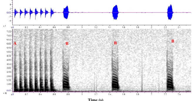
From the 15 males studied, 10 spontaneously emitted vibrational signals 6-80 s after being placed on the leaf, that we named “calling phrase”. The phrase was composed of two elements, a tapping sequence and a series of male pulses (Figure 13). Males produced this phrase and then remained silent or jumped off the leaf, but those who remained on the leaf and were stimulated with the “short protocol” emitted signals soon after. During the emission of the calling phrase males remained stationary.
Figure 13: Male calling phrase of O. ishidae composed of the tapping sequence (A) and three male pulses (B). Above: oscillogram; below: spectrogram
Slika 13: Pozivna fraza samca japonskega škržatka (O. ishidae), sestavljena iz zaporedja "trkov" (A) in treh
samčevih pulzov (B). Zgoraj: oscilogram; spodaj: spectrogram
The calling phrase typically started with the tapping sequence (although in two cases it started with a pulse), with a recognisable rhythmic pattern. This sequence was composed by several “taps” with a mean duration of about 5 ms (Standard deviation (SD) = ±0,5 ms; Number of signals (N) = 50) and were separated by a 3 ms interval (SD = ±1 ms; N = 50). If the male emitted the calling phrase spontaneously, the number of taps in the sequence would be between 2-6, however, in response to the short stimulation protocol (in which the tapping sequence was composed of five taps), the number of taps was typically as high as in the playback, or larger, up to 12 per sequence (Figure 14).
A B B B F re q u en cy (k Hz ) Time (s)
Figure 14: Male calling phrase emitted after short stimulation. Note the high number of taps in the tapping sequence. Above: oscillogram; below: spectrogram
Slika 14: Samčeva pozivna fraza, oddana po kratki stimulaciji. Opazno je večje število "trkov" v zaporedju.
Zgoraj: oscilogram; spodaj: spektrogram
After the tapping sequence, followed a silent interval of 60-300 ms, after which, 2-12 pulses were emitted. The duration of a single pulse was between 80-130 ms, depending on the individual, with an average duration of 100 ms (SD = ±20 ms; N = 221). Repetition time (RT) was usually around 800 ms between the first two pulses, but RT’s in the range of 360-1200 ms were recorded, values differing highly between individuals. We observed that the RT slightly increased after every pulse, with a recorded maximum of four seconds. The male pulse was a frequency modulated harmonic signal, with dominant frequency around 616 Hz (it varied between 559 and 690 Hz, depending on the male). The fundamental frequency of this signal was around 300 Hz. We noted that at the beginning of the pulse the strongest energy component was between 732-602 Hz and at the end of the signal the dominant frequency decreased to 646-387 Hz (Figure 15). Both in the pulse and the taping sequence, most energy was contained in the frequency range below 4000 Hz.
Several calling phrases could be emitted one after another and we recorded up to four phrases in a sequence, separated on average by a silent interval of 540 ms (SD = ±230 ms; N = 37)
Short stimulation F re q u en cy (k Hz ) Time (s)
Figure 15: Decreasing frequency during the course of the male pulse in the calling phrase. Order refers to each of the 18 intervals in which we divided the pulse, from the beginning to the end
Slika 15: Zmanjševanje frekvence samčevega pulza v pozivni frazi. Pulze smo razdelili na 18 intervalov, izmerki frekvenc teh intervalov so prikazani po zaporedni številki
4.1.2 Female pulses
From the 15 females analysed in individual behaviour experiments, three emitted sporadic signals (Figure 16) and the rest replied to the “stimulation protocol”. Those who signalled spontaneously, emitted between two and four pulses after staying on the leaf for 20-96 seconds. From the females that replied the playback stimulation, most replies were emitted in the last section that contained the complete male calling phrase (Figure 17). When the female replied in the first two sections of the stimulation protocol, she emitted one or two pulses and then remained silent until the last section.
Figure 16: Female pulses spontaneously emitted by an O. ishidae female. Above: oscillogram; below: spectrogram
Slika 16: Spontano oddani signali samice japonskega škržatka (O. ishidae). Zgoraj: oscilogram; spodaj: spektrogram
Figure 17: Female replies (red arrows) during the last section of the stimulation protocol. Above: oscillogram; below: spectrogram
Slika 17: Samičini odgovori (rdeče puščice) med zadnjim odsekom »Stimulation protocol«. Zgoraj: oscilogram; spodaj: spektrogram
Female pulse was a vibrational signal with a harmonic structure and with most energy contained in the range below 3000 Hz (Figure 18). From the signals analysed in our mating behaviour experiments, the dominant frequency was around 590 Hz (SD = ±255 Hz; N = 685, excluded peak frequencies of 0). Up to seven harmonics could be observed. Female pulse duration varied between 57-610 ms (mean ± SD = 190±96 ms; N = 720). Like in the male pulse, female pulse was frequency modulated (Figure 19). We also noted that longer pulses were produced in the first reply of the female to a stimulus (either a protocol or a live male). F re q u en cy (k Hz ) F re q u en cy (k Hz ) Time (s) Time (s)
Figure 18: Female pulses. Above: oscillogram; below: spectrogram. The red arrows mark the dominant frequency around 600 Hz in A and 300 Hz in B
Slika 18: Samičina pulza. Zgoraj: oscilogram; spodaj: spektrogram. Rdeči puščici označujeta dominantno
frekvenco okoli 600 Hz v A in 300 Hz v B B A
F
requency
(kH
z)
Time (s)Figure 19: Decreasing frequency during the course of the female pulse. Order refers to each of the 18 divisions to which we divided the pulse, from the beginning to the end
Slika 19: Zmanjšanje frekvence samičinega pulza. Pulze smo razdelili na 18 intervalov, izmerki frekvenc teh
intervalov so prikazani po zaporedni številki
4.2 MATING BEHAVIOUR
From the ten mating experiments analysed, in five males spontaneously emitted vibrational signals 30-100 s after the start of the trial, while in other five signalling had to be induced by playback of “short stimulation protocol”. In one trial, the female emitted the first recorded signal, a single pulse, three s after the start of the trial, in this case the male did not emit his calling phrase until 50 s later. As our individual behaviour experiments suggested, vibrational interaction between a male and a female started with emission of the male calling phrase, to which female replied by coordinating the emission of her pulses with the male signals. To better clarify, as well as simplify the whole mating sequence of O. ishidae, we divided the reproductive behaviour into three main phases. The total duration of our mating behaviour experiments ranged from 6-15 min.
4.2.1 Phase 1: Recognition duet
Males of O. ishidae emitted calling phrases, with the tapping sequences composed of 2-12 taps, as observed in individual behaviour trials, and high-amplitude pulses. Typically, females replied after the first male pulse and we observed that after the first female reply, all subsequent male signals had lower amplitude.