258
Progressive Neurological Deficit in Adult Idiopathic Syringomyelia (IS):
Case Report and Literature Review
Farid Yudoyono, Anugrah O. Widhiatmo, Beny A. Wirjomartani
Department of Neurosurgery, Faculty of Medicine
Padjadjaran University
–
Hasan Sadikin Hospital Bandung
Jl. Pasteur no 38, Bandung, Jawa Barat, Indonesia 40161
Email: [email protected]
Abstract
A syringomyelia is a clinical entity of any tubular fluid-filled cavity within the spinal cord that causes slowly but relentlessly progressive symptoms as well as expansion of the cavity, which is most commonly associated with Chiari malformation Type I, which due to advancement of imaging techniques has resulted in more incidental idiopathic syringes that are not associated with tumor, trauma, or postinfectious causes. Idiopathic syringomyelia (IS) is a pathological entity in which no overt etiology is evident for a syrinx. In this study, we describe a case in a 45 year-old woman presented with progressive difficulties in walking and also had myelopathic signs evidenced by hyperreflexia in the lower extremities and underwent foramen magnum decompression and C1 lamiectomy. Idiopathic syringomyelia is a pathological entity in which no overt etiology is evident for a syrinx. It can be managed succesfully by conservative treatment but if there is a progression of neurological deficit, surgical decompression is a mandatory.
259
Defisit Neurologis Progresif pada
Idiopathic Syringomyelia
(IS)
Dewasa: Laporan Kasus dan Tinjauan Pustaka
Farid Yudoyono, Anugrah O. Widhiatmo, Beny A. Wirjomartani
Departemen Bedah Saraf, Fakultas Kedokteran
Universitas Padjadjaran – Rumah Sakit Hasan Sadikin Bandung Jl. Pasteur no 38, Bandung, Jawa Barat, Indonesia 40161
Email : [email protected]
Abstrak
Syringomyelia adalah kelainan berupa terbentuknya rongga berisi cairan tubular berlebih dalam medulla spinalis yang dapat menyebabkan defisit neurologis progresif, keadaan ini erat hubungannya dengan Chiari malformasi tipe I, dengan kemajuan teknik pencitraan memiliki kemampuan memdeteksi Chiari malformasi yang bukan disebabkan oleh tumor, trauma, atau penyebab postinfectious. Idiopathic Syringomyelia (IS) adalah keadaan patologis dimana tidak ada etiologi yang jelas untuk terbentuknya syrinx. Pada kasus ini, digambarkan seorang wanita usia 45 tahun, dengan kesulitan defisit neurologis progresif dengan gangguan berjalan disertai hyperreflexia di ekstremitas bawah kemudian dilakukan foramen magnum dekompresi dan lamiectomy C1. IS merupakan suatu keadaan patologis dengan penyebab yang belum diketahui, pada beberapa kasus IS disebabkan karena gangguan aliran LCS (Liquor Cerebro Spinal). Penting untuk diketahui pada kasus tertentu IS dapat dilakukan pengelolaan konservatif namun untuk keadaan dengan kelainan neurologis yang progresif dapat dilakukan pembedahan.
260
Introduction
Syringomyelia is a chronic disorder involving the spinal cord occurs in some cases
without a Chiari malformation or other obvious cause. When the clinical evaluation reveals no
evidence of tumor, trauma, or another cause of the syringomyelia, the condition is classified as
idiopathic syringomyelia (IS). There are few hypothesis address the pathophysiology of IS.
Depending on the source of origin syringomyelia evidence into 3 categories: CSF Entrance from
the fourth ventricle, CSF Entrance from the subarachnoid space, extracellular fluid origin and
the lates that unifying principles of all types syringomyelia was intramedullary pulse pressure
that caution cord distension and subsequent cavitation.1Another results showed that IS most
frequently associated with Chiari Malformation Type I patients.2 Some patients with IS with
progression symptoms are selected for craniocervical decompression or direct decompression of
the syrinx, depending on the clinical findings and the surgical experience and judgment of the
surgeon.1,2,4,5 IS deteriorates progressively if untreated.
Case
This 33-year-old woman who was otherwise healthy, presented with progressive
difficulties in walking. on physical examination that were evidenced by all motoric disturbance
(3/5) sensoric disturbance, hyperreflexia in the upper and lower extremities and urinary urgency.
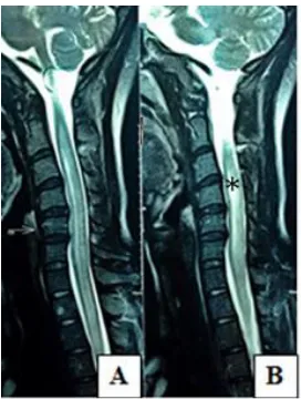
MRI T2WI revealed syringomyelia at cervical level with etiology is unclear (Fig1). She
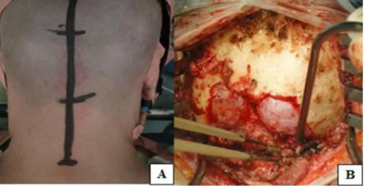
underwent craniocervical decompression, which consisted of the following procedures: 1) wide
foramen magnum decompression 3 cm x 3 cm via suboccipital craniectomy, 2) peeling of the
of the dura mater, 3) opening of the arachnoid membrane and dissection of arachnoid adhesions
over and around the foramen of Magendie and confirmation of sufficient CSF flow and brain
pulsation, 4) duraplasty in which we used a lyophilized dura allograft. (Fig 2).6 This patient did
well and showed improvement of upper motoric (5/5) and lower motoric (4/5), urinary urgency
261
Fig 1. A,B Idiopathic Syringomyelia on T2WI MRI (asterisk)
Discussion
The prevalence of syringomyelia is 8.4 cases per 100,000 population. Approximately
21,000 Americans have syringomyelia, a disorder in which a cyst forms within the spinal cord,
resulting in myelopathy. Various theories include the cerebellar piston theory, intramedullary
pulse pressure theory, and increased spinal subarachnoid pressure. Idiopathic syringomyelia and
Chiari type-I syringomyelia are associated with a small PF with narrow CSF spaces. The
etiology of CSF flow disturbances and syringomyelia in idiopathic and Chiari I-type
syringomyelia appears to be Posterior Fossa (PF) underdevelopment and neuraldisplacement.
A PF with decreased compliance and narrow CSF spaces promotes the development of
accentuated pulsatile CSF subarachnoid pressure waves that promote the development of
syringomyelia. Continued progression of symptoms, however, could be approached using
decompressive strategies such as C1 laminectomy, lysis of adhesions, and craniocervical
decompression, depending on the level of pathology.1,4,10,11
The role of CSF flow in the development of IS has not been clarified. Currently, CSF
flow obstruction is presumed to cause the accumulation of fluid in the spinal cord. Most
research revealed that the syrinx is subject to continuous pulsatile pressure from outside the
spinal cord and to sudden rises in pressure associated with Valsalva maneuvers. Cord cavitation
causes slow but relentlessly progressive symptoms and signs, depending on its location, extent,
and frequent asymmetry.7,9 The early symptoms usually consist of any combination of motor
and sensory dysfunction. Disassociated sensory findings of impaired pain and temperature with
262
presentation. Sensory changes usually affect the hands, but a careful examination may disclose a
similar deficit affecting the neck, shoulders, upper chest, and back.7
This is particularly important because of the good prognosis and excellent chance of
recovery if patients are properly managed. The reduced symptoms in patients with IS after
cranio-occipital decompression support this presumption.2
Fig 2. A) Linear Skin Incision B) Foramen Magnum Decompression and C1 Laminectomy
The strategy for treatment patients with IS varies according to the extent of disease
progression. Some patients show no signs or symptoms during disease progression for many
years and/or the syrinx dimension of some patients may resolve without surgical intervention or
remain stable.8,9 This patient did well and showed improvement of upper motoric (5/5) and
lower motoric (4/5), urinary urgency and no hipesthesia and hyperreflexia 10 months following
surgery, slight weakness in right lower extremity resulting in possibility of progressive atrophy
the lower extremities musculature.
Conclussion
Idiopathic syringomyelia is a pathological entity in which no overt etiology is evident
for a syrinx. It can be managed succesfully by conservative treatment but if there was
263
Reference
1. Roy AK, Slimack NP, Ganju A. Idiopathic syringomyelia: retrospective case series, comprehensive review, and update on management. Neurosurg Focus. 2011;31(6):E15,
2. Struck AF, Haughton VM. Idiopathic syringomyelia: phase- contrast mri of cerebrospinal fluid flow dynamics at level of foramen magnum1.Radiology. 2009;253(1).
3. Shaffer N,Martin BA, Rocque B,Madura C, Wieben O, Iskandar BJ, Dombrowski S et al. Cerebrospinal fluid flow impedance is elevated in type I chiari malformation. Journ Of Biomech Eng. February 2014. 136.
4. Heiss JD, Snyder K, Peterson MM, Patronas NJ, Butman JA, Smith RK, et al. Pathophysiology of primary spinal syringomyelia. J Neurosurg.2012;17:367-380
5. Magge SN, Smyth MD, Governale LS, Goumn erova L, Mads J, Munro B. Idiopathic syrinx in the pediatric population: a combined center experience. J Neurosurg Pediatrics.2011;7:000–000.
6. Kumar A, Bhattacharjee S and Sahu BP. Importance of C1 laminectomy in foramen magnum decompression surgery: A technical note. Asian J Neurosurg. 2014;9(4): 235.
7. Chen JK, Chen CH, Lee CL, Chen TW, Weng MC, Huang MH. Acute idiopathic syringomyelia: a case report, Kaohsiung J Med Sci. 2000; 20:404–9.
8. Ataizi S, Çanakçi Z, Baloglu M, Çerezc A. Spontaneously resorbed idiopathic syringomyelia: a case report. Turkish Neurosurgery. 2007;17(4): 247-50.
9. Lin JW, Lin MS, Lin CM, Tseng CH, Tsai SH, Kan IH, Chiu WT. Idiopathic syringomyelia: case report and review of the literature. Acta Neurochir Suppl.2006; 99: 117–120.
10. El Shazly AA, Mashaly HA. Surgical treatment of idiopathic syringomyelia on the basis of intramedullary pulse pressure theory: a report of nine cases with clinical and radiological outcomes. Egyptian Journal of Neurosurgery. 2013;28(4).