Series: Mechanical Engineering Vol. 1, N 10, 2003, pp. 1 - 2
SURFACE TREATED AND FIBRIN COATED ELECTROSPUN
POLYACRYLONITRILE FIBER FOR ENDOTHELIAL CELL
GROWTH AND PROLIFERATION
Nur Syazana Rashidi1, Irza Sukmana2,*, Agung Mataram3, Noor Jasmawati1, Mohd Ramdan Mohd Rofi1, Mohammed Rafiq Abdul Kadir1
1
Medical Devices and Technology Group (MEDITEG), Faculty of Bioscience and Medical Engineering, Universiti Teknologi Malaysia, 81310 Johor Bahru, Malaysia
2
Department of Mechanical Engineering, Faculty of Engineering, University of Lampung, Bandar Lampung 35143, Indonesia
3
Department of Mechanical Engineering, Faculty of Engineering, University of Sriwijaya, Ogan Ilir, South Sumatera, Indonesia
Abstract. Polyacrylonitrile (PAN) is a synthetic biocompatible polymer that has been utilized as filtering membrane in hemodialysis and enzyme immobilization purposes. However, its potential usage in other medical applications is limited due to its poor hydrophilicity. To overcome this problem, two groups of electrospun PAN
fibers were treated with sodium carbonate (Na2CO3) and sodium hydroxide (NaOH)
respectively at 100oC for 5 minutes. Fibrin gel was then coated to the treated samples
before seeding with human umbilical vein endothelial cells (HUVECs) for 1 and 3 days. X-ray diffraction results showed increased crystallinity of PAN fibers when
treated with Na2CO3 and NaOH. Contact angle measurements showed that the
hydrophilicity of Na2CO3 treated and NaOH treated samples improved from 115° to
88° and 64° respectively. Fourier transform infrared spectroscopy confirmed their
hydrophilicity was due to the existence of carboxyl and hydroxyl groups. However, tensile strength of PAN fibers reduced by 34% when treated with Na2CO3 and 42% when treated with NaOH. Cytotoxicity tests showed increased absorbance in day 3 for both treated samples. The absorbance value for NaOH treated PAN fibers maintained
until day 7 while Na2CO3 treated PAN fibers showed a slight increase in absorbance
on day 7. In vitro tests showed increased cell adhesion and proliferation after 3 days of culture. PAN treated fibers coated with fibrin are therefore proven to attract HUVEC cells and promote endothelialization.
Key words: Polyacrylonitrile fibre, surface treatment, scaffold, endothelial cells ________________________________
Received:
Corresponding author: Irza Sukmana
Affiliation: Department of Mechanical Engineering, Faculty of Engineering, University of Lampung, Jl. Prof. Soemantri Brojonegoro No. 1, Bandar Lampung 35143, Indonesia
1. INTRODUCTION
Due to its high biomechanical strength, thermal stability, tolerance to solvents as well as being resistant to bacteria and photo irradiation effects, Polyacrylonitrile (PAN) has been used extensively to produce various manufactured materials [1]. These include products such as commercial carbon fibers, filtration [2], adsorption [3], as well as weaved products such as blankets, carpets and clothes [4]. PAN is also proven as a biocompatible material and it is suitable for biomedical purposes such as tissue engineered bioartificial skin [5], hemodialysis [6], and biocatalysts immobilization [7].
However, due to its poor hydrophilicity, the use of PAN without surface treatment appears limited. Surface treatments to overcome this issue have been previously described. These include methods such as plama treatment [8-10], plasma initiated graft polymerization [11,12], and photoinduced grafting [13]. These methods nevertheless have inherent limitations. Compounded by the high cost involved in the treatment process, many of these methods have been deemed impractical for large scale manufacturing purposes [14]. It has been suggested that the use of Na2CO3 and NaOH solutions in
Millipore-purified water can be used to provide similar effects without involving exorbitant cost [15,16].
The promotion of endothelial adhesion and vascularization to prevent thrombosis as well as intimal hyperlasia of small diameter vascular graft has been tested to the PAN fibres. Previous studies showed that without the fibrin coating process, cell differentiation onto the material surfaces would be non-existence thus rendering the material less useful for vascularization of an engineered tissue substitute [17]. The role of fibrin actively directs cellular responses through specific receptor-mediated interactions with endothelial cells of the vessel wall [18]. Therefore, the use of fibrin as a coating active matrix onto a bioactive scaffold to support endothelialization is of interest. However there is no study yet showing electrospun PAN fibers with Na2CO3 and NaOH surface treatment followed
by fibrin coating can form endothelial cells monolayer for blood vessel.
Surface treatment using NaOH nevertheless is already established for PAN materials by authors mostly in ultrafitration and haemodylisis applications [19,20]. However, the effects of sodium carbonate treatment on the PAN mechanical and physical properties have not been intensively studied compared with NaOH surface treatment. Research on Na2CO3 surface treatment onto PAN only covered hydrolysis part while biocompatibility
was not addressed before [15,20]. The present study aims to analyze hydrophilicity of treated PAN fibers with Na2CO3 and NaOH. PAN treated surfaces then were coated with
2. MATERIALS AND METHODS
2.1 Materials
Polyacrylonitrile (PAN; Mw=150,000; 53.06 g/mol), N,N-dimethylformamide (DMF; 73.10 g/mol) and acrylamide (AM; 71.08g/mol) were bought from Aldrich Chemical and were used without further purification. Other materials are Sodium hydroxide (NaOH; Sigma Aldrich, USA), Sodium bicarbonate (Na2CO3; Sigma Aldrich; USA), Fibrinogen
and Thrombin (Sigma Aldrich; USA).
2.2 PAN fiber preparation
The random PAN fibers were developed through electrospinning under optimum conditions. Briefly, 14% (w/v) of PAN was dissolved in a DMF solution. This solution was loaded into a 5 mL glass syringe and the flow rate of 2 mL.h−1 was controlled by syringe pump. A high voltage (21 kV) was applied using a high voltage regulated DC power supply (Model ES 30P-5W, Gamma High Voltage Research, Ormond Beach, FL). A piece of aluminum foil was placed towards the tip at a distance of 12 cm as grounded collector. Afterwards, the electrospun fibers were peeled off from the aluminum foil and the residual solvent was released in an oven at 40oC. The dried electrospun fibers were then stored in desiccator prior to further processes.
Electrospun PAN fibers mat was cut by 2 cm x 2 cm. 1% w/v aqueous solution of Na2CO3 was prepared. PAN fiber was dropped into boiling Na2CO3 at temperature 100oC
for 5 minutes and next was rinsed with plenty of water until pH~7. The substrate then was dried at ambient temperature (T~25oC) for 24 hours. This step was repeated for NaOH surface treatment. PAN fiber was sterilized by immersing the fiber in 10% penicillin-streptomycin under ultraviolet light (UV) for 15 minutes. Next, the substrate was dried under UV for 1 hour.
In 24 well plates, fibrin gels were prepared using 500 μl/well of fibrinogen solution (2.0 mg.mL-1) made in phosphate buffered saline (PBS). This solution was directly mixed with 1 mg / 500 μl (100 U.mL-1
in PBS) for polymerization process of fibrinogen into fibrin. PAN fiber after treated with Na2CO3 and NaOH was transferred and deposited in
the prepared fibrin gel. Fibrin-coated PAN fiber was then stored in incubator at 4oC overnight. The substrate then was dried on filter paper under room temperature in sterile condition.
Human umbilical vein endothelial cells (HUVECs) were cultured in endothelial cell growth (Promo Cell) supplemented by 1% antibiotic penicillin-streptomycin (Gibco, USA). The cells were replaced twice a week and cultures were maintained at 37oC in incubator containing 5% CO2. HUVEC between passages 3 and 5 were used in all
experiments. After coating process, sterile fibrin-coated PAN fibers treated with Na2CO3
and NaOH were seeded with HUVECs at a density of 3x10⁴ cells/cm2 and observed for day 1 and day 3.
2.3 Surface characterization
microscope (FESEM; ZEISSSupra 35VP), operated at an acceleration voltage of 10 kV. The fibers diameters were measured from the SEM (JEOL, JSM-6390LV) micrographs measured randomly within an average of 50 fibers. Composition of PAN fiber before and after surface treatment and coating was determined from Energy dispersive X-ray spectroscopy (EDS).
Fourier transformed infrared (FTIR) spectra of the fibers were collected on Perkin– Elmer Spectrum 2000 to analyze the chemical structure of electrospun PAN fiber over a range of 400-4000 cm-1 with typically 32 scans at a resolution of 4 cm-1. Also, the crystallinity of sample was characterized by x-ray diffraction (XRD) (D5000, Siemens, Germany) in a thin film mode at 40 kV and 40 mA. The data were recorded in 2θ range using CuKα radiation of 1.5406 Å.
Water contact angle of PAN fibers before and after surface treatment were measured using VCA Optima (AST Product, Inc.) mounted with a CCD camera. The fiber was placed on the sample stage and a drop of ultrapure water (Milli-Q, 2 μL) was dropped to the surface for contact angle measurement. Five points per fiber were measured to determine the mean value of the water contact angle (n = 3).
2.4 Mechanical properties
Mechanical properties of the electrospun fibers were verified using a tensile machine (Instron 8845) with a load cell capacity of 10N. The appropriate specimen gripping is required to prevent the fibers from breaking or slipping at the grips. According to ASTM D882, samples were mounted and had a 25 mm gage length. Samples were prepared for testing at a crosshead speed of 5 mm/min at ambient conditions. The initial modulus, ultimate strength and elongation at ultimate strength were measured.
2.5 Endothelial cell responses
3. RESULTS
3.1 Morphology of untreated and treated PAN fiber
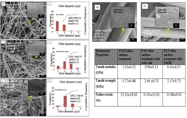
FESEM images of electrospun PAN fibers are to investigate the effect of surface treatment towards fiber morphology structure presented in Figure 1.
Fig. 1 FESEM micrograph and distribution of untreated PAN fiber (a, b); fiber treated with soda ash (c, d) as well as with NaOH (e, f). Soda ash (f) and NaOH (g) treated fiber then coated with fibrin. Table shows mechanical properties of the electrospun PAN fibers
before and after surface treatment.
Fig. 1(a) shows the micrographs of untreated PAN fibers obtained by electrospinning have bead-free and homogenous morphology. The PAN fiber surfaces also seem featureless and smooth as shown in greater detail in the figure. The morphology of PAN after surface treatment with Na2CO3 is shown in Fig. 1(c) has an uneven surface after
treated. As indicated by the arrows, PAN fiber after NaOH as shown in Fig. 1(e) surface treatment reveals ripple and is rougher compared with Na2CO3 surface treatment. Surface
treatment processes were proved to result PAN fibers with different diameter range before surface treatment (AFD (average fiber diameter): 2.06 ±0.54; MaxFD (maximum fiber diameter): 4.15 and MinFD (minimum fiber diameter):1.66). Next, PAN fiber after surface treatment with Na2CO3 become (AFD (average fiber diameter): 2.44 ± 0.79;
increases 15% as a result of surface treatment with Na2CO3 and decreases 18% after
treated with NaOH.
Fig. 1(g-h) showed that PAN fiber after treated by Na2CO3 and NaOH was fully
coated with fibrin gel. EDS results reported the existence of fibrin with significant changes of nitrogen and oxygen element of PAN fiber after coating process. Increasing in carbon composition confirms the existence of fibrin. The coating is fully covering the cellular fibers due to the structure of fibrinogen transformed into fibrin gel, deposited on the surface and formed a mesh of fibrils obviously in-between of the junction fibers.
3.2 Mechanical properties of untreated and treated PAN fiber
Fig. 1(i) summarizes the tensile properties of electrospun PAN fibers before and after reaction with Na2CO3 and NaOH. It is noted that there are significant differences between
PAN fibers before and after surface treatment with Na2CO3 and NaOH for tensile strength
and tensile modulus. After surface treatment, overall there were decreases in tensile strength, tensile modulus and strain to failure due to loss of chain orientation. The decrease of tensile strength after Na2CO3 and NaOH surface treatment of PAN fiber is as
much as 34% and 42%, respectively. Tensile modulus of substrate decreases from 1.35 GPa to 0.9 GPa and 0.41 GPa while strain decrease 9% and 4% after treated with Na2CO3
and NaOH, respectively.
3.3 Surface characterization of untreated and treated PAN fiber
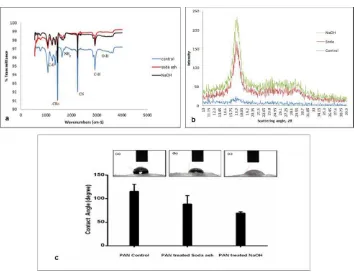
Surface characterization of treated and untreated PAN fiber is presented in Fig. 2. FTIR spectra of the PAN fibers, before and after treated are included in Fig. 2(a). The control PAN molecule consists of functional groups such as methyl (CH3) and nitrile
(C≡N). It is found that surface treatment in the presence of Na2CO3 and NaOH includes
the stage of carboxyl, amide and aldehyde.
PAN fiber shows a characteristic peak at 2260-2210 cm-1 before and after treating with NaOH and Na2CO3 indicating the likely and expected presence of acrylonitrile due
to nitrile stretch. The alkali reaction of Na2CO3 and NaOH will attack chain of nitrile to
Fig. 2 (a) FTIR, (b) XRD, and (c) Contact angle results of PAN fiber, PAN treated with soda ash and PAN treated with NaOH.
Briefly, the peak at 1645 cm-1 is assigned to carbonyl stretching, but the peak broadened after treated into 1652 cm-1 for NaOH and 1649 cm-1 for Na2CO3 surface
treatment. In conjunction with the peaks at 1320 cm-1 and 1000 cm-1 range (due in part to C-O stretch), 1760-1665 cm-1 (due to C-O-C bend), and broad absorption around 3300 cm-1 and 2500 cm-1 (due to -OH stretch) the carbonyl peak seems to confirm the presence of acrylate (H C=C(CH)CO R) as a co-monomer for NaOH surface treatment. This carboxylic acid formation will lead to the formation of secondary amines in which hydroxyl group has been replaced by amine. However, in the presence of Na2CO3 surface
treatment, it does not include the stage of amide formation for second degree and does not result in the complete exhaustion of nitrile groups in a PAN considering that Na2CO3 is
not really strong reducing agent like NaOH and will not fully attack and hence will not harm the substrate.
system [21]. The weak peak of PAN control showed from the diffraction pattern with the value of 2θ at 17.0o
. This proves that fibers fabricated using electrospinning giveslimited crystallinity. This low crystallinity leads to stretched PAN chains solidified rapidly after elongation, preventing crystal formation in the electrospun PAN fibers.
In contrast, PAN with Na2CO3 and NaOH surface treatment increase in crystallinity
even after short timing treatment showed two diffraction peaks indexed with values of 2θ of 17.0o and 29.5o. The crystallinities of the NaOH-treated fiber are higher than PAN control and PAN after treated with Na2CO3. The main factor depends on the alkali
concentrations selected for the treatment and the type of fiber studied [22]. Crystallinity factor will lead to the structural changes of PAN fibers after surface treatments, thus will influence the course of their entire degradation process.
The contact angle of PAN-modified surfaces treated via Na2CO3 and NaOH as well as
the control is shown in Figure 2 (c). It indicated that PAN-treated by Na2CO3 and NaOH
were more hydrophilic (significant lower contact angle) than found in untreated PAN. The contact angle of the PAN surface of the PAN was reduced from 115° to 88° and 64° after treated with Na2CO3 and NaOH, respectively. These results clearly indicate that untreated
PAN fibers had the least attraction toward deionized water. However, this was improved upon by surface treatment. The water contact angle of the PAN surface gradually decreased after treating with Na2CO3 and NaOH. It is noteworthy that receding contact
angle decreases for all PAN fibers after surface treatment.
3.4 Endothelial cell adhesion and proliferation
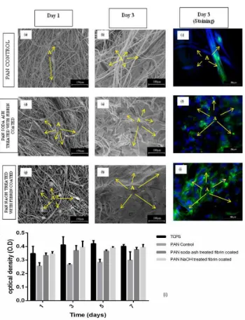
Once HUVECs were seeded onto both untreated and surface treated and coated PAN fibrous scaffolds in presence of fibrin, cell attachment and proliferation were evaluated at day 1 and day 3. Endothelial cell adhesion and proliferation to PAN fiber is presented in Figure 3. After day 1, HUVECs cultured on PAN fibers were rounded in phenotype for all fibers (Fig. 3 (a,d,g)). This is due to the short timing of culture. HUVECs seemed to be securely attached and spread on the surface in a flatten formation, regardless of the surface treatments and surface coating after day 3. It was evident that cells were more actively proliferating across the modified surfaces (Fig. 3 (e) and (h)) while cells remain separately on PAN control even for day 3. It suggests that HUVECs adhered to PAN treated with Na2CO3 and NaOH then coated with fibrin surface were higher than that
found on the PAN control at the time points of 1 and 3 days. This was proven by staining after day 3 culture (Fig. 3(f,i)) which confirmed that not only confluent HUVECs layer was formed, but also cells oriented completely along the aligned fiber direction and exhibited an elongated morphology for both surface treated fibers coated with fibrin.
Absorbance of PAN treated with Na2CO3 and NaOH coated with fibrin increase by
increasing time as shown in Fig. 3(i). However, PAN control increases absorbance slowly because of reduced toxic of fluorine coming from DMF which is PAN solution during fabrication of fibers. The MTT absorbance of PAN after treated with Na2CO3-fibrin
Fig. 3 SEM and images of HUVECs cultured on a untreated fiber as control (a,b,c); soda ash treated and fibrin coated (d,e,f); and NaOH treated and fibrin coated (g,h,i) after 1 and 3 days of study. Letters “A” indicate HUVEC cells. MTT test on cytotoxicity of PAN
4. DISCUSSIONS
PAN fiber has been used for membrane applications [7]. In this study, PAN fiber was prepared to promote endothelial cell adhesion and proliferation. Surface treatment and bioactive coating of polymer fiber may lead endothelial cell proliferation thus encourages endothelial network or endothelialization, as presented elsewhere [23]. We have studied the effect of bioactive coating on PAN fiber to HUVEC responses, complimented previous study of Groth et al. that used PAN fiber for fibroblast cell for wound healing application [5].
It has been previously shown that the use of Na2CO3 and NaOH removes the natural
surface wax thereby exposing the underlying texture of substrates [24]. This improves the wetting and wicking properties of the material which in turn increases the material surfaces for better surfacing coating [25]. In this experiment, it is observed that PAN fiber showed increased hydrophilicity after treating with Na2CO3 and NaOH thus improve
coating of fibrin gel on PAN fibers. Bioactive protein; in this case was fibrin, has become positive surface charged at neutral pH and this will attract negative charge on the treated PAN sample [19]. Fibrin coating is able to attract HUVECs on PAN fiber since fibrin has directs cellular responses through specific receptor [26].
Many studies have already discussed about NaOH surface treatment on various substrates [14,24]. Afra et al. showed porous surface and rough morphology on PET substrate after surface treatment of 2 hours with NaOH [13]. A higher concentration of NaOH and long period of surface treatment will cause degradation of surface fiber [19]or grooved serrated surfaces. It has been reported that substrate with very high surface roughness decreases cell adhesion onto its surface [27]. Therefore, in this study the lower concentration for both treatments were used. There are no significant differences in morphology on the PAN surface after treating with both NaOH and Na2CO3 since the
concentration for both treatments as low as 0.1 and 0.2 molar of Na2CO3and NaOH
respectively.
Even though the morphology of PAN fiber after surface treatment gives not much difference but the diameter of fibers is affected. This is due to the transformation of nitrile to carboxylic group of the PAN surface may result in a decrease of fiber diameter. However incomplete exhaustion of nitrile group for Na2CO3 surface treatment gives an
advantage to Na2CO3 compared to NaOH surface treatment in terms of mechanical
properties since fiber diameter is strongly related with strength of the fiber [28]. Tensile strength of PAN fiber after treated with Na2CO3 is higher than NaOH surface treatment
but still lower than PAN before surface treatment. This is because exposure the fiber to the heated surface treatment makes the fiber gives an emission and auto exhaustion to the environment [29]. In theory, tensile modulus decreases because of the random PAN fiber would become gradually aligned during uniaxial tensile test. This also leads to decreasing PAN fiber diameter after surface treatment, causing decrease in tensile strength and the fiber will easily fracture.
Alteration in physical characteristics of fibers will influence tensile properties due to increasing shrinkage and density of fibers [14]. The decreasing result for tensile modulus of PAN after treated with Na2CO3 and NaOH shows that PAN surface is relatively ductile
presence of a strong alkali. Result of mechanical properties depends on the material used. Instead of synthetic fiber, previous study used natural fiber for base material [29]. Alteration in ductility of specific material after removal of impurities modified mechanical result [28]. However, tensile strength in this experiment before and after surface treatment is still in the range of coronary artery which is from 1.40 MPa to 11.14 MPa [23].
Cytotoxicity test on PAN fibers after surface treatment with Na2CO3 and NaOH shows
these fibers are suitable for growth of HUVECs, attaining a significant level of increase in cell viability until day 7 of culture. This shows that the fibers with fibrin assisted increase proliferation of HUVECs. The SEM micrographs of HUVECs on PAN control and PAN treated with Na2CO3- fibrin coated and NaOH scaffolds obtained on day 1 of culture
showed a normal morphology of cell growth on the fibers. HUVEC cells disconnects to individual cells, round phenotype and non-proliferating for all fibers. This is due to the shorten time of culture. This cell behavior is same with previous report [8], where fibers have low surface density and large inter fiber which did not encourage adhesion of the cells across the nearest fibers in short time seeding. HUVECs attach and spread more on PAN substrate after surface treatments than PAN control by day 3. In particular, the HUVECs were numerous and well-spread, forming monolayer cell on the PAN treated with NaOH scaffolds while reaching sub confluence. Some cells aggregated along the fibers on PAN treated with Na2CO3 as confirmed by double-staining with Alexa Fluor
488 for the cell cytoskeleton (i.e., actin filaments) and with Hoechst 33258 for nuclei. These observations suggested significance increase of HUVECs adhesion due to better hydrophilicity after PAN fiber surface treatment process subsequently coated with fibrin. It also shows that fibrin was immobilized on the PAN, which is important for strong cell adhesion [30]. Fibrin contains many integration-binding sites and this reason makes it easier for cell to adhere on coated PAN fiber [31]. Formation of focal adhesions only occurs if the ligands can withstand cell contractile forces as actin stresses fibers and focal adhesions are critical for cell survival. This indicates culturing cell on native substrates proves that cell attachment, spreading and growth enhanced, depending on the substrate, which has been reported elsewhere [32]. SEM and staining micrographic observations support the trend of HUVECs proliferation quantified by the MTT assays.
5. CONCLUSIONS
Acknowledgement:This work was supported by Malaysian Ministry of Higher Education (MOHE) grant (vote# 4F124) and Universiti Teknologi Malaysia Tier-1 Research grant (vote # 03H12). I. Sukmana is supported by Hibah Kompetensi Y2017grant from Kemenristekdikti, Indonesia.
REFERENCES
1. Houa, D., Huanga, X., Weia, A., 2011, Surface modification of electrospun PAN nanofibers and its application for adsorption of lead ions, Journal of Fiber Bioengineering & Informatics, 4, pp.383-8. 2. Panapoy, M., Dankeaw, A., Ksapabutr, B., 2008, Electrical conductivity of PAN-based carbon
nanofibers prepared by electrospinning method, Thammasat Int J Sc Tech, 13, pp. 11-7.
3. Yu, X., Xiang, H., Long, Y., Zhao, N., Zhang, X., Xu, J., 2010, Preparation of porous polyacrylonitrile fibers by electrospinning a ternary system of PAN/DMF/H 2 O, Materials Letters, 64, pp. 2407-9.
4. Farsani, R.E., Raissi, S., Shokuhfar, A., Sedghi, A., 2009, FT-IR study of stabilized PAN fibers for fabrication of carbon fibers, World Academy of Science, Engineering and Technology, 50, pp. 430-3.
5. Groth, T., Seifert, B., Malsch, G., Albrecht, W., Paul, D., Kostadinova, A., Krasteva, N., and Altankov, G., 2002, Interaction of human skin fibroblasts with moderate wettable polyacrylonitrile– copolymer membranes, Journal of biomedical materials research, 61, pp. 290-300.
6. Wang, Z.-G., Wan, L.-S., Xu, Z.-K., 2007, Surface engineerings of polyacrylonitrile-based asymmetric membranes towards biomedical applications: An overview, Journal of Membrane Science, 304, pp. 8-23.
7. Kang, Y., Ahn, K., Jeong, S., Bae, J., Jin, J., Kim, H., H.G., Hong, S.W., and Cho, C.R., 2011, Effect of plasma treatment on surface chemical-bonding states and electrical properties of polyacrylonitrile nanofibers, Thin Solid Films, 519, pp. 7090-4.
8. Shi, Q., Vitchuli, N., Nowak, J., Caldwell, JM., Breidt, F., Bourham, M., M., Zhang, X., and McCord, M., 2011, Durable antibacterial Ag/polyacrylonitrile (Ag/PAN) hybrid nanofibers prepared by atmospheric plasma treatment and electrospinning, European Polymer Journal, 47, pp. 1402-9.
9. Fleming, R., Pardini, L., Brito, Jr, C., Oliveira, Jr, M., Alves, N., Massi, M., 2011, Plasma treatment of polyacrylonitrile/vinyl acetate films obtained by the extrusion process, Polymer Bulletin, 66, pp. 277-88.
10. Zhao, Z.-P., Li, J., Wang, D., Chen, C.-X., 2005, Nanofiltration membrane prepared from polyacrylonitrile ultrafiltration membrane by low-temperature plasma: 4. grafting of N-vinylpyrrolidone in aqueous solution, Desalination, 184, pp. 37-44.
11. Zhao, Z.-P., Li, J., Zhang, D.-X., Chen, C.-X., 2004, Nanofiltration membrane prepared from polyacrylonitrile ultrafiltration membrane by low-temperature plasma: I. Graft of acrylic acid in gas, Journal of Membrane Science, 232, pp. 1-8.
12. Ulbricht, M., Oechel, A., Lehmann, C., Tomaschewski, G., Hicke, H.G., 1995, Gas‐phase photoinduced graft polymerization of acrylic acid onto polyacrylonitrile ultrafiltration membranes, Journal of applied polymer science, 55, pp. 1707-23.
13. Hadjizadeh, A., Ajji, A., Bureau, M.N., 2010, Preparation and characterization of NaOH treated micro-fibrous polyethylene terephthalate nonwovens for biomedical application, Journal of the mechanical behavior of biomedical materials, 3, pp. 574-83.
14. Liu, W., Cai, M., He, Y., Wang, S., Zheng, J., Xu, X., 2015, Development of antibacterial polyacrylonitrile membrane modified with a covalently immobilized lysozyme, RSC Advances, 5, pp. 84432-8.
15. Dyatlov, V.A., Grebeneva, T.A., Rustamov, I.R., Koledenkov, A.A., Kolotilova, N.V., Kireev, V.V., and Prudskov, B.M., 2012, Hydrolysis of polyacrylonitrile in aqueous solution of sodium carbonate, Polymer Science Series B, 54, pp. 161-6.
17. Herrick, S., Blanc-Brude O., Gray A., Laurent, G., 1993, Fibrinogen, the International Journal of Biochemistry & Cell Biology, 31, pp. 741-6.
18. Wang, J., Yue, Z., Ince, J.S., Economy, J., 2006, Preparation of nanofiltration membranes from polyacrylonitrile ultrafiltration membranes, Journal of Membrane Science, 286, pp. 333-41. 19. Chiu, H., Lin, J., Cheng, T., Chou, S., 2011, Fabrication of electrospun polyacrylonitrile
ion-exchange membranes for application in lysozyme, Express Polymer Letters, 5, pp. 308-17.
20. Hou, X., Yang, X., Zhang, L., Waclawik, E., Wu, S., 2010, Stretching-induced crystallinity and orientation to improve the mechanical properties of electrospun PAN nanocomposites, Materials & Design, 31, pp. 1726-30.
21. Hou, C., Qun, W., Qu, R., Wang, C., 2006, Interaction between polyacrylonitrile and alkalis, Journal of applied polymer science, 102, pp. 272-5.
22. He, W., Ma, Z., Yong, T., Teo, W.E., Ramakrishna, S., 2005, Fabrication of collagen-coated biodegradable polymer nanofiber mesh and its potential for endothelial cells growth, Biomaterials, 26, pp. 7606-15.
23. Nilghaz, A., Wicaksono, D.H., Gustiono, D., Majid, F.A.A., Supriyanto, E., Kadir, M.R.A., 2012, Flexible microfluidic cloth-based analytical devices using a low-cost wax patterning technique, Lab on a Chip, 12, pp. 209-18.
24. Basu, S., Yang, S-T., 2005, Astrocyte growth and glial cell line-derived neurotrophic factor secretion in three-dimensional polyethylene terephthalate fibrous matrices, Tissue engineering, 11, pp. 940-52.
25. Xu, C., Yang, F., Wang, S., Ramakrishna, S., 2004, In vitro study of human vascular endothelial cell function on materials with various surface roughness, Journal of biomedical materials research Part A, 71, pp. 154-61.
26. Liu, C-K., Sun, R-J., Lai, K., Sun, C-Q., Wang, Y-W., 2008, Preparation of short submicron-fiber yarn by an annular collector through electrospinning, Materials Letters, 62, pp. 4467-9.
27. Van, de, Weyenberg, I., Chi Truong, T., Vangrimde, B., Verpoest, I., 2006, Improving the properties of UD flax fibre reinforced composites by applying an alkaline fibre treatment, Composites Part A: Applied Science and Manufacturing, 37, pp. 1368-76.
28. Keun, Kwon, I., Kidoaki, S., Matsuda, T., 2005, Electrospun nano- to microfiber fabrics made of biodegradable copolyesters: structural characteristics, mechanical properties and cell adhesion potential, Biomaterials, 26, pp.3929-39.
29. Adamczak, M., Scislowska-Czarnecka, A., Genet, M.J., Dupont-Gillain, C.C, Pamula, E., 2011, Surface characterization, collagen adsorption and cell behaviour on poly (L-lactide-co-glycolide), Acta of Bioengineering & Biomechanic, 13.
30. Mwaikambo, L.Y., Ansell, M.P., 2002, Chemical modification of hemp, sisal, jute, and kapok fibers by alkalization, Journal of applied polymer science, 84, pp. 2222-34.
31. Hadjizadeh, A., Doillon, C.J., Vermette, P., 2007, Bioactive polymer fibers to direct endothelial cell growth in a three-dimensional environment, Biomacromolecules, 8, pp. 864-73.