1 Muh. Ardi Munir, Cubital Tunnel Syndrom... CUBITAL TUNNEL SYNDROME
Oleh:
Muh. Ardi Munir
Department of Orthopedic and Traumatology - Faculty of Medicine Tadulako University Palu
Introduction
Prior to 1957, ulnar neuropathy was felt to be a "stretch neuritis" caused by cubitus valgus. In fact, the predominant cause of ulnar neuropathy was secondary to elbow injuries. Osborne proposed the concept of compression of the ulnar nerve in 1957, with Feindel and Stratford defining the "cubital tunnel" the following year. Since the advent of more successful orthopedic management of complex elbow injuries, the more frequent cause of ulnar nerve entrapment has become idiopathic or related to a "susceptible" patient. Nonetheless, it appears that ulnar nerve entrapment is increasing in prevalence (although no data are available), with pain often being a predominant morbidity. In 1898, Curtis performed the first published case of management for ulnar nerve neuropathy at the elbow, which consisted of a subcutaneous anterior transposition. (1,2)
It is hoped that the reader may develop an increased awareness of cubital tunnel syndrome and its place in the differential diagnosis of the painful upper extremity. An overview of the diagnostic and treatment options are presented, along with complications that may occur. (1,2)
Kata kunci :
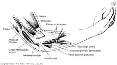
Surgical Anatomy of The Ulnar Nerve
The ulnar nerve is formed from
the medial cord of the brachial plexus,
which originates from the C8 and T1
cervical roots along the lateral wall of the
axilla. As it passes through the proximal
arm, it maintains a posteromedial
relationship to the brachial artery,
initially lying between the
coracobrachialis and triceps muscles. In
the middle one third of the arm, the ulnar
nerve accompanies the superior ulnar
collateral artery posteriorly through the
intermuscular septum to lie on the
anterior aspect of the medial head of the
2 Muh. Ardi Munir, Cubital Tunnel Syndrom... posterior surface of the intermuscular
septum medial to the humerus, to reach
the elbow. It traverses the elbow region
bounded medially and anteriorly
(superiorly) by the medial humeral
epicondyle, laterally by the olecranon and
by a connective tissue roof spanning the
two bony prominences-the "epicondylar
groove". The nerve then enters the
"cubital tunnel" by passing deep to the
arcuate ligament (Osborne's ligament),
which connects the ulnar and humeral
heads of the flexor carpi ulnaris (FCU)
muscle. The nerve then passes between
the two heads of the FCU and passes
deep to the deep flexor pronator
aponeurosis. It then travels through the
forearm between the FCU and flexor
digitorum profundus (FDP), giving off
motor branches to the FDP of the small
and ring fingers. The nerve enters the
wrist through Guyon's canal; a
fibro-osseous canal, extending 4 cm from the
palmar carpal ligament to the fibrous
edge of the hypothenar muscles. This is
also a common site of ulnar nerve
entrapment. (3)
The ulnar nerve is primarily a
motor and sensory nerve for the hand. As
such, it does not give off any branches in
the arm (except small sensory articular
branches to the elbow). It supplies the
FCU muscle and the ring and small FDP
muscles in the elbow/ proximal forearm
region. Proximal to the wrist, it gives off
the dorsal sensory branch to the ulnar
aspect of the hand. In the hand, it is the
primary innervation to the intrinsic
muscles (except for some of the thenar
muscles; superficial head of the flexor
pollicis brevis, opponens pollicis, and
abductor pollicis brevis), the index and
long finger lumbricals, and the sensation
to the ulnar 1.5 digits.
The ulnar is also well known to
have anomalous connections
(Martin-Gruber anastomosis) to the median nerve,
primarily occurring in the forearm, which
3 Muh. Ardi Munir, Cubital Tunnel Syndrom... nerves (primarily the intrinsic motor
supply). This is important when assessing
for distal weakness caused by proximal
median or ulnar nerve lesions. (3,4)
O'Driscoll (1991) believes that the
roof of the cubital tunnel, or Osborne
ligament, is a remnant of the anconeus
epitrochlears muscle. He also identified a
retinaculum at the proximal edge of the
arcuate ligament in all but 4 of 25
cadaveric specimens. He classified this
retinaculum as 1 of 4 types, as follows:
1. An absent retinaculum
2. A thin retinaculum that becomes tight
with full flexion without compressing
the nerve
3. A thick retinaculum that compresses
the nerve between 90° and full flexion
4. An accessory anconeus epitrochlears
muscle
Upon entering the cubital tunnel,
the ulnar nerve gives off an articular
branch to the elbow. It then passes
between the humeral and ulnar heads of
the FCU, the next potential site of
compression. The nerve then descends
into the forearm between the FCU and
the FDP muscles. (4,5)
About 5 cm distal to the medial
epicondyle, the ulnar nerve pierces the
flexor pronator aponeurosis, the fibrous
common origin of the flexor and pronator
muscles. The flexor-pronator aponeurosis
is another point of possible compression,
with compression of the ulnar nerve
beneath the muscle belly of the FCU.
The ligament of Spinner is an
additional aponeurosis between the flexor
digitorum superficialis (FDS) of the ring
finger and the humeral head of the FCU.
This septum is independent of the other
aponeuroses and attaches directly to the
medial epicondyle and medial surface of
the coronoid process of the ulna. This
structure was found in 4 of 20 specimens
in one study, and it is important to
recognize and to release with anterior
transposition of the ulnar nerve to prevent
kinking. (6)
In the forearm, the ulnar nerve
extends motor branches to the FCU and
the FDP of the ring and small fingers.
The ulnar nerve may extend as many as 4
branches to the FCU, ranging from 4 cm
above to 10 cm below the medial
epicondyle. Proximal dissection of the
4 Muh. Ardi Munir, Cubital Tunnel Syndrom... ulnar nerve may be performed up to 6.7
cm proximal to the medial epicondyle,
facilitating anterior transposition of the
nerve.
An aberrant muscle, the anconeus
epitrochlears, has been found in 3-28% of
cadaver elbows and in as many as 9% of
patients undergoing surgery for cubital
tunnel syndrome. This muscle arises from
the medial humeral condyle and inserts
on the olecranon, crossing superficially to
the ulnar nerve, where it may cause
compression.
The arcade of Struthers must be
differentiated from the ligament of
Struthers, which is found in 1% of the
population and extends from a
supracondylar bony or cartilaginous spur
to the medial epicondyle. This
supracondylar spur can be found on the
anteromedial aspect of the humerus, 5 cm
proximal to the medial epicondyle, and it
can often be seen on radiographs. The
ligament of Struthers may occasionally
cause neurovascular compression. This
compression generally involves the
median nerve or the brachial artery;
however, the ulnar nerve can also be
compressed by this structure.
Posterior branches of the medial
antebrachial cutaneous nerves cross the
ulnar nerve anywhere from 6 cm
proximal to 4 cm distal to the medial
epicondyle. These branches are often cut
when making the skin incision for a
cubital tunnel release, creating an area of
dysesthesia or resulting in potential
neuroma formation.
Extrinsic blood supply to the
ulnar nerve is segmental and involves 3
vessels. These include the superior ulnar
collateral artery, the inferior ulnar
collateral artery, and the posterior ulnar
recurrent artery. Typically, the inferior
ulnar collateral artery (and often the
posterior ulnar recurrent artery) is
sacrificed with anterior transposition. At
the level of the medial epicondyle, the
inferior ulnar collateral artery is the sole
blood supply to the ulnar nerve. In an
anatomic study, no identifiable
anastomosis was found between the
superior ulnar collateral artery and the
posterior ulnar recurrent arteries in 20 of
22 arms. Instead, communication
between the 2 arteries occurred through
proximal and distal extensions of the
5 Muh. Ardi Munir, Cubital Tunnel Syndrom... Intrinsically, the blood supply is
composed of an interconnecting network
of vessels that run along the fascicular
branches and along each fascicle of the
ulnar nerve itself. The surface
microcirculation of the ulnar nerve has
been shown to have an anastomotic
stepladder arrangement. The inferior
ulnar collateral artery is consistently
found 5 mm deep to the leading edge of
the medial intermuscular septum on the
surface of the triceps.
Finally, acute ulnar neuropathy
may have a sex predilection. This
perioperative condition is found 3-8 times
more frequently in men than in women.
Contreras et al (1998) revealed that the
medial aspect of the elbow has 2-19 times
more fat content in women than in men.
In men, the coronoid tubercle is
approximately 1.5 times larger. He
suggests that the coronoid process may
be a potential site for ulnar nerve
compression in men, and the increased
subcutaneous fat around the ulnar nerve
in women may provide a protective
advantage against acute ulnar neuropathy.
The most common potential sites
of compression of the ulnar nerve at the
elbow are the medial intermuscular
septum, the arcade of Struthers, the
retrocondylar groove, the cubital tunnel,
and the deep flexor-pronator aponeurosis.
The 2 most common sites of compression
are the retrocondylar groove and the true
cubital tunnel, where the ulnar nerve
passes between the 2 heads of the FCU.
Frequency
The elbow is the most common site of
compression of the ulnar nerve. Cubital
tunnel syndrome is the second most
common compressive neuropathy (after
carpal tunnel syndrome). Cubital tunnel
syndrome affects men 3-8 times as often
as women.
Etiology
Cubital tunnel syndrome may be caused
by constricting fascial bands, subluxation
of the ulnar nerve over the medial
epicondyle, cubitus valgus, bony spurs,
hypertrophied synovium, tumors, ganglia,
or direct compression. Occupational
activities may aggravate cubital tunnel
syndrome secondary to repetitive elbow
flexion and extension. Certain
occupations are associated with the
6 Muh. Ardi Munir, Cubital Tunnel Syndrom... however, a definite relationship with
occupational activities is not well
defined. (1,2)
Pathophysiology
Cubital tunnel syndrome is the
common term used for ulnar compressive
neuropathies at the elbow (from the mid
arm to the mid forearm). Ulnar
entrapment neuropathy develops because
of the predisposing anatomy of the elbow
region and the biomechanics of the ulnar
nerve at the elbow; it is based on
compressive, traction, and frictional
forces, with the possible association of a
nerve at risk. Systemic diseases such as
diabetes, chronic alcoholism, renal
failure, and malnutrition may predispose
the patient to compressive neuropathy
(i.e., a nerve at risk). Ultimately, the
cumulative effect on the nerve is to cause
a region of ischemia and inflammation
resulting in ulnar nerve dysfunction or
cubital tunnel syndrome. (3,4)
Compression of the ulnar nerve at
the elbow may be idiopathic, but often
there is a component of extrinsic
compression aiding in the entrapment.
Moreover, the compression of the nerve
may be dynamic or static. The dynamic
anatomy and biomechanics of the cubital
tunnel dramatically affect the uInar
nerve, resulting in relative regional
ischernia to the nerve. Dynamic
impingement tends to occur early in the
clinical course of the disease (i.e.,
position-dependent, intermittent).
Although initially reversible, fixed
structural changes occur over time,
leading to static compression. Static
compression also can develop from
structural abnormalities of osseous
architecture or space occupying lesions
(Table 1).
With flexion of the elbow, the
aponeurosis covering the cubital tunnel
stretches, changing the cross-sectional
geometry of the cubital tunnel from
smooth and round to flattened and
triangular. This both decreases the
volume of the tunnel by 55%, and
significantly increases the intraneural
pressure, therefore putting the nerve at
risk of ischernia. Intraneural pressure can
be increased up to 600% with shoulder
abduction, elbow flexion, and wrist
extension. Moreover, contraction of the
7 Muh. Ardi Munir, Cubital Tunnel Syndrom... on the ulnar nerve. Normally, the uInar
nerve at the cubital tunnel is known to
elongate 4.7 mm during elbow flexion.
Should the nerve be tethered by
perineural fibrosis (e.g., postoperative,
post trauma), it can no longer elongate
and may experience up to doubled
intraneural pressures. (7)
The ulnar nerve has five common
sites of compression in the elbow region
(Fig. 1). The most common sites are at
the level of the elbow. As the nerve
passes by the medial epicondyle (or
epicondylar groove) and into the cubital
tunnel (passing deep to the aponeurosis
linking the two heads of the FCU), it is at
its greatest risk of compression. These are
the primary sites of idiopathic disease.
The other three tend to be sites of
secondary or iatrogenic compression
(often because of inadequate release).
The most proximal site is the arcade of
Struthers, a fascial band 8 cm proximal to
the medial epicondyle, extending from
the medial head of the triceps to the
medial intermuscular septum. (Note: This
is different from the ligament of
Struthers, which extends from a
supracondylar process to the medial
humeral epicondyle and is involved
primarily with proximal median nerve
compression. (7,8)
Table 1. Causes Of Cubital Tunnel Syndrome
Idiopathic
Trauma
Acute compression or direct injury
Entrapment in distal humeral fracture or elbow
dislocation
Compression secondary to deformity--cubitus
valgus, cubitus varus, malunion, nonunion
Heterotopic ossification-post traumatic, secondary
to burns, secondary to head injury
Aberrant or abnormal musculature
anconeus epitrochlearis reverse flexor carpi
ulnaris, triceps brachii (snapping)
Cumulative trauma disorders-keyboard operator,
baseball pitcher
Arthritides
Osteoarthritis-secondary to osteophytes, loose
bodies, synovial cysts
Inflammatory arthritis-synovitis
Synovial chondromatosis
Vascular bands--often branches of uInar artery
Iatrogenic-postanesthetic palsy
Space-occupying lesions of the cubital tunnel and
epicondyle region--e.g., lipomata
Although Struthers described the
deep fascia of the arm, Spinner coined
the term arcade when he noted that it
appeared as a distinct entity in revision
8 Muh. Ardi Munir, Cubital Tunnel Syndrom... anterior transposition). This structure is
often indistinguishable from the deep
fascia at the primary surgery, however.
The strong, taut intermuscular septum,
normally not a structure to entrap the
nerve, runs anterior to the nerve, inserting
throughout the length of the medial
epicondyle. Following anterior
transposition, the intermuscular septum
acts secondarily as a point of local
entrapment if its distal 3 cm is not
excised. Finally, the last site is at the
deep flexorpronator aponeurosis.
Although uncommon as a primary cause,
it is often a secondary compression site
following transposition.
There is increasing evidence that
cumulative or repetitive trauma work
disorders are a cause of cubital tunnel
syndrome. Keyboard operators, for
instance, appear to be at increased risk. It
is postulated that poor seating is a
significant ergonomic factor; the
keyboard placed too high and too close to
the operator causes shoulder flexion,
elbow flexion, wrist extension, and,
therefore, traction on the ulnar nerve.
Figure 1. Five potential levels (boldface type) of
compression of the uInar nerve in the region of
the elbow. (From Amadic, PC: Anatomical basis
for a technique of uInar nerve transposition. Surg
Radiol Anat 8:158, 1986; by permission of the
Mayo Foundation.)
When considering the painful
upper extremity, reflex sympathetic
dystrophy (RSD) often comes to mind.
Although often reported as a
posttraumatic or postoperative condition,
it can occur without insult. In a series of
35 patients with RSD, 30 were noted to
have peripheral nerve entrapment (single
or multiple). Grundberg and Reagan also
noted that, in their study of 93 cases of
RSD, patients did not respond to standard
treatment. Further investigation of those
demonstrated carpal tunnel syndrome in
and cubital tunnel syndrome in 5.
Surgical decompression in those 27
patients led to complete resolution of
[image:8.612.359.548.94.200.2]9 Muh. Ardi Munir, Cubital Tunnel Syndrom... pertinent painful manifestation of
upper-limb peripheral nerve compression
syndromes such as cubital tunnel
syndrome. (3,6)
Clinical Presentation
Patients with cubital tunnel
syndrome tend to present with a pain,
often aching or lancinating, primarily in
the region of the elbow. It may radiate
proximally or distally. The most frequent
complaint, however, is of paresthesias in
the ring and small fingers, initially
intermittent and position-related, often
waking them at night. Objective sensory
loss usually occurs later, with progression
of the disease (Table 2). A clue to the
location of proximal uInar nerve
entrapment is the presence of dorsal
sensory loss. There is usually no sensory
loss along the medial forearm because it
is supplied by the medial antebrachial
cutaneous nerve, a branch of the medial
cord of the brachial plexus. Most
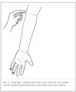
importantly, examining for Tinel's sign
helps localize the entrapment. (8)
Figure 2. Percussion test (Tinel sign): Tapping
over the ulnar nerve at the elbow causes a
reproduction of symptoms.
Patients often note they have "weak
or clumsy hands," often dropping objects
or being unable to open jars. Muscle
weakness or atrophy often occurs and, in
many patients, in the absence of any
objective sensory change. The fascicles
to the intrinsic ulnarly supplied hand
muscles are more susceptible because of
their superficial topographic location
within the ulnar nerve at the cubital
tunnel (FCU and FDP often are spared).
Muscle weakness may be demonstrated
by clawing of the hand, Wartenberg's
sign or Froment's sign although these
indications are not helpful in localizing
the lesion. 23,41 Because of the common
anomalous connections between the
[image:9.612.387.516.89.245.2]10 Muh. Ardi Munir, Cubital Tunnel Syndrom... of Martin-Gruber anastomosis) that
primarily affect the intrinsic innervation,
however, weakness from significant
cubital tunnel syndrome may be absent.
Histologically,severedemyelination
of the nerve may occur in ulnar
neuropathy. Demyelination may be
located in the bulbous swelling just
proximal to the entry of the nerve into the
cubital tunnel.
McGowan (1950) established the
following classification system:
1. Grade I - Mild lesions with
paresthesias in the ulnar nerve
distribution and a feeling of
clumsiness in the affected hand; no
wasting or weakness of the intrinsic
muscles
2. Grade II - Intermediate lesions with
weak interossei and muscle wasting
3. Grade III - Severe lesions with
paralysis of the interossei and a
marked weakness of the hand
Diagnosis
The elbow flexion test, a
provocative test, is analogous to Phalen's
test for carpal tunnel syndrome. Patients
are positioned with their arms at their
side, and elbows flexed approximately
120 degrees. The patient maintains that
position for 3 minutes in an attempt to
reproduce their symptoms (by
compression or tension on the nerve).
Rayan found this test positive in 10% of
the control group and therefore
questioned its usefulness. Novak and
Mackinnon recently demonstrated that
elbow flexion combined with pressure
over the ulnar nerve for 30 seconds was
the most sensitive and specific
provocative test for cubital tunnel
syndrome. (6,7)
Routine radiographs are often
desirable to establish the presence of
abnormal skeletal anatomy,
malalignment, or compressive osseous
structures. On specialized plain
radiographs of the elbow, approximately
20% to 29% of patients with cubital
tunnel syndrome had abnormalities,
compared with 6% of controls.
Imaging Studies
Radiography
Obtain a cubital tunnel projection
11 Muh. Ardi Munir, Cubital Tunnel Syndrom... arthritis to exclude medial trochlear lip
osteophytes.
If a supracondylar process on the
medial aspect of the humerus is
suspected, obtain an elbow radiograph
5 cm proximal to the medial
epicondyle.
Obtain a chest radiograph if the patient
has a history of smoking and
symptoms in the ulnar nerve
distribution to exclude a Pancoast
tumor in the apical lung.
Magnetic resonance imaging (MRI)
MRI is both sensitive and specific in
the diagnosis of ulnar nerve
entrapment at the elbow. It may be
useful if the patient has previously
undergone an anterior transposition of
the ulnar nerve. On MRI, increased
signal intensity is better than
enlargement of the nerve for detecting
ulnar nerve entrapment. A
disadvantage of MRI in diagnosing
cubital tunnel syndrome is its expense.
Britz et al (1996) examined the use of
MRI in diagnosing cubital tunnel
syndrome using a short tau inversion
recovery sequence. They studied 31
elbows with documented ulnar nerve
entrapment and found increased signal
intensity in the ulnar nerve in 97% of
their cases and enlargement of the
ulnar nerve in 75%.
High-resolution ultrasonography
High-resolution ultrasonography has
been used to evaluate the morphologic
changes in the ulnar nerve at the
cubital tunnel in ulnar nerve
neuropathy.
Using high–resolution
ultrasonography, Chiou et al (1998)
found that the mean value of the area
of the ulnar nerve at the level of the
medial epicondyle in symptomatic
patients was significantly larger than
that of the control group and that of
the unaffected, contralateral side.
Their P value was less than 0.001.
Their conclusions were that if the area
of the ulnar nerve was greater than
0.075 cm2, at the level of the medial
epicondyle, the patient probably had
12 Muh. Ardi Munir, Cubital Tunnel Syndrom... Electromyography
An electromyograph (EMG) is not
essential when the diagnosis of cubital
tunnel syndrome is obvious on clinical
examination, as a false test result can
be misleading; however, it is
important to perform an EMG when
the diagnosis of cubital tunnel
syndrome is unclear or to determine
the efficacy of conservative treatment.
EMG findings are considered positive for cubital tunnel syndrome when the
motor conduction velocity across the
elbow is less than 50 m/s or when the
difference between the motor
conduction velocity across the elbow
and that below the elbow is greater
than 10 m/s. During the test, it is
important to stimulate the nerve over
2-cm intervals to precisely localize the
area of entrapment. Compression of
the ulnar nerve is probably at the level
of the retrocondylar groove when the
point of maximum conduction delay
and drop in amplitude of the
compound muscle action potential is
at or just proximal to the medial
epicondyle. In contrast, compression is
probably in the cubital tunnel when
the point of maximum conduction
delay and drop in amplitude of the
compound muscle action potential is 2
cm distal to the medial epicondyle.
Unfortunately, false-positive results
are obtained in 15% of cases. (5,7)
Table 2. Staging of Ulnar Nerve Compression
Mild
Sensory, Intermittent paresthesias; vibratory
perception increased
Motor, Subjective weakness, clumsiness, or loss
of coordination
Tests, Elbow flexion test or Tinel's sign may be
positive
Moderate
Sensory, Intermittent paresthesias; vibratory
perception normal or decreased
Motor, Measurable weakness in pinch or grip
strength
Tests, Elbow flexion test or Tinel's sign is
positive; finger crossing may be abnormal
Severe
Sensory, Persistent paresthesias; vibratory
perception decreased; abnormal two-point
discrimination (static >6 mm, moving >4 mm)
Motor : Measurable weakness in pinch and grip
plus muscle atrophy
Tests : Positive elbow flexion test or positive
Tinel's sign may be present; finger crossing
13 Muh. Ardi Munir, Cubital Tunnel Syndrom...
Electrophysiologic Studies
Electrophysiologic investigations
are very helpful in the diagnosis of
cubital tunnel syndrome, particularly in a
difficult diagnostic problem, such as the
patient with multiple complaints or
diffuse upper-extremity pain. In some
circumstances, however, the studies may
prove to be normal in a clinically
diagnostic case of cubital tunnel
syndrome (often mild and intermittent).
One review noted nerve conduction study
abnormalities ranged from 23% to 93%
in classic cases of cubital tunnel
syndrome. Moreover, the severity of the
clinical manifestations does not always
correlate with the objective
electrodiagnostic changes .
Determination of exact location of
entrapment may be difficult and, more
importantly, -the accuracy of these
studies is user-dependent. The most
commonly described abnormality is a
decrease of 10 meters/second in
conduction velocity over the region of the
elbow. Raynor et a recently found that
surface-recorded sensory and mixed
nerve studies are more sensitive than
motor studies in the diagnosis of cubital
tunnel syndrome, particularly in cases
with subtle clinical involvement. (7,8)
Many do not believe that nerve
conduction studies are necessary if the
diagnosis of cubital tunnel syndrome is
obvious. In fact, Mackinnon and Dellon
stated: "It is clear that there is no reason
to deny surgery to a patient with a normal
preoperative electrical study, or to require
all patients to have such a study if the
history and physical examination of the
patient are themselves consistent with
uInar nerve compression at the elbow."
Ideally, however, most upper-extremity
surgeons believe that an electrodiagnostic
abnormality should be present to
establish a diagnosis of cubital tunnel
syndrome. Moreover, some have stated
that a normal electrophysiologic study is
a contraindication to surgery.
Electrodiagnostic studies
postoperatively do not have a strong
correlation with the recovery the patient
experiences. There is a high rate of
velocity and latency recovery following
surgery, but the nerve conduction
recovery does not always correlate with
the functional recovery, particularly the
14 Muh. Ardi Munir, Cubital Tunnel Syndrom...
Differential Diagnosis of Cubital
Tunnel Syndrome
The diagnosis of cubital tunnel
syndrome is based on the clinical
symptoms and signs, along with the
objective electrophysiologic findings.
Reaching the correct diagnosis in a
timely fashion is difficult but plays an
important role in outcomes. Chan noted
that, if treated within 1 year of onset of
symptoms, 88% of patients with cubital
tunnel had a good result, compared with
only 67% showing improvement in those
treated longer than 1 year after onset. A
delay in diagnosis therefore puts the
patient at increased risk of chronic
neuritis and pain caused by the prolonged
and increasing injury. (2,4)
Awareness of the differential
diagnosis is important to be aware of to
arrive at the correct diagnosis in a timely
manner. Compression of the ulnar nerve
at the cubital tunnel is most often
confused with other sites of ulnar nerve
(or its fibers) compression, proximal or
distal to the elbow.
1. Cervical disc disease-in particular,
that of the C8 root (C7-T1 level)
often causes paresthesias in the
small and ring fingers, sometimes
with associated weakness of the
intrinsic muscles of the hand. It
often has associated painful, limited
cervical motion, however.
2. Thoracic outlet syndrome is a
compression of the medial
structures of the brachial plexus,
primarily the medial cord, as they
exit the thoracic outlet. It often
mimics cubital tunnel syndrome
(pain, paresthesias, and weakness).
The clinical diagnosis can be made
or at least suspected with
provocative testing ("hands up" test
90 degree shoulder abduction,
external rotation while making a
fist, often replicates the symptoms).
Electrodiagnostic studies are often
not reliable.
3. Compression of the ulnar nerve at
Guyon's canal, at the level of the
wrist, is often very similar in
presentation. The ulnar nerve may
have sensory, motor, or combined
dysfunction, depending on the level
15 Muh. Ardi Munir, Cubital Tunnel Syndrom... The discriminating clinical findings are a
lack of sensory changes on the dorsum of
the hand and more distal pain (variable).
Uncommon sites of compression
may occur proximally, such as by a
Pancoast tumor. So called double crush
syndromes may also occur, whereby the
nerve is compressed or irritated at two
sites, with the effect being additive.
Examples include thoracic outlet
syndrome plus ulnar nerve compression
at the elbow and compression of the
cubital tunnel along with Guyon's canal.
Indeed, we have observed a number of
cases in which clinical or
electrodiagnostic examination suggested
pathology at both the cubital tunnel and
Guyon's canal. Our practice has been to
decompress both sites (with good results)
when we were unable to identify a single
predominant site preoperatively. (1,5)
Errors or delay in the diagnosis of
cubital tunnel syndrome may have a
profound impact on the outcome. With
continued neural dysfunction caused by
any unusual repetitive stress, load, or
anatomical abnormality in the region,
chronic compression may lead to
irreversible neural inflammation and
fibrosis. The longer the injury to the
nerve continues without treatment, the
higher the likelihood the nerve will not
recovery completely. In fact, Gabel and
Amadio noted the time from onset of
symptoms to index treatment in their
group of failed decompressions averaged
35 months (range 8-180 months). A delay
in diagnosis therefore puts the patient at
increased risk of chronic neuritis and pain
because of the accumulating injury.
Systemic- Diabetes, renal disease,
multiple myeloma, amyloidosis, chronic
alcoholism, malnutrition, leprosy, others
Treatment of Cubital Tunnel
Syndrome
Progress in the management of
ulnar nerve syndromes has been affected
by : (1) delayed diagnosis and (2)
treatment that is often based on personal
bias rather than scientific results. The
principles of treatment of ulnar nerve
compression and chronic arm pain are:
1. Identification of the presenting signs
and symptoms
2. Reaching the correct diagnosis and
ruling out other common differential
16 Muh. Ardi Munir, Cubital Tunnel Syndrom... 3. Treating the patient appropriately to
optimize recovery of neural function
and lower the risk of postoperative
complications (neuroma, neuritis,
inadequate release)
Nonoperative Treatment
As discussed, idiopathic ulnar
neuropathy often initially manifests as
intermittent or mild symptoms. At the
early, mild stage, the process may be
reversible if treated prior to the onset of
chronic neural changes.The goals of
conservative treatment are to reduce the
inflammatory state of the perineural
tissues, to enhance vascular perfusion of
the nerve, and, indirectly, to restore
normal axonal transport. The results are
quite encouraging for those with mild
symptoms. Dimond and Lister reported
on 73 patients with cubital tunnel
syndrome, treated with long arm splints.
Dellon et all noted that, with more
progressive clinical and
electrophysiologic findings, it is more
likely that the patient will require
surgery. Specifically, 89% of those with
only mild, intermittent disease were
successfully treated nonoperatively,
whereas only 38% of those with
moderate disease (persistent paresthesias,
muscle weakness, abnormal two-point
discrimination [< 10 mm]) were
successfully managed conservatively.
Moreover, if the neuropathy was
posttraumatic in nature, there was an
increased chance of failure with
conservative treatment. (1)
To date, no guidelines have been
widely agreed upon as to whom should
be treated conservatively. Urbaniak
believes patients with the following traits
should undergo a trial of conservative
treatment:
1. Early symptoms, intermittent
episodes
2. Mild paresthesias without significant
pain
3. Minimal physical findings (slight
numbness), with normal motor
examination
It is well recognized that those
with severe findings of weakness,
decreased two-point discrimination, and
electromyographic evidence of
denervation potentials should undergo
operative exploration without a trial
17 Muh. Ardi Munir, Cubital Tunnel Syndrom... The principles of conservative
treatment are: (1) avoiding aggravating
factors (i.e., pressure on the elbow,
repetitive flexion/extension), (2) avoiding
full flexion (particularly at night), and (3)
optimizing the environment (nutrition,
decrease inflammation). Effective
measures include the avoidance of
pressure with elbow pads, splints
(particularly at night) to prevent elbow
flexion, avoidance of repetitive
movements, and ergonomic workplace
modification. The use of nonsteroidal
anti-inflammatory drugs, local
corticosteroids, and vitamin B6 may be
beneficial. Patients should be followed at
1 to 3 month intervals until improvement
is noted and maintained. If no
improvement occurs or if the symptoms
progress, repeat electrodiagnostic studies
are warranted, particularly in
noncompliant patients. (3,5)
Operative Treatment
If the symptoms are moderate or
severe, or if the patient has failed
nonoperative treatment, surgical
decompression is the next treatment
option. Most authors agree that the
patient should have an electrodiagnostic
abnormality to be considered an operative
candidate.
Local procedures to decompress
the u1nar nerve aim to restore a favorable
environment for the nerve but maintain
its normal anatomic position, whereas
transposition procedures reposition the
uInar nerve into an uninvolved
environment. Understanding the effects
of surgery on the nerve is critical to
deciding which procedure to undertake.
Ogata et all have shown that the
segmental blood supply of the ulnar
nerve is significantly reduced for 72
hours following transposition, whereas
neither of the local procedures alter its
vascularity. Conversely, if there is a
disease process in the region of the
cubital tunnel, a transposition procedure
removes the nerve from the pathology.
Urbaniak and Gabel make the following
recommendations about primary
procedures :
1. If decompressed in situ or by medial
epicondylectomy, there should be no
evidence of abnormalities within the
cubital tunnel (i.e., no structural
changes, inflammatory arthritis, or
18 Muh. Ardi Munir, Cubital Tunnel Syndrom... 2. If the nerve is transposed, the surgeon
should take care to ensure no new foci
of compression are created. (1)
Local Procedures
Local decompression of the ulnar
nerve can be achieved in situ by either
simple decompression or by medial
epicondylectomy. In situ decompression
involves the release of the deep fascia
overlying the nerve in the epicondylar
groove and of the FCU aponeurosis. In
one review, 90% of those with mild
symptoms had relief with any surgical
procedure, but local decompression was
rarely successful for moderate disease.
Bednar recommends that this procedure
be limited to those with compression
isolated to the FCU aponeurosis. Ferlic
recommends decompression alone only
when (1) symptoms are mild or
intermittent, (2) there is no subluxation or
instability of the uInar nerve (anterior to
the epicondyle), (3) there is absence of
pain, (4) osseous architecture of the
elbow is normal, and (5) the findings at
surgery are consistent with compression
in the cubital tunnel. In situ
decompression is therefore recommended
for those with a short history, normal
architecture of the cubital tunnel and
osseous structures, and in whom the ulnar
nerve is compressed by the FCU arcade
(Osborne's ligament). (4,5)
The most common complication
of in situ decompression is a failure to
relieve symptoms. Anterior subluxation is
a problem that tends to become a
complication only if it is not detected at
surgery (and treated with anterior
transposition).
Medial epicondylectomy has been
advocated by many for treatment of all
stages (mild to severe) of ulnar nerve
compression at the elbow. Dellon's
review,13 however, noted good results in
90% of those with mild disease but in
only 50% of those with moderate disease.
Moreover, the procedure had the highest
rate of recurrence of all procedures
reviewed. Heithoff et all found that
extensive or complete epicondylectomy
was associated with improved results but
controversy surrounds the possibility of
medial collateral ligament (MCL)
instability by excessive resection of the
epicondyle (and MCL origin). With only
a single report of instability following
19 Muh. Ardi Munir, Cubital Tunnel Syndrom... reasonable but remains theoretical.
O'Driscoll et all reported the precise
anatomy of the origin of the MCL and
described an oblique osteotomy of the
epicondyle to preserve its origin (Fig. 2A,
B). We believe this technique provides
excellent decompression of the nerve, at
the same time preserving stability. Other
possible complications include failure to
fully decompress the nerve and
inadequate resection, with persistent
symptoms. (8)
We believe that local
decompression with or without
epicondylectomy should include
neurolysis proximal (to include the
arcade of Struthers) and distal into the
FCU (aponeurosis and flexor pronator
aponeurosis). We have had good success
employing an extensive local
decompression combined with medial
epicondylectomy. Care must be taken to
preserve the origin of the MCL with an
oblique osteotomy of the epicondyle, to
repair the flexor-pronator origin, and to
cover the exposed bone with fascia and
muscle to prevent nerve adherence to
bone.
Figure 3. A, Medial part of the distal humerus
removed by orthogonal osteotomies. On right 2
mm thick coronal slice through the widest part of
the AMCL origin and the tip of the epicondyle.
(Mean values are shown.) B, As the AMCL origin
is anterior and inferior on the medial epicondyle,
an osteotomy in a plane between the sagittal and
coronal planes would permit removal of more
bone while minimizing violation of the AMCL
origin. (From O'Driscoll, et al: Origin of the
medial ulnar collateral ligament. J Hand Surg.
17(A): 165-166, 1992; with permission.)
Transposition Procedures
Anterior transposition procedures
move the ulnar nerve anterior to the axis
of elbow motion and thereby theoretically
decrease the traction and compressive
forces upon the nerve. There are three
types of transposition-subcutaneous,
submuscular, and intramuscular.
Subcutaneous transposition moves the
nerve into the subcutaneous plane and
[image:19.612.394.509.94.235.2]20 Muh. Ardi Munir, Cubital Tunnel Syndrom... Submuscular transposition moves the
nerve deep to the flexor pronator mass.
Intramuscular transposition places the
nerve in a channel within the flexor
pronator mass without damaging the
muscle origin. (5)
Gabel and Amadio have observed
that "anterior transposition can remove
the nerve from the environment of the
cubital tunnel and medial epicondyle, but
the surgeon may overlook or even create
other levels of mechanical impingement."
Does the decreased blood flow caused by
anterior transposition predispose the
nerve to fibrosis? The results of anterior
subcutaneous transposition were 90%
good for mild disease, 70% good for
moderate disease, and 50% good for
severe disease. The results of
submuscular and intramuscular transfer
were equally good for mild and moderate
disease, but the intramuscular
transpositions had the worst results of
any treatment of severe disease. The risk
of scar formation and perineural fibrosis
appears to be equal with both
intramuscular and submuscular
transposition, according to an in vivo
study by Dellon et all. (7)
Internal Neurolysis
The question has arisen as to
whether decompression should include
internal neurolysis. The recommendation
for internal neurolysis is based on
anecdotal results, with no studies
demonstrating its efficacy in uInar
neuropathy. Moreover, internal
neurolysis may incite prolonged pain
along the ulnar nerve.
External neurolysis tends to cause
minimal trauma to the nerve whereas
internal neurolysis has been shown to be
detrimental in both clinical and
experimental studies 1, It disrupted the
segmental as well as the longitudinal
blood supply over the neurolysed
segment. Gabel and Amadic, reported
that two of six patients who underwent
internal neurolysis at the index procedure
experienced permanent loss of
motor/sensory function upon repeat
decompression. Very extensive external
neurolysis may also be harmful and it
should be limited to the extent needed to
21 Muh. Ardi Munir, Cubital Tunnel Syndrom... Complications of Treatment of Cubital
Tunnel Syndrome
Complications of Nonoperative
Management
1. Poor outcome of treatment of
moderate or severe compression
2. Poor outcome in the noncompliant
patient
3. Failure to recognize progression of
symptoms
Complications of Operative Treatment
Complications of operative management
are primarily:
1. Injury to the medial antebrachial
cutaneous nerve
2. Failure to completely decompress the
nerve
3. Formation of new points of
constriction
Injury to the medial antebrachial
cutaneous nerve (MACN) is the most
common complication of uInar nerve
surgery. In fact, in one study, 7 of 14
failed decompressions had painful
neuromata of the MACN. Understanding
the anatomy of the MACN is the key to
preventing this painful complication.
Masear et al46 noted at least one branch
of the MACN (from the posterior
division) crossing the region of the
classic cubital tunnel incision in 40% of
cadavers. Most have a branch at the level
of the medial humeral epicondyle. These
branches, one to three in number, can lie
from 6 cm proximal to 6 cm distal to the
humeral epicondyle. If transected, the
manifestations of injury to the MACN are
hypesthesia, painful scar "neuroma pain,"
or hyperalgesia. Hypesthesia is usually of
little concern. The painful "neuromatous"
scar and hyperalgesic nerve are often
extremely symptomatic complications.
The pain, localized to the elbow and the
scar, is often aggravated by movement or
touching the region. It may overshadow a
successful restoration of u1nar nerve
function. The diagnosis is usually by
MACN block and treatment is initially
nonoperative, with a 6-month course of
desensitization. Surgery is reserved for
those who fail therapy and have a
well-localized, tender scar. (1,2,4)
Surgical treatment is varied. If
injury to the MACN is noted at the time
of initial surgery, then the nerve may be
22 Muh. Ardi Munir, Cubital Tunnel Syndrom... and buried in muscle. Re-exploration for
a neuroma usually involves resecting the
neuroma and burying the nerve end deep
into muscle, away from the scar tissue.
Awareness, recognition, and prevention
are the keys to successful treatment.
Failed Treatment of Cubital Tunnel
Syndrome
"The patient's best opportunity for
full, permanent relief of symptoms is at
thefirst operation. Undoubtedly, no
matter how diligent the surgeon, there
will always be patients who fail their
initial management procedure. Although
Dellon noted that approximately 20% of
all patients treated had fair or poor results
(prior to 1987), very little is reported on
the failure and management of failed
ulnar nerve decompression.
Typically, treatment failures are
attributable to "Pain." Rogers et all noted
that the primary complaint leading to
repeat surgery in all of their patients was
unremitting pain. Gabel and Amadio,
found that of pain and sensory and motor
dysfunction, on average, pain was the
most severe finding in their 30 patients
failing initial treatment. (3,7)
Unfortunately, very little is
reported in the literature on why these
patients have painful outcomes following
their initial surgery. Gabel and
Amadio,25 reported in their series that, in
the surgeon's opinion, on average, 2.2
levels were found to be compressed
(from the arcade of Struthers to the flexor
pronator aponeurosis) at revision
neurolysis, whereas, on average, only one
level was involved at the initial surgery.
The failure to adequately release the
nerve or the creation of new levels of
entrapment by transposing over a short
distance at the initial decompression
certainly contribute to poor and typically
painful outcomes. Although 20 of 30
patients for revision neurolysis did not
experience improvement following their
index procedure, 10 patients initially had
complete resolution of symptoms, only to
relapse after approximately 5 months.
This latter group, hypothetically, may
have developed fibrosis around and,
possibly, within the nerve. At repeat
decompression, they noted tremendous
fibrosis at the level of the epicondyle
(both medially and anteriorly) in 80% of
23 Muh. Ardi Munir, Cubital Tunnel Syndrom... Our experience is similar with
respect to re-explorations of ulnar nerves.
The primary surgeon may have never
looked for a second site of involvement
(i.e., double crush syndrome with
involvement usually at Guyon's canal) or
did not release extensively enough,
especially into the FCU. We have seen
secondary compression by the arcade of
Struthers in a few instances. Although
failures may have sensory or motor
deficits, perhaps worse than
preoperatively, it is usually pain that
brings the patient back for further
treatment. (3,4,8)
When we find a clear site(s) of
entrapment at the primary operation, the
outcome is usually very good. Pain and
sensory recovery are consistent and even
motor deficits recover a significant
percentage. If, at the primary surgery,
however, the nerve is not found to be
particularly compressed, but rather is
inflamed (indicative of intrinsic neuritis
or neuropathy), the results are often poor
or, at least, recovery is slow to occur.
The indications for repeat ulnar
nerve decompression at the elbow have
not been well established. Broudy and
colleagues believed significant pain or
progressive motor or sensory loss after
surgery, unresponsive to conservative
measures, warranted repeat
decompression. One must be diligent,
however, to ensure another cause is not
the source of complaints. In particular,
the double crush syndrome, proximal or
distal ulnar nerve entrapment, and
MACN injury may have similar
complaints. Repeat electromyography is
warranted in all cases. (3)
The best method of treating a
failed ulnar nerve decompression remains
a matter of discussion. Rogers et all used
submuscular transposition in all cases,
whereas Kleinmann advocates
intramuscular transposition as his
procedure of choice. Dellon noted that
most repeat decompressions are done
using submuscular transposition to
correct failures of other procedures. In
one of the few reports on the treatment of
failed u1nar nerve decompression,
Urbaniak and Gabel recommend that the
key elements of revision surgery involve
(1) re-exploration and decompression of
each level of the ulnar nerve from the
24 Muh. Ardi Munir, Cubital Tunnel Syndrom... missed and second degree or iatrogenic
entrapment). And (2) at reoperation,
placing the nerve in a healthy
environment, preferably void of scar
tissue (i.e., if initially decompressed in
situ, then transpose anteriorly to healthy
tissue. If subcutaneously transposed, then
transpose intra- or submuscularly). (1)
Repeat decompression of the
ulnar nerve achieves its objective in
many cases. Broudy et all reported all of
their patients had a diminution of pain
and all were improved clinically. Rogers
and associates 65 noted that repeat
procedures were highly successful for
relief of pain (all 14) and paresthesias,
but recovery of motor function and return
of sensibility were variable and
unpredictable. Gabel and Amadio state
that revision ulnar nerve decompression
gave the greatest benefit in relief of pain,
moreso than sensory and motor
improvement. In fact 25 of 30 were
improved, 3 were unchanged, and only 2
were worse following repeat surgery. But
motor and sensory recovery were not
improved as reliably. They identified the
following factors associated with a poor
outcome :
1. Previous submuscular transposition
2. Age greater than 50 years at index
procedure
3. Denervation on electromyography
Amadio and Gabel suggest that
medial epicondylectomy is ineffective as
a revision procedure (because of
fibrosis), but we have had success in a
few cases. We agree, however, that the
presence of significant inflammation or
fibrosis of the nerve or its bed mandates
transposition to a healthy bed. In
situations of multiple reoperations or post
trauma, the local tissue bed may be too
compromised even for sub- or
intramuscular transposition. In these
cases, there are few other good
alternatives. We, as others (Masear VR,
personal communication, 1987) have had
some success in a small number of cases
using autogenous saphenous vein
wrapping. Allograft and xenograft vein
wrapping are also reported in small series
or anecdotally but they may incite
increased fibrosis (Masear VR, personal
communication). Few local flaps are
available to cover a fibrotic or ischemic
nerve bed. In some cases, an island
25 Muh. Ardi Munir, Cubital Tunnel Syndrom... rotated on the ulnar artery pedicle. In the
case of extensive or proximal damage
post trauma, we have had success rotating
or freely transferring a latissimus dorsi
flap to provide healthy soft-tissue
coverage. Recent reports note
improvement of chronic neuritic pain
with free or pedicle tissue transfers to
circumferentially wrap the fibrotic or
ischemic nerve. (2)
Authors' Preferred Surgical
Approach. When planning a
decompression of the u1nar nerve at the
elbow, the method of treatment is decided
upon with consideration given to the
patient, the degree of compression, the
architecture of the elbow, the soft-tissue
bed, and, often most importantly, the
findings at the time of surgery. With this
in mind, our preferred surgical approach
is:
1. External neurolysis alone for very
localized compression and a stable
nerve
2. External neurolysis with medial
epicondylectomy for more extensive
compression, subluxation of the nerve,
or excessive nerve tension when the
elbow is flexed
3. Anterior transposition for an
environment compromised by fibrosis
of the tissue bed, joint pathology,
architectural abnormality of the elbow
region, or extensive entrapment or
fibrosis of the nerve. This is usually
reserved for revision or posttraumatic
procedures. (1)
Summary
Cubital tunnel syndrome is the
secondmost-common compressive
neuropathy. With the increasing
prevalence of entrapment neuropathies,
the presentation of ulnar nerve
compression with a painful upper
extremity appears to be more common.
Although our knowledge and
understanding of this disease are
increasing, the principles of management
remain constant. We are obliged to reach
a timely and appropriate diagnosis to
minimize the extent of neurologic injury
and institute an appropriate treatment
regimen to preserve and restore normal
neural function. Although there are many
ways to reach these goals, the avoidance
of complications is paramount to achieve
a reliable and pain-free outcome.
26 Muh. Ardi Munir, Cubital Tunnel Syndrom... antebrachial cutaneous nerve, complete
release of all sites of compression, and
avoidance of creating new compressive
sites are the keys to this end.
References
1. Pedro KB, David JB : Cubital Tunnel
Syndrome, Nerve, Review of Hand
Surgery, An Imprint of Elsevier,
Saunders 2008, pp 88-89.
2. Miller MD : Cubital Tunnel
Syndrome, Hand and Microsurgery,
Review of Orthopaedics, 4th Ed, 2004, pp 370-371.
3. Marc T, David RP : Cubital Tunnel
Syndrome and The Painful Upper
Extremity, Simmons orthopaedic &
spine associates, 2007.
http://www.simmonsortho.com/inquir
y/inquiry.html
4. James RV, Andrew KP : Cubital
Tunnel Syndrome, Updated : February
9, 2007,
http://emedicine.medscape.com/
5. Dawn ML : Cubital Tunnel Syndrome,
5-Minute Orthopaedic Consult, 2nd
Edition - Frassica,Lippincott Williams
& Wilkins, 2007.
6. Harry BS : Ulnar Neuropathy, Cubital
Tunnel Syndrome , Current Diagnosis
& Treatment in Orthopedics, Fourth
Edition, The McGraw-Hill
Companies, Inc. 2006.
7. Edward DW, Lawrence CH : Elbow
and Forearm - Chapter 15, Netter's
Orthopaedics, 1st Ed., Saunders, An Imprint of Elsevier 2008.
8. Louis S, David W, Selvadurai N :
Peripheral Nerve Injury, Ulnar Nerve
Compression, Apley’s System of