Four developmental stages identified by genetic dissection of pea
(
Pisum sati
6
um
L.) root nodule morphogenesis
Elena V. Morzhina, Viktor E. Tsyganov, Alexey Y. Borisov *, Vladimir K. Lebsky,
Igor A. Tikhonovich
All-Russia Research Institute for Agricultural Microbiology,Podbelsky Chaussee 3,Pushkin 8,St. Petersburg 189620, Russia
Received 30 September 1999; received in revised form 31 December 1999; accepted 19 January 2000
Abstract
Root nodule structural organisation of nine pea Fix−mutants representing seven symbiotic loci has been studied. This set of
mutants has revealed lesions at four developmental stages in the pea –Rhizobium symbiosis. (i) Mutant RisFixA is affected in infection thread differentiation in nodule tissue, infection droplet formation, bacteroid differentiation and nodule persistence. (ii) Allelic mutants RisFixL and RisFixO (sym32) are blocked in bacteroid differentiation. (iii) Mutants RisFixM (sym26), RisFixN, RisFixQ (sym27), RisFixT (sym26) show premature degradation of symbiotic structures. (iv) Mutant RisFixV shows similar defects in nodule persistence but, in addition, this mutant is characterised by an abnormal and severe increase in the thickness of infection thread walls during the process of infection thread ‘maturation’ and senescence. Combining our new data with previously published studies of pea mutant phenotypes has allowed us to create an integrated scheme for the sequential functioning of the late pea symbiotic genes identified and characterised to date. © 2000 Elsevier Science Ireland Ltd. All rights reserved.
Keywords:Plant – microbe interactions; Legume –Rhizobium symbiosis;Pisum sati6um L.; Pea mutants; Symbiotic genes; Nodule ultrastructure;
Sequential functioning of genes
www.elsevier.com/locate/plantsci
1. Introduction
Legume –Rhizobium symbiosis is a complex
pro-cess requiring the expression of numerous symbi-otic genes of both partners in an integrated pattern of development. In order to analyse this process it is necessary to use the process of genetic
dissection to identify discrete developmental
stages, which are under the control of one or a small group of genes. For this purpose, numerous collections of symbiotically defective mutants have been created in different legume species [1], but the
collection of pea (Pisum sati6um L.) symbiotic
mutants is currently the most extensive available. It consists of more than 200 independently ob-tained mutants that were isolated using seven
dif-ferent genotypes [2 – 8], (Duc, Sagan, personal communication, Tsyganov et al., unpublished re-sults). Many of these mutants have been used for complementation analysis and, as a result, more than 40 symbiotic loci have been identified in pea [9,10] (Duc, Sagan, personal communication).
Comparative morphological analysis of a series of symbiotic mutants has revealed specific stages of symbiotic development that are blocked in dif-ferent pea mutants [6,10 – 19]. As a result, eight discrete sequential stages of nodule development have been revealed. To classify those stages, a system of phenotypic codes has been developed [10,20,21]: Hac, root hair curling; Iti, infection thread growth initiation; Ith, infection thread dif-ferentiation inside root hair cells; Itr, infection thread differentiation inside root cortical cells; Itn, infection thread differentiation inside nodule tis-sues; Idd, infection droplet differentiation; Bad, bacteroid differentiation; Nop, nodule persistence.
* Corresponding author. Tel.: +7-812-4705100; fax: + 7-812-4704362.
E-mail address:[email protected] (A.Y. Borisov)
Four stages (from Hac to Itr) are associated with early steps of symbiosis development. Mutants
blocked at these stages have Nod− or Nod+ / −
phenotype. Mutants blocked at the following four
stages (from Itn to Nop) have Fix− phenotype.
This study was aimed at a morphological
analy-sis of a series of uncharacterised Fix− mutants
obtained in cv. Finale [3]. The observed pheno-types were then integrated into a more general survey of how the sequence of nodule developmen-tal stages is blocked by specific mutations.
2. Material and methods
2.1. Plant material
Fix− mutants RisFixA, RisFixK, RisFixL,
Ris-FixO, RisFixM, RisFixN, RisFixQ, RisFixT and RisFixV [3] and initial cv. Finale used in this study were kindly provided by K.J. Engvild, Agricul-tural Research Department, Riso National
Labo-ratory, Roskild, Denmark. These mutants
represent seven different symbiotic genes: RisFixL,
RisFixO –sym32; RisFixM, RisFixT –sym26;
Ris-FixQ –sym27 (Duc, Sagan, personal
communica-tion); whereas mutants RisFixA, RisFixK,
RisFixN and RisFixV, which were nonallelic to each other nor to any of the mutants above, represent four additional symbiotic genes, but
these have no assigned gene symbols yet
(Tsyganov et al., unpublished results).
2.2. Plant growth conditions and collection of the material for analysis
Plants were grown in growth cabinets Heraeus
Vo¨tch HPS2000 (day/night 16/8 h, temperature
21/19°C, relative humidity of 75%, photon
irradi-ance of 490 mE/m2 per s). Nutrient solutions and
methods of seed inoculation were described previ-ously [22]. The commercial effective strain CIAM
1026 of Rhizobium leguminosarum bv. 6iciae [23]
was used as the inoculant. For microscopy the nodules were collected from the roots when plants were at the stage of early flowering (approximately 28 days after inoculation coinciding with plant-ing). At this stage wild type nodules were mature enough and had no signs of degradation. The nodules were collected from certain zone of the root below the cotyledons to have nodules of
approximately the same age. About five nodules from each of five plants of each genotype were collected in three independent experiments.
2.3. Microscopy
The techniques used for microscopy have been described previously [10].
3. Results
3.1. C6. Finale
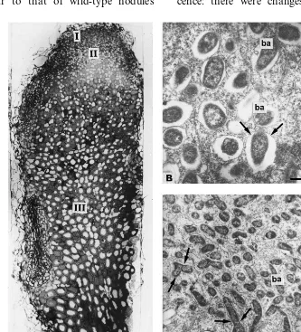
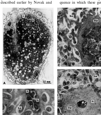
The nodules of cv. Finale demonstrated the histological zonation and ultrastructural organisa-tion (Fig. 1A – C) characteristic for typical wild-type pea symbiotic root nodules [10,14,19,22,25]. In zone III [26], each mature symbiosome con-sisted of one differentiated pleomorphic Y- or X-shaped bacteroid surrounded by symbiosome membrane (Fig. 1B and C).
3.2. Mutant RisFixA
Nodules of mutant RisFixA were characterised by alterations of histological differentiation (Fig. 2A). Zones I, II, were abnormally enlarged. There was no distinct interzone II – III. Nodules con-tained hypertrophied infection threads that fre-quently occupied the most part of a profile of the host cells in zone II (Fig. 2B). Endocytosis of bacteria was ‘delayed’ (as manifested by an ex-tended zone II) but was otherwise apparently nor-mal, i.e. one by one (Fig. 2C). After endocytosis, the bacterial matrix was less electron-opaque than that of bacteria in infection threads, but these ‘juvenile’ bacteroids did not enlarge in size (Fig. 2C). Symbiosomes containing several bacteroids were frequently observed in infected cells in the central part of the nodule (Fig. 2D). Nodules of this mutant were characterised by ‘early senes-cence’, as manifested by the premature appearance of zone IV with degrading symbiotic structures compared to the nodules of the initial line Finale (Fig. 2E).
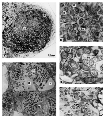
3.3. Mutants RisFixL and RisFixO
II – III and a true zone III was lacking. Infected cells were significantly vacuolised (Fig. 3A), which is not characteristic for effective pea nodules [6]. Infection threads appeared normal and were indis-tinguishable from those in wild-type nodules (Fig. 3B and C). Bacteria were endocytosed one by one and ‘juvenile’ symbiosomes generally contained a single bacteroid (Fig. 3B). Symbiosomes contain-ing several bacteroids were mainly observed in infected cells in the central part of the nodules (Fig. 3C). Degradation of the infected cells (zone IV) began with disintegration of symbiosome membranes (Fig. 3D).
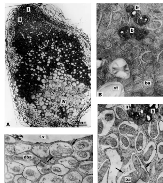
3.4. Mutants RisFixK, RisFixM (sym26) RisFixN, RisFixQ (sym27) and RisFixT (sym26)
Nodule histological differentiation of these mu-tants was similar to that of wild-type nodules
[10,14,19,22,24,25], except for zone IV that was abnormally enlarged (Fig. 4A). Bacteroids demon-strated morphologically pronounced differentia-tion (Fig. 4B and C), however symbiosomes could only be found in narrow zone correspond-ing to zone III. In zone IV symbiosomes were transformed into the lysosome-like com partments containing the ‘ghosts’ of bacteroids (Fig. 4D).
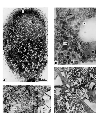
3.5. Mutant RisFixV
All morphological traits characteristic for the previous group of mutants were found in the nodules of this mutant (Fig. 5A and B). A distin-guishing feature of this mutant, however, was the process of infection thread ‘maturation’ and senes-cence: there were changes in the structure and
Fig. 2. Nodule structural organisation of mutant RisFixA: (A) longitudinal nodule section; (B) plant cells containing infection threads in histological zone II; (C) bacteria endocytosed from infection thread; (D) infected cells of the central part of the nodule containing symbiosomes with several bacteroids; (E) lysosome-like compartments containing degrading bacteroids in nodule histological zone IV. I, II nodule histological zones; it, infection thread; b, bacteria; ba, bacteroids; v, vacuole; st, starch granules; dba, degrading bacteroids; arrows, peribacteroid membranes and membranes of lysosome-like compartments. Bars in electron micrographs, 1 mm.
thicknesses of plant cell walls surrounding infec-tion threads. Initially thickening of the infecinfec-tion thread wall could be observed in zone III (Fig. 5C). In zone IV, this process apparently continued and, in addition, the bacteria inside infection thread showed signs of degradation (Fig. 5D).
4. Discussion
Morphological analysis of nodules from nine
Fix− mutants has shown that they are blocked at
four different stages of nodule morphogenesis, as discussed below.
4.1. Mutant RisFixA
The mutant gene identified in line RisFixA ap-parently controls several stages of nodule develop-ment: Itn, Idd, Bad and Nop (for description, see introduction). A similar mutant phenotype was
described for mutant SGEFix−-1 (sym40) [10].
The only ultrastructural difference is that in nod-ules of RisFixA the bacteroids were of the same size and shape as bacteria inside infection threads
whereas, in nodules of mutant SGEFix−-1
(sym40), enlarged pleomorphic bacteroids could
also differences in the number of root nodules
formed by these two mutants: mutant SGEFix−-1
(sym40) showed an increased number of nodules
(relative to wild type), which is a common host
plant reaction to the formation of Fix− nodules
[10]. However, in mutant RisFixA, a decreased number of nodules was described [27]. In addition to differences in mutant phenotypes, it was also shown that these mutants are not allelic to each other (Tsyganov et al., unpublished results). All the data represented above suggest that gene
sym40 and the symbiotic gene identified in line
RisFixA control very close (if not the same) nod-ule developmental stages.
4.2. Mutants RisFixL and RisFixO
In nodule infected cells of the allelic mutants RisFixL and RisFixO, the only sign of bacteroid differentiation is a decreased electron density of the matrix. Bacteroids remain rod-shaped with no increase in size. As a rule, several bacteroids are surrounded by peribacteroid membrane. The phe-notype of these allelic mutants can be described as
Bad−. Such an ultrastructural organisation of
bac-teroids is very similar to that of mutant line
Sprint-2Fix− (sym31) [6,22]. These data confirm
the results, which were obtained by Novak et al. [19] except that, in the present study,
Fig. 4. Nodule structural organisation of mutants RisFixK, RisFixM (sym26) RisFixN, RisFixQ (sym27) and RisFixT (sym26): (A) longitudinal nodule section; (B) plant cells containing infection threads and ‘juvenile’ bacteroids; (C) symbiosome containing differentiated bacteroid in central part of the nodule; (D) degrading symbiosomes in nodule histological zone IV. I, II, IV nodule histological zones; it, infection thread; b, bacteria; ba, bacteroids; v, vacuole; st, starch granules; dba, degrading bacteroids; arrows, peribacteroid membranes and membranes of degrading symbiosomes. Bars in electron micrographs, 1 mm.
phied infection threads were not observed for the mutant RisFixO. These differences in nodule ul-trastructure could perhaps be explained by the different strains used for inoculation or by varia-tions in plant growth condivaria-tions. Finally, taking into account certain differences in the patterns of
senescence between line Sprint-2Fix− (sym31)
(which shows no signs of nodule premature
degra-dation) and allelic mutants RisFixL (sym32) and
RisFixO (sym32) (which have an increased
histo-logical zone IV) one can conclude that pea
symbi-otic genes sym31 and sym32 control very close (if
not the same) stages of nodule development.
4.3. Mutants RisFixK, RisFixM (sym26) RisFixN, RisFixQ (sym27) and RisFixT (sym26)
In mutants RisFixM and RisFixT (sym26),
Ris-FixN, RisFixQ (sym27), abnormalities of
nodules could begin to function properly. This mutant phenotype has been described previously in pea and other legumes [13,14,19,28,29]. Because
the Nop− phenotype can be found quite
fre-quently among legume symbiotic mutants, it sug-gests that many legume genes are involved in stabilisation of the nitrogen-fixing symbiosis and the avoidance of premature senescence.
4.4. Mutant RisFixV
Although the mutant phenotype of RisFixV was very similar to the group of ‘early senescent’ mu-tants discussed above, the difference in infection thread development made it possible to place this
mutant in a different class (phenotype Itn−, Nop−).
Abnormal development of infection threads in this mutant was described earlier by Novak and
colleagues [19]. In this study it was discovered that cell walls around infection threads became much thicker during the process of infection thread ‘maturation’ and senescence. Further comparative study of mutant RisFixV will help to reveal the sequential functioning of the genes controlling the stages associated with Nop.
5. Conclusions
Combination of the results of the present study for mutants obtained in cv. Finale [3] with the
data on morphological analysis of pea Fix−
mu-tants published previously [6,10,13,14,19,22] has
made it possible to arrange all the pea Fix−
mu-tants in order, according to the hypothetical se-quence in which these genes function in the late
stages of symbioitic development. Where a muta-tion was found to induce developmental abnor-malities at several stages, we have applied the principle of the ‘first morphological abnormality observed’ in order to define the position of each mutant in this scheme. According to this principle, the earliest nodule developmental stage affected by the mutation represents the first point at which the expression of the gene function is required.
The predicted order of sequential gene function-ing for late nodule development is as follows.
Phenotype (i) Itn−, Bar−: the first position in the
sequence is occupied by the mutant SGEFix−-2
(sym33) [10]. Phenotype (ii) Itn−, Idd−, Bad−,
Nop−: mutants SGEFix−-1 (sym40) [10] and
Ris-FixA are located in the second position.
Pheno-type (iii) Bad−: mutants Sprint-2Fix− (sym31)
[22], RisFixL (sym32) and RisFixO (sym32), with
no differentiation of bacteroids occupy the third
position. Phenotype (iv) Itn−, Nop−: mutant
Ris-FixV is located in the next position, having abnor-malities in infection thread ‘maturation’ and senescence along with premature degradation of symbiotic structures in infected cells. Phenotype
(v) Nop−: this position is occupied by the largest
group of mutants (FN1 [13], E135f (sym13) [14],
RisFixK, RisFixM (sym26) RisFixN, RisFixQ
(sym27) and RisFixT (sym26), characterised by
premature degradation of the nodules. Thus, as a result of the present study, seven more pea symbi-otic genes have been classified in accordance with their sequential functioning.
This hypothetical scheme for the sequential functioning of pea symbiotic genes is based only on morphological analysis. Further confirmation of the order of the genes could be achieved by the creation of double mutants carrying mutations in genes with adjacent positions on the scheme derived from phenotypic analysis. This approach has already proved to be very fruitful in the case
of Sym13 and Sym31 where the sequential
func-tioning of these genes was recently confirmed [22].
Similarly, the analysis of double Fix− mutants
created for all identified and classified pea late symbiotic genes will confirm the sequential func-tioning identified to date.
Acknowledgements
For VET, AYB and IAT this work was
sup-ported by the Russian Foundation for Basic
Re-search (98-04-49883), Volkswagen-Stiftung,
Germany (I/72 935), NATO Scientific and
Envi-ronmental Affairs Division (HTECH.LG 971210). The authors are very grateful to N.J. Brewin for
fruitful discussion and critical reading the
manuscript and to L.E. Dvoryaninova and V.M. Semyonov for their excellent technical assistance.
References
[1] D.A. Phillips, L.R. Teuber, Plant genetics of symbiotic nitrogen fixation, in: G. Stacey, R. Burris, H.J. Evans (Eds.), Biological Nitrogen Fixation, Chapman and Hall, New York, 1992, pp. 200 – 235.
[2] E. Jacobsen, Modification of symbiotic interaction of pea (Pisum sati6um L.) and Rhizobium leguminosarum by
induced mutations, Plant Soil 82 (1984) 427 – 438. [3] K.J. Engvild, Nodulation and nitrogen fixation mutants of
pea (Pisum sati6um), Theor. Appl. Genet. 74 (1987)
711 – 713.
[4] G. Duc, A. Messager, Mutagenesis of pea (Pisum sati6um
L.) and the isolation of mutants for nodulation and nitrogen fixation, Plant Sci. 60 (1989) 207 – 213. [5] T.A. LaRue, N.F. Weeden, The symbiosis genes of pea,
Pisum Genet. 24 (1992) 5 – 12.
[6] A.Y. Borisov, E.V. Morzhina, O.A. Kulikova, S.A. Tch-etkova, V.K. Lebsky, I.A. Tikhonovich, New symbiotic mutants of pea (Pisum sati6umL.) affecting either nodule
initiation or symbiosome development, Symbiosis 14 (1992) 297 – 313.
[7] K.K. Sidorova, P. Uzhintseva, Use of mutants to detect genes controlling symbiotic characteristics in the pea, Soviet Genet. 28 (1992) 494 – 500.
[8] V.E. Tsyganov, A.Y. Borisov, S.M. Rosov, I.A. Tikhonovich, New symbiotic mutants of pea obtained after mutagenesis of laboratory line SGE, Pisum Genet. 26 (1994) 36 – 37.
[9] N.J. Brewin, M.J. Ambrose, J.A. Downie, Root nodules,
Rhizobium and nitrogen fixation, in: R. Casey, D.R. Davies (Eds.), Peas: genetics, molecular biology and bio-technology, CAB International, Wallingford, 1993, pp. 237 – 291.
[10] V.E. Tsyganov, E.V. Morzhina, S.Y. Stefanov, A.Y. Borisov, V.K. Lebsky, I.A. Tikhonovich, The pea (Pisum sati6um L.) genes sym33 and sym40 control infection
thread formation and root nodule functioning, Mol. Gen. Genet. 259 (1998) 491 – 503.
[11] T. Degenhardt, T.A. LaRue, F. Paul, Investigation of non-nodulating cultivar ofPisum sati6um, Can. J. Bot. 54
(1976) 1633 – 1636.
[12] J.G. Postma, E. Jacobsen, W.J. Feenstra, Three pea mutants with an altered nodulation studied by genetic analysis and grafting, J. Plant Physiol. 132 (1988) 424 – 430.
Phenotypical description and bacteroid activity, Plant Sci. 68 (1990) 151 – 161.
[14] B.E. Kneen, T.A. LaRue, A.M. Hirsch, C.A. Smith, N.F. Weeden,Sym13 — a gene conditioning ineffective nodulation in Pisum sati6um, Plant Physiol. 94 (1990)
899 – 905.
[15] F.C. Guinell, T.A. LaRue, Light microscopy study of nodule initiation inPisum sati6umL. cv. Sparkle and in
its low-nodulating mutant E2 (sym5), Plant Physiol. 97 (1991) 1206 – 1211.
[16] C.P. Markwei, T.A. LaRue, Phenotypic characterization ofsym8 andsym9 genes conditioning non-nodulation in
Pisum sati6um ‘Sparkle’, Can. J. Microbiol. 38 (1992)
548 – 554.
[17] A.Y. Borisov, S.M. Rozov, V.E. Tsyganov, O.A. Ku-likova, A.N. Kolycheva, L.M. Yakobi, A.O. Ovtsyna, I.A. Tikhonovich, Identification of symbiotic genes in pea (Pisum sati6umL.) by means of experimental
muta-genesis, Russ. J. Genet. 30 (1994) 1284 – 1292.
[18] M. Sagan, T. Huguet, G. Duc, Phenotypic characteriza-tion and classificacharacteriza-tion of nodulacharacteriza-tion mutants of pea (Pisum sati6umL.), Plant Sci. 100 (1994) 59 – 70.
[19] K. Nova´k, K. Pesina, J. Nebesa´rova´, V. Skrdleta, L. Lisa´, V. Nasinec, Symbiotic tissue degradation pattern in the ineffective nodules of three nodulation mutants of pea (Pisum sati6umL.), Ann. Botan. 76 (1995) 303 – 313.
[20] J.M. Vincent, Factors controlling the legume –Rhizobium
symbiosis, in: W.E. Newton, W.H. Orme-Johnson (Eds.), Nitrogen Fixation, vol. 2, University Park Press, Balti-more, 1980, pp. 103 – 129.
[21] G. Caetano-Anolles, P.M. Gresshoff, Plant genetic
con-trol of nodulation, Annu. Rev. Microbiol. 45 (1991) 345 – 382.
[22] A.Y. Borisov, S.M. Rozov, V.E. Tsyganov, E.V. Morzhina, V.K. Lebsky, I.A. Tikhonovich, Sequential functioning ofSym-13 and Sym-31, two genes affecting symbiosome development in root nodules of pea (Pisum sati6umL.), Mol. Gen. Genet. 254 (1997) 592 – 598.
[23] V.I. Safronova, N.I. Novikova, Comparison of two methods for root nodule bacteria preservation: lyophilization and liquid nitrogen freezing, J. Microbiol. Methods 24 (1996) 231 – 237.
[24] W.E. Newcomb, A correlated light and electron micro-scopic study of symbiotic growth and differentiation in
Pisum sati6um root nodules, Can. J. Bot. 54 (1976)
2163 – 2186.
[25] N.J. Brewin, Development of the legume root nodule, Ann. Rev. Cell Biol. 7 (1991) 191 – 226.
[26] J. Vasse, F. de Billy, S. Camut, G. Truchet, Correlation between ultrastructural differentiation of bacteroids and nitrogen fixation, J. Bacteriol. 172 (1990) 4295 – 4306. [27] K. Nova´k, V. Skrdleta, M. Nemkova´, L. Lisa´, Symbiotic
traits, growth, and classification of pea nodulation mu-tants, Rostlinna´ Viroba (Plant Production) 39 (1993) 157 – 170.
[28] C.P. Vance, L.E.B. Johnson, Plant determined ineffective nodules in alfalfa (Medicago sati6a): structural and
bi-chemical comparisons, Can. J. Bot. 61 (1983) 93 – 106. [29] D. Purdom, A.T. Trese, Morphological and molecular
characteristics of host-conditioned ineffective root nod-ules in cowpea, Plant Physiol. 109 (1995) 239 – 244.