Anticancer Property of Protein
Isolated from Thermophilic Bacteria
Against Breast T47D Cancer Cell Lines
Evy Yulianti
1, Anna Rakhmawati
1, Kartika ratna Pertiwi
1Biology Education Dept, Faculty of Mathematics and Science, Yogyakarta State University [email protected]
Abstract— Many pharmaceutical agents have been discovered by screening natural products from plants, animals, marine organisms and microorganisms. Cationic Antimicrobial Peptides (AMPs) in the protein isolated from bacteria, are an example of an anticancer agent originating from thermophilic bacteria. The aim of this study was to investigate the anticancer activity of proteins isolated from six isolates of thermophilic bacteria (113a, 94b, 153, 104c, 83, 110a). Bacteria was fermented in NB medium with 1% glucose in 55⁰C for 24 hr. Proteins isolated from cell free extract by salting out method with 60% ammonium sulphate and continued with dyalisis. Anticancer activity was conducted by cytotoxic test using MTT assay. The results of this study showed that proteins isolated from thermophilic bacteria which had anticancer activity toward T47D cell lines were proteins from isolat D153. Proteins from isolat D153 showed the IC50 value with 2,35 μg/mL concentration. Proteins from the other isolates (D83, D94b, D110a, D104c, and D113) showed the IC50 value with
436,5 μg/mL, 954,99 μg/mL, 629,5 μg/mL, 371,5 μg/mL, 501,1 μg/mL, and D153
2,35 μg/mL consentration respectively for T47D cell lines.
Keywords:anticancer, protein, thermophilic bacteria , T47D, breast cancer
I. INTRODUCTION
Cancer is a disease that is characterized by continuous cell division, uncontrolled and can spread to other tissues located nearby. Uncontrolled cell growth is caused by damage to DNA from mutations of genes that control cell division. Mutations can be caused by exposure to carcinogenic agents. This damage begins in the cells then multiply and gain additional changes are favorable for survival between neighboring cells are normal. The abnormal cells multiply to produce cancer-forming cells (MacDonald and Ford, 1997, Lodish et al., 2000, Kokha et al., 2004). Cancer is the result of long-term processes that lead to the effects of genetic drift and molecular changes that the process of change runs gradually. It usually takes a long time to change from normal to increased levels peak, dysplasia, which is phenotypically invasion and metastasis. The accumulation of genetic and molecular changes in a long time provides an opportunity for intervention field clinic for the prevention of cancer initiation and action before premalignant lesions (Crowel, 2005).
In 2002, breast cancer is the most common cancer in women in Indonesia, and in 2005 is the leading cause of death in women in Indonesia. The cause of breast cancer is lifestyle, environmental and genetic factors. The pattern of life include fatty foods, alcohol and little kosumsi fruits and vegetables.
Besides a positive family history of breast cancer have a high risk
(http://www.who.int/infobase/cancer.aspx). In breast cancer occurs upregulation of ligand-dependent activation of estrogen receptor (ER), which resulted in an anti-estrogen resistance. Some changes in the breast cancer is the abnormal upregulation and regulation of growth factor pathway (EGFR, IGFR, HER2) and signal transduction of intracellular molecules (Ras, Raf, MAPK, Akt). The growth factors such as IGF and EGF can activate ER in a state of availability of estrogen through estrogen receptor phosphorylation mechanism (Rennie et al., 2005). Approximately 80% of breast cancer cases known to have overexpression of Bcl-2. Some apoptotic proteins such as XIAP inhibitors, cIAP1, cIAP2, and survivin is also a breast cancer biomarker (Parton et al., 2001).
A thermophilic organism, is a microorganisms that are able to adapt grows optimally at high temperatures. Termofil microorganisms have been isolated from both terrestrial and marine habitats with high temperatures, for example the area of the volcano and hot springs (Bertoldo and Antranikian, 2002). This grup of bacteria is one of the microorganism that kan produce antimicrobial peptides. Antimicrobial peptides are chemical compounds produced by eukaryotic cells to fight bacteria, protozoa, and viruses
function. These compounds can also be found in bacteria, fungi, plants and animals (Hoskin, D. W. and Ramamoorthy, A., 2008).
Differences in biochemical and biophysical properties of the microbe with host cells and their settings which cells become the target of a peptide provides the basis selective toxicity of AMPs. Some AMPs provides a dynamic mechanism in working on a phospholipid bilayer, thereby providing rapid and potent effect (Yeaman, R. and M. Yount, N. Y, 2003).
Some studies had showed that cationic antimicrobial peptides (AMPs), had a toxic property toward bacteria but not to normal mammalian cells, and this substances are toxic to cancer cells with cytotoxic activity in wide spectrum. Naturally, AMPs are substances wich produced by eukaryotic cells to kill bacteria, protozoa, fungi and virus. This substances also found in bacteria, fungi, plant and animal. Some studies had showed that synthetic or natural AMPs, could increase the immune system and potential as antibiotic. The electrostatic bond between bacterial component or cancer cells wich have negative charge and AMPs with positive charge was believed had an important role to make a strong and selective bonding to destroy bacterial cell membrane and cancer cells. Peptides have the ability to neutralize lipopolysaccharide (LPS) and increasing adaptive immune reponses (Hoskin, D. W. and Ramamoorthy, A., 2008).
Cancer treatment in general is based on the cancer tissue sampling or by killing the cancer cells and minimize the effect of treatment toward normal cells in the surounding. The current cancer treatment is trough surgery, radiotherapy and chemotherapy, but the third type of treatments have its drawbacks. Operations will be succes in several tumor that has been growing, but it is difficult to treat in the early stages of metastasis (Lodish et al., 2000). Treatment with radiation capable of killing a local tumor, but radiation will also kill the surrounding normal cells. Most chemotherapy drugs such as taxol, 5-fluorouracil (5-FU) and adriamycin have a target on cell division (Boyer and Tannock, 2005), but chemotherapy can cause diarrhea and hair loss. Chemotherapeutic agents are also not effective in cells that mutated p53. So it is necessary to develop new agents for cancer treatment that is safe (Lodish et al., 2000). Chemopreventive is an innovative area in cancer research for developing pharmacology, biology, and nutrition interference, to prevent, repair, and retard carsinogenesis. These actions include preclinical agents and molecular identification, screening in vitro and in vivo, effects of pharmacological and chemical synthesis (Crowel, 2005). Identification and use of effective chemopreventive agent in cancer is an important issue in public health research. To identify potential chemopreventive in cancer some reseach do in vitro test that is relevant for in vivo prevention (Gerhäuser, 2002).
The use of natural materials as a cure for cancer is very desirable, because natural materials have no harmful side effects when compared with chemotherapy that has high toxicity and side effects. Secondary metabolites in biological natural ingredients can be used as traditional medicine against some disease. Chemopreventive agent itself can be defined as compounds that can inhibit and suppress the process of carcinogenesis in humans so that the growth of cancer can be prevented (Kakizoe, 2003). The purpose of this research is to determine the cytotoxic effect of protein on cancer cells T47D
II. RESEARCHMETHOD
This study is exploratory and experimental research using thermophilic bacteria isolated from water and sand river Gendol Merapi as research objects.
A. Tools and materials
Thermophilic bacteria, NB Medium, NA Medium, PDA Medium, PD Broth Medium, dimethyl sulfoxide (DMSO), distilled water, methanol, breast cancer cells T47D, fetal bovine serum (FBS) 10%, Fungizone 0 , 5%, 2% Penstrep, Dulbecco's Modified Eagle Medium (DMEM), SDS 10% in 0,01N HCl, Phosphate Buffer Saline (PBS), trypsin, 3- [4,5-dimethylthiazol-2-yl] -2, 5-diphenyl-tetrazolium bromide (MTT), acridine orange / ethidium bromide (100 mcg / ml acridine orange (bio-Rad) in PBS and 100 ug / ml ethidium bromide (bio-Rad) in PBS), ammonium sulphate, LAF, bottle Flash cultures, tubes konikel , pipette, micropipette, blue tip, tissue, erlenmeyer tube, incubator, inverted microscope, hemocytometer, 96 well plate, an analytical balance, ELISA reader, CO2 incubator, cover slips (Nunc), microplate 96 wells,.
B. Inoculum preparation
and incubation performed again at 55 ° C for 24 hours. One ose isolate transferred to NB medium from NA medium.
C. Batch fermentation
NB medium with 1% glucose in 1.5 liters of distilled water is used as the production medium. Inoculum (10%) trasferred into 200ml NB medium. And incubated again at 55 ° C. After 24 hours, the sample is added to 15 mL tubes, centrifuged for 30 minutes at 3,000 rpm to obtain a supernatant containing no cells.
D. Protein Isolation
Protein precipitation was done by using 60% ammonium sulfate followed by dialysis to remove residual ammonium sulfate.
E. Cytotoxic test to cancer T47D cell lines
The extract is diluted to concentrations of 50%, 25%, 12.5%, 6.25%, 3.125%. Further tested on cancer
T47D cell lines. The data obtained is calculated IC50. Cytotoxic test was done by adding the 100μL extract
per well. Then incubated at 37 °C for 24 hours. After sufficient incubation, the media was removed and
added 100μL new media and 10 mL MTT 5 mg/mL per well. Then incubated 4-6 hours. MTT reaction was
stopped by providing a stopper in the form of 10% SDS in 0,01N HCl as 100μL per well and incubated
overnight. Results was performed by Elisa reader at a wavelength of 595 nm.
III. RESULTANDDISCUSSION
Large protein concentration was measured using a spectrophotometer at a wavelength of 595 nm. And the results obtained are shown in Table 1.
Table 1. extracellular protein concentration of each isolate
Isolate Protein concentration between the concentration of protein and % of living cells is illustrated as follows.
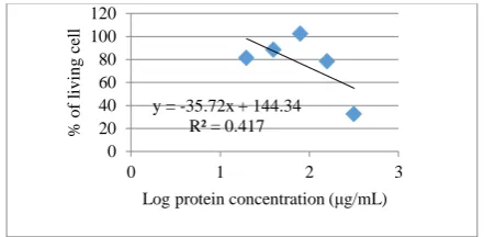
Figure 1. The relationship between the concentration of protein and % of living cells According to the graph, the IC50 values obtained from isolates D83 is 436.5 µg / mL
y = -35.72x + 144.34
Figure 2. The relationship between the concentration of protein and % of living cells
According to the graph, the IC50 values obtained from isolates D94b which kill 50% of cancer cell lines is 954,99 μg/mL.
Figure 3. The relationship between the concentration of protein and % of living cells
According to the graph, the IC50 values obtained from isolates D110a protein concentration which can cause death of 50% of cancer cells, is 629.5 µg / mL.
Figure 4. The relationship between the concentration of protein and % of living cells
According to the graph, the IC50 values obtained from isolates D104 protein concentration which can cause death of 50% of cancer cells, is 371.5 µg / mL.
Figure 5. The relationship between the concentration of protein and % of living cells
According to the graph, which shows the IC50 values obtained from isolates D113, protein concentration which can cause death of 50% of cancer cells is 501.1 µg/ mL.
Figure 6. The relationship between the concentration of protein and % of living cells
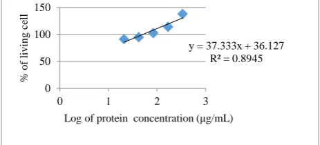
According to the graph, which shows the IC50 values obtained from isolates D153, protein concentration which can cause death of 50% of cancer cells is 2.35 µg/ mL. Here is a picture of a cell after being treated and MTT.
Several studies have shown that AMP, either synthetic or natural can boost the immune system and potentially as a potential antibiotic. Their electrostatic bonding between cancer cells which are negatively charged and positively charged of AMP can damage cell membranes of cancer cells. Most of
y = -106.29x + 347.39
Log of protein concentration (μg/mL)
y = -114.36x + 343.84
Log of protein concentration (μg/mL)
y = -83.502x + 275.72
Log of protein concentration (μg/mL)
y = 37.333x + 36.127
AMP have anticancer and antiviral activity. The peptides were found have the ability to enhance the adaptive immune response (Hoskin, D. W. and Ramamoorthy, A., 2008).
The sequence of amino acids in different AMPs are highly heterogeneous and has a very large variation in the secondary structure. AMPs are generally cationic (have a charge at neutral pH between +2 to +9) and are amphipathic, which allows it to interact with membran peptide and damaging the membrane lipids. Most AMPs have a short structure, consisting only of 5 to 40 amino acid residues, and many of these have length to more than 40 residues. The residue has a positive charge such as Lys and Arg and most have a hydrophobic residue (~ 30% or more) are usually found on these peptides (Hoskin, D. W. and Ramamoorthy, A., 2008).
IV. CONCLUSIONANDSUGGESTION
A. Conclusion
Proteins isolated from thermophilic bacteria which had anticancer activity toward T47D cell lines were proteins from isolat D153 (with IC50 value 2,35 μg/mL). Proteins from the other isolates (D83, D94b,
D110a, D104c, and D113) showed the IC50 value with 436,5 μg/mL, 954,99 μg/mL, 629,5 μg/mL, 371,5 μg/mL, 501,1 μg/mL, and D153 2,35 μg/mL consentration respectively for T47D cell lines.
B. Suggestion
Further tests can be carried out on the structure of proteins and the mechanism of action of anti-cancer
REFERENCES
[1.] Boyer, M.J., and Tannock, I.F., 2005, The Basic Science of Oncology: Cellular and Molecular Basis of Drug Treatment for Cancer, Mc Graw Hill Compay, forth edition, New York.
[2.] Brem, S., MD, 1999, Angiogenesis and Cancer Control: From Concept to Therapeutic Trial, Moffitt Cancer Center & Research institute, (http://www.medscape.com/cancercontrol/1999/v06.n02.brem/cc0605.02.brem-01.html)
[3.] Chevile NF. 1999. Introduction to Veterina ry Pathology. 2nd. Ed. Iovastate University Press/AMS.
[4.] Crowel, J.A. 2005. The chemopreventive agent development research program in the Division of Cancer Prevention of the US National Cancer Institute: An overview. European Journal of Cancer, v.41, p.1889–1910,.
[5.] Dorai ,T., Aggarwal, B.B. 2004 Role of chemopreventive agents in cancer therapy. Cancer Lett. Nov 25;215(2):129-40 [6.] Folkman, J., 1976, The Vascularization of Tumors, Sci Am, 6:59-73
[7.] Flora SD, Ferguson LR. 2005. Overviuw of mechanisms of cancer chemopreventive agents. Mutation reseach 591: 8-15. [8.] Gerhäuser C. et al. 2005. Mechanism-based in vitro screening of potential cancer chemopreventive agents. www.elsevier
.com/locate/molmut.
[9.] Gondhowiardjo, S., 2004, Proliferasi Sel dan Keganasan, Majalah Kedokteran Indonesia, 54 (7): 289-299
[10.]Hoskin, D. W. and Ramamoorthy, A. 2008. Studies on Anticancer Activities of Antimicrobial Peptides. Biochim Biophys Acta. 2008 February ; 1778(2): 357–375.
[11.]Jadav SJ, Nimbakar SS, Kalkuni AD, dan Madhavi. 1996. Lipid oxidation in biological and food systems. Food Antioxidant. Marchel Dekker Inc.
[12.]Kakizoe, T., 2003, Chemoprevention of Cancer Focusing on Cinical Trial, Nationa Cancer Center, Jpn.J.Clin.Oncol., 33(9): 421-442
[13.]King, R. J. B., 2000, Cancer Biology, 2nd ed, Pearson Eduation Limited, London.
[14.]Macdonald,F.,Ford,C.H.J.,1997, Molecular Biology of Cancer, BIOS Scientific Publishers Ltd. [15.]Matter, A., 2001, Tumor Angiogenesis as a Theurapeutic Target, DDT, Vol.6, No.19, Hal. 1005-1020.
[16.]Montgomery, R.,Dryer, L. R., Conway, W. T., Spector,A.A.,1993. Biokimia suatu pendekatan Berorientasi Kasus, Gadjah Mada University Press, Yogyakarta, 1071 – 1106
[17.]Mulyani, Sri.,1992, Pedoman Kuliah Karsinogenik dan Antineoplastik, PAU-Boteknologi UGM, Yogyakarta [18.]Parton, Martina., Dowsett, Mitchel., and Smith, I., 2001, Studies of Apoptosis in Breast Cancer, BMJ, 322: 1528-1532 [19.]Pan, M.H., Chen, W.J., Lin, S., Ho, C.H., Lin, J.K., 2002, Tangeretin Induces Cell Cycle Through Inhibiting Cyclin
Dependent Kinase 2 & 4 Activities As Well As Elevating Cdk Inhibitor p21 in Human Colorectal Carcinoma Cells, Carsinogenesis, Oxford University Press, 23: 1677-1684
[20.]Poeloengan dan Soeripto. 1998. Pengaruh Putih Telur Terhadap Pertumbuhan Gram Positif Dan Gram Negatif Secara In Vitro. Media kedokteran Hewan Institute Pertanian Bogor. Bogor.
[21.]Steele VE, Kellof GJ. 2005. Development of cancer chemopreventive drugs based on mechanistic approaches. Muta Res (591): 16-23
[22.]Surh, Y. J., 1999, Molecular Mechanism of Chemopreventive Effect of Selected Dietary and Medicinal Phenolic Substances, Mutation Res. 428: 305-327.