Formulation of Medium Viscosity Chitosan-Pectin –MJ Protein
Nanoparticles Conjugated with Anti-Ep-CAM and Its Cytotoxicity Against
T47D Breast Cancer Cell Lines
Anggun Feranisa
1,3, Dewa Ayu Arimurni
1, Hilda Ismail
2, Ronny Martien*
2,
Sismindari*
1,21Biotechnology Study Program, Graduate School, Universitas of Gadjah Mada, Yogyakarta, Indonesia 2Faculty of Pharmacy, Universitas Gadjah Mada, Yogyakartaa, Indonesia
3Faculty of Dentistry, Islamic Universitas Sultan Agung, Semarang, Central Java, Indonesia
Abstract
Chitosan nanoparticle could become potential formula to protect protein degradation during therapy, since chitosan nanoparticles have “proton sponge hypothesis” mechanism on its protection. Chitosan and pectin is used as basic formula of drug delivery because of its biodegradable and biocompatible properties. Chitosan-pectin nanoparticles can be formulated by polyelectrolit complex. EpCAM showed excessive expression in epithelial cancer cells thus can be used as a therapeutic biomarker. MJ protein, a Ribosome-Inactivating Proteins (RIPs) isolated from Mirabilis jalapa L had a higher cytotoxicity on malignant cells than normal cells. MJ protein need to be formulated to protect from proteosome degradation in endosome. The aim of this research was to develop MJ protein-chitosan-pectin nanoparticles and conjugated with anti EpCAM for breast cancer therapy. Mj protein was extracted from M.jalapa leaves. RIPs activity was assayed by supercoiled DNA cleavage. MJ protein were loaded into chitosan nanoparticles using medium viscous chitosan and pectin as cross-linker with polyelectrolit complex method. Anti EpCAM was conjugated to MJ protein-chitosan-pectin nanoparticles by carbodiimide reaction and characterized for its entrapment effi ciency, morphology by transmission electron
microscope, particles size, and zeta potential. MJ protein nanoparticles conjugated anti EpCAM and without anti EpCAM were cytotoxicity assayed toward T47D and Vero cell lines. MJ protein was able to cleave the supercoiled DNA into linear and nicked-circular ones. The nanoparticles optimal concentration of medium viscous chitosan: MJ protein: pectin was 0.01%: 0.01%: 1% (m/v). A high entrapment effi ciency of MJ protein
nanoparticles was 98.97 ± 0.07%. Morphology nanoparticles showed an amorphic structure with 200.00 nm particles size. The nanoparticles conjugated anti EpCAM showed average particles size 367.67nm, polydispersity index 0.332, and zeta potential +39.97mV. MJ protein-chitosan-pectin nanoparticles conjugated anti EpCAM and unconjugated both had higher cytotoxicity with the IC50 57.64 μg/mL and 46.84 μg/mL respectively against T47D and 99.38 μg/mL and 111.34 μg/mL against Vero cell lines compared to MJ protein with IC50 of 3075.61 μg/mL against T47D and 3286.88 μg/mL against Vero cell lines. Both MJ protein-nanoparticles could increase the cytotoxicity effects about 50 times compared to the unformulated MJ protein activity, however had less specifi city toward T47D and Vero cell lines.
Keywords:Mirabilis jalapa L., RIPs, nanoparticle, pectin, chitosan, anti EpCAM
*Corresponding author:
Sismindari
Department of Biotechnology, Graduate School, Universitas Gadjah Mada, Email: sismindari@ugm. ac.id
Ronny Martien
Faculty of Pharmacy, Universitas Gadjah Mada Email: [email protected]
Introduction
it can protect proteins from degradation in endosome (Taira et al., 2005). Chitosan and pectin are biocompatible, biodegradable, muchoadhesif (Morris et al., 2010; Jana et al., 2011), and low toxic materials (Yuan et al., 2009), hence its can be used as drug delivery system.
Carcinogenesis is started from normal cells which then become uncontrolly divide without differentiate (King and Robins, 2006). Chemotherapy is the main choice for cancer treatment (McCartney and Turkington, 2002), however, it generally have toxic side effect toward normal cells and not specifi c only
for cancer cells (Liao et al., 2013). Therefore, delivering drug into the cell target will be necessary. Ribosome-inactivating proteins (RIPs) are nature cytotoxic compounds (De Virgilio et al., 2010). One of them is MJ protein, a RIP isolated from Mirabilis
jalapa L. leaves. MJ protein have shown in
vitro cytotoxic activity against cancer cells (Ikawati et al., 2003; Ikawati et al., 2006). It also had cytotoxic activity against breast-cancer cell-lines, T47D (Sismindari et al., 2010). It found that MJ protein had higher cytotoxic effect against cancer cells than normal cells (Stirpe and Battelli, 2006; Sismindari et al., 2010). In addition, MJ protein had the ability to delay the onset of tumorigenesis for 2 weeks and reduced the incidence of tumor and the tumor multiplicity by 40% and 30% respectively (Hussaana et al., 2009). However, MJ protein will be unstable when enter the cells, and easy being degradated. MJ protein need to be formulated as nanoparticle with the delivery system to raise it’s stability and specifi city toward cancer cells (De Virgilio
et al., 2010).
Cell Adhesion Molecules (CAM) are glycoprotein membrane or transmembrane that mediated communication among cells or attached cells with its substrates (Vleminckx, 2001). Breast cancer cells interact among each other by cell adhesion proteins. In normal physiologis condition, its have a role to control cells polarity and restraint epithelial cells. When the cells are mutated in its side,
it can be a triger to adhesion, invasion, and migrasion (metastatic) in cancer cells (Offi ah
et al., 2012). Point mutation are happened
in E-caderin gene that code CAM. In this case, E-cadherin mutation are most found in lobular breast cancer (Gullick, 2001). Epithelial Cell Adhesion Molecule (EpCAM) is cell adhesion protein/molecules that very potent to use as targeted therapy in breast cancer treatment since it is overexpressed in this cells (Osta et al., 2004). Nevertheless, based from immunofl uorescence analysis,
it was reported that anti EpCAM in T47D didnot express AUA-1 subtype (Sterzynska
et al., 2012).
Self-assembled Polyelectrolyte Complex Nanoparticles Formed from chitosan and pectin nanoparticles as wound dress had been characterized. Chitosan-pectin nanoparticles could also be formulated by polyelectrolyte complex method (PEC) (Birch and Schiffman, 2014). Cytotoxicity effects of chitosan-pectin nanoparticles conjugated antibody toward cells needed to be identifi ed in order to reach
the specifi c target. In this study, cytotoxic
activity of MJ proteins nanoparticles was analyzed against T47D and Vero cell lines.
Materials and Methods
Materials
This study used red flower Mirabilis
jalapa L. leaves from Pogung, Sleman, DIY
and pET28 plasmids from Microbiology Laboratory, Department of Fisheries, Faculty of Agriculture, UGM. Medium viscous chitosan (Aldrich) and pectin from apple (Sigma) were used as polymers to formulate the nanoparticles. AUA-1 subclone Anti EpCAM antibody (Abcam) were used as biomarker. T47D and Vero cell lines (ATCC, LPPT UGM) were used in cytotoxic assayed.
M. jalapa leaves were sliced and
grounded into a powder and homogenized in the 5 mM sodium phosphate buffer pH 7.2 with 0.14 M sodium chloride. The solution was fi ltered and subsequently centrifuged in
The protein was precipitated using acetone 1:1 (v/v) and centrifugated 9000 x g for 45 min at 4 °C. The protein pellet was dissolved in 5mM phosphate buffer pH 6.5 and mixed gentlely (modificated from Ikawati et al., 2003). Proteins concentration were measured by Bradford assay.
Proteins concentration were measured by formula as follows (Nobel, 2000):
Proteins concentration = concentration x dilution
Protein
concentration slope
(average of
absorbance intercept x dilution =
-RIPs Activity Assay
One μg of DNA plasmid (pET28) was incubated with various amount of MJ proteins (0.05; 0.1; 0.5 mg/mL) at a final volume of 10 μL containing 1xTMN buffer at 30 oC for 1 hour. In the end of reaction, 4μL
loading buffer were added. Electrophoresis was done using 0.8% agarose in 1x TBE buffer. DNA bands were visualized using ethidium bromide (Ikawati et al., 2006).
Formulation of Nanoparticles
Optimization of nanoparticles formula (1:1 w/v) were done at various concentration of chitosan and pectin (Chitosan: 0.01%; 0.05%; 0.1%; 0.5% and 1%; Pectin: 0.01%; 0.05%; 0.1%; 0.5%; and 1%). Each of chitosan-pectin nanoparticles formula were mixed for 20 sec using vortex (modifi cated from Birch
and Schiffman, 2014). The best formula was opact turbidity and did not clot.
Optimization of BSA nanoparticles (BSA as proteins model) were done using optimum formula of chitosan-pectin nanoparticles with various concentration of BSA concentration, 0.01%; 0.05%; 0.1%; 0.5%; and 1% (v/v) with 1:1 (w/v) ratio. Optimum formulas were selected from opaque and did not clot formulas.
MJ proteins nanoparticles were formulated based on the optimum formula of BSA nanoparticles (0.01% chitosan; 1%
pectin). Various RIPs concentration were used in this formulas, 0.05 mg/mL; 0.1 mg/ mL; and 0.2 mg/mL. This nanoparticles were formulated in 1:1:1(w/v) ratio of chitosan, MJ protein and pectin. The optimum formula of this nanoparticles were dialysed overnight.
A n t i E p C A M a n t i b o d y w e r e conjugated with optimum formula of MJ proteins nanoparticles using 1-Ethyl-3-(3-dimethylaminopropyl)carbodiimide (EDC) reaction. Ten microliters of Anti EpCAM antibody 1 mg/mL were reacted with 10 μL of 1 mg/mL EDC in the addition of 80 μL MOPS pH 6. Fifty microliters of this antibody suspension were reacted with 3 mL MJ proteins nanoparticles (modifi cated
from Hoganson et al., 1998), and was dialysed overnight at 4 °C.
Nanoparticles Characterization
E n t r a p m e n t e f f i c i e n c y o f M J proteins concentration in chitosan-pectin nanoparticles, was analyzed by measuring the concentration of un-entraped protein at nanoparticles using spectrophotometry. Chitosan-pectin nanoparticles integrated MJ proteins and without MJ proteins (as blanko) were centrifuged with 15.000 x g speed in 35 min to separate nanoparticles and it’s solvent. Supernatan were taken to spectrofotometry assayed. Proteins concentration were measured using Bradford assay.
Effi ciency concentration of un-entraped
MJ proteins in chitosan-pectin nanoparticles (EE) was measured as follows (Ranjan et al.,
Amount of RIPs residue in nanoparticles suspension were measured by formula as follows (Ranjan et al., 2012):
Morphology character of nanoparticles surface were observed by Transmission Electron Microscope (TEM). A droplet of poly-ion complex nanoparticles suspension were droped on a layer of carbon fi lm on
copper net. It droplet were coloured by Sodium (K) Phosphotungstate (PTA). Then, that droplet were observed by TEM (Jang and Nah, 2003).
The nanoparticles were mixed in deionized water. It was meant to measured the diameters and particles distribution (polydispersity index) that were measured by photon corelated in spectroscopy by Particle Size Analyzer. Particles charge were qualifi ed as zeta potential by anemometri
laser Doppler. All of this measured were done in triple measured (Kocbek et al., 2007).
Cytotoxicity Assay
Following starvation, 5x10⁴ cells/mL of
T47D and Vero cell lines were treated with 100 μL of conjugated anti EpCAM-chitosan-pectin-MJ protein nanoparticles (33.33 μg/ mL); chitosan-pectin-MJ protein nanoparticles (33.33 μg/mL); unformulated MJ proteins (1000 μg/mL; and 100μL) and Doxorubicin (10 μg/mL). Cells were incubated at 37
oC for 24 hours in 5% CO
2. After 24 hours
incubated, cells were washed and added with 100 μL of fresh media contained 5 mg/ mL MTT ((3-(4,5-dimethyltiazol-2-il)-2,5-diphenyl tetrazolium bromide) reagen. Cells were then incubated for 3 hours at 37 oC in
CO2 incubator, followed by the addition of 100μL of stopper solution. The formation of dark broun-purple colour were measured on 550 nm. IC50 values were calculated by probit analysed. Percetage of death cells were measured with formula as follows (Ikawati
et al., 2006):
%death cells
A
A A x100
control cells control cell cel treated
=
-Cells morphology were observed by inverted microscope. Cytotoxic effect were observed by curve of % death cells and IC50.
Inhibitory concentration were measured with probit analysed by SPSS programme (Ikawati
et al., 2006).
Results and Discussion
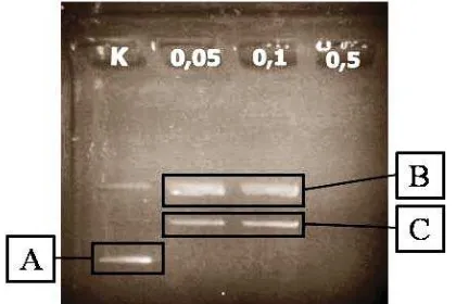
Ribosome-inactivating proteins (RIPs) extraction from M.jalapa L leaves obtained about 40% (123.25 mg) of MJ protein. proteins. RIPs activity assays in MJ proteins were assayed by supercoil (DNA pET-28) cleaved methods. Proteins concentrations that were used for analysis were 0.05 mg/ mL; 0.1 mg/mL; and 0.5 mg/mL. The results demonstrated that MJ protein was able to cleave the supercoiled DNA into nick sircular and linear form as shown at Figure 1. Therefore, it was indicated that the protein was in an active form. At higher MJ proteins concentration (0.5 mg/ mL), had a higher RIPs cleavage activity, which resulted in the pDNA degradation. This results supported the previous work, where at low concentration, RIPs was able to cleave supercoiled DNA into a nick circular. At the increasing RIPs concentration, the supercoiled DNA form will be disappeared and the linear form produced followed by the degradation of DNA at higher concentration (Sismindari et al., 1998)
Nick circular DNA migrate slower than linear DNA, and both of them migrate slower than control, supercoiled DNA (Figure 1). MJ proteins showed good condition
Figure 1. Electrophoregram of RIPs activity assayed (K
and contained active RIPs, then it could be further formulated in chitosan-pectin nanoparticles.
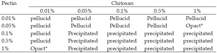
Optimization of nanoparticle formula was carried out with a various chitosan concentration as matrixes and pectin as crosslinked at the same concentrations, range from 0.01% to 1% (w/v). Each concentration were mixed at 1:1 (v/v) ratio to fi nd out a
stable nanoparticles. The results indicated that there were two optimum nanoparticles formulas, that were at the chitosan and pectin concentration of 1% : 0.05% and 0.01% : 1% respectively (Table 1). This result based on the opact turbidity of suspensions observation, which indicated the nanoparticles formation as a micro or macroparticle.
Polyelectrolyte complex (PEC) is formed between chitosan polycationic that bring positive charge (NH3+) and pectin polyanionic
that bring negative charge (-COO-). On the
appropriate concentration ratio, chitosan and pectin would form nanoparticles that signed as opact turbidity. When the concentration was too low, the suspension will be pellucid, it would not be formulated as polyelectrolyte complexes. Whereas, when the concentration is too high, the polyelectrolyte complexes would become more viscous, so it would be formed as micro or macroparticles that was signed as precipitation in suspension.
Optimization of BSA concentrations as proteins model for BSA-nanoparticles were formulated according to the candidate formula (Table 1), at chitosan:pectin concentration of 0.01% : 1 and 1% : 0.05%. BSA is often used as drug model in drug delivery systems because of it’s stable structure and
thermostable characteristic (Odunuga and Shazhko, 2013). The BSA concentration that were used in this formula, were 0.2% to 1%. The result indicated that nanoparticle formula with 1% BSA was precipitated after 3 days storage. Pellucid suspension was formed at all of chitosan:pectin concentration 1% : 0.05% formulas. There were not formed PEC in all of BSA concentration (0.2% and 1%), it meant that it were not formed nanoparticles. It probably caused by the used of high methoxyl pectin. This pectin has more metoxyl groups than carboxyl groups, which could reduced the negative charges of pectin chains. If pectin concentration was less than chitosan, so that carboxyl groups of pectin will less and the amine groups in chitosan chains will over abundant. Hence, chitosan would not be able to react with pectin, then it could not form PEC. Based on the result, the chosen formula would be chitosan:pectin concentration of 0.01% : 1% with the BSA concentration of 0.2%. In this pectin’s concentration that over abundant (1%) than chitosan’s concentration (0.01%), both of its polymers would form stable PEC. This results was then used as a basic for formulating the MJ protein-chitosan-pectin nanoparticle.
MJ protein at various concentration, 0.05 mg/mL, 0.1 mg/mL, and 0.2 mg/ mL was formulated using the candidate nanoparticle formula, with the chitosan-pectin concentration at 0.01% : 1%. The result demonstrated that these formulas with the 1:1:1 ratio (v/v) showed similar turbidity.
Particles dispersion shown opact turbidity after its were formulated. Protein
Table 1. Optimization chitosan-pectin nanoparticles formula.
Pectin Chitosan
0.01% 0.05% 0.1% 0.5% 1%
0.01% pellucid pellucid Pellucid Pellucid Pellucid 0.05% pellucid Pellucid Pellucid Pellucid Opact* 0.1% pellucid Precipitated precipitated precipitated precipitated 0.5% pellucid Precipitated precipitated precipitated precipitated 1% Opact* Precipitated precipitated precipitated precipitated
concentrations that was chosen were the highest concentration that could be formulated into stable nanoparticles. MJ protein concentrations that was chosen was 0.1 mg/mL, since at the MJ protein concentration of 0.2 mg/mL, the formula was precipitated after one week storage in 4 to 10o C. Morphology of MJ protein
nanoparticles was observed by TEM. It shown amorphic structure of nanoparticles with 200.0nm diameter (Figure 2).This concentration was expected to form stable nanoparticle when conjugated with anti EpCAM. The MJ protein nanoparticles were stable in 4 to 10 °C during 2 to 3 weeks storage.
MJ protein nanoparticles and anti EpCAM were conjugated by carbodiimede reaction with EDC as space linker. EDC
activated carboxyl groups in pectin, thus it can bind to amin groups of anti EpCAM. Entrapment efficiency of MJ protein nanoparticles was 98.97 ± 0.07%.
Figure 2. Visualization of MJ protein nanoparticle’s
morphology
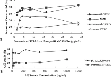
Figure 3. Cytotoxic effect of nanoparticle formulated and un-formulated MJ protein against
T47D and Vero cell lines.
Both cells-lines (5x10⁴ cells/mL) were treated with formulated and un-formulated MJ-protein nanoparticle at various concentration with the incubated at 37oC for 3 hours. The cell death was
visualized by measuring the dark broun-purple colour using ELISA reader at 550 nm. A. Formulated MJ protein nanoparticle and MJ-protein immuno-nanoparticle
B. Un-formulated MJ protein
0 5 10 15 20 25 30 35
Konsentrasi RIP dalam Nanopartikel CSM-Pec (μg/mL)
nanoAb T47D
nano T47D
nanoAb VERO
nano VERO
Persentase Kematian Sel (%)
50
40
30
20
10
0
B
0 200 400 600 800 1.000 1.200
Cell Death (%)
MJ Protein Concentration ( g/mL)
Protein MJ T470 Protein MJ VERO 40
30
20
10
0
Following immuno-nanoparticles formulation, diameter of nanoparticles were measured by particle size analyzer (PSA). Diameter of MJ protein nanoparticles conjugated anti EpCAM was 367.67 nm, which was longer than the diameter of unconjugated MJ-nanoparticle. This diameter was similar to polylactic-co-glycolic acid (PLGA) nanoparticles, which was 320-360nm (Kocbeck et al., 2007). It was indicated that the conjugated MJ nanoparticle was obtained.
Further analysis was carried out by measuring the polydispersity index (P.I). It was indicated that the average P.I of immuno-nanoparticles was 0.332. The value was close to zero, so that it refers to more monodispers (Ranjan et al., 2012). Zeta potential of MJ protein immune-nanoparticles was +39,97 mV, it mean that this nanoparticles had a high positive charge, so that, this nanoparticles could attach the negatively charge of the cells membrane and enter the cells easily.
The cytotoxicity analysis indicated that the MJ-protein-nanoparticle and MJ-protein immuno-nanoparticle had more cytotoxic against T47D cells than Vero cells (Figure 3A). At the concentration of 2.85 μg/mL, MJ-protein-nanoparticle and MJ-protein immuno-nanoparticle had cytotoxic effects of 34.71%, 35.29% respectively against T47D and 9.02%, 20.87% respectively against Vero cells line (Figure 3B). Similar pattern was also occurred on MJ protein, it had higher cytotoxicity against T47D (31% cell death
at 125 μg/mL) compared to Vero cell lines (11.40% at 125 μg/mL).
Calculated IC50 (Figure 4) clearly demonstrated that either formulated MJ-protein nanoparticle had slightly higher cytotoxicity (IC50 of 46.84 μg/mL) compared to MJ-protein immuno-nanoparticle (IC50 of 57.64 μg/mL) and both of them had higher cytotoxicity, compared to un-formulated MJ protein with the IC50 of 2902.70 μg/mL against T47D. Similar results, both MJ-protein nanoparticle and MJ-protein immuno-nanoparticle had higher cytotoxicity against Vero cells with the IC50 of values 99.38 μg/mL and 111.34 μg/mL respectively, compared to un-formulated MJ protein with the IC50 of 3286.88 μg/mL (Figure 4).
Unfortunately, there were no difference cytotoxic activity between MJ-protein-nanoparticle and MJ-protein immuno-nanoparticle, as indicated by the slightly differences between their IC50 values. It probably caused by no interaction process between antibody anti EpCAM type AUA-1 with EpCAM of T47D cells. It seem that EpCAM of T47D could strong interact with antibody anti EpCAM type EBA-1 and moderate interaction with the type 9C4 (Sterzynska et al., 2012). On the other side, the higher cytotoxicity of these formulas against Vero cells were suspected that it has EpCAM receptor. EpCAM expression in ductal ren were 71% (Went et al., 2008). It probable affected a higher cytotoxicity of MJ protein
Figure 4. Calculated IC50 of formulated MJ protein and un-formulated MJ protein against
T47D and Vero cell lines
nanoparticles conjugated anti EpCAM in Vero cells than it nanoparticles without anti EpCAM.
In addition, although the ideal nanoparticle sizes for nanotherapy are range from 1 to 100nm for easy internalization into the cells, however, for drug delivery system the size could be tolerated to 400 nm (Rao et al. 2010), and the MJ-protein nanoparticles size was 200 nm, so that it could still pass the capiler safely, as indicated by the high cytotoxicity (low IC50 value) against T47D and Vero cells-line.
The higher IC50 values obtained from un-formulated MJ protein compared to previous work (111 μg/mL) that was done by Sismindari et al. (2010), could probable be caused by the period length of incubation. In this experiment the incubation was done for 3 hours, whereas at previous work was 48 hours. In addition, it was found that the activity was also depended on the size of the nanoparticle. Formulated MJ protein using low viscosity-alginate which produced smaller nanoparticles size (130.73 nm) with zeta potential of +26,36 mV, had an IC50 of 14.87 μg/mL against T47D cell line and 27.84 μg/mL against Vero cell line (Wicaksono, 2014). Smaller size of this nanoparticle probable affected it to endosite easierly, thus it’s IC50 values could be fulfi lled.
Conclusion
Medium viscousity Chitosan-pectin nanoparticles could be formulated with MJ proteins (MJ protein-nanoparticle) had EE value of 98.97 ± 0.07%, diameter 200.00 nm, and amorphic structure. Conjugated the MJ protein medium viscousity-chitosan-pectin nanoparticles with anti EpCAM (MJ protein-immuno-nanoparticle) produced nanoparticle with a diameter of 367.67 nm, PI 0.332, and zeta potensial +39.97 mV. The formulated MJ protein-nanoparticles or MJ protein-immuno-nanoparticle could increase the cytotoxicity effects about 50 times compared to the unformulated MJ protein activity. However, there were less specifi city
of anti EpCAM AUA-1 MJ-protein immuno-nanoparticle cytotoxic activity between T47D and Vero cell lines.
References
Birch, N.P., and J.D. Schiffman, 2014, Characterization of Self-assembled Polyelectrolyte Complex Nanoparticles Formed from Chitosan and Pectin,
Langmuir 30: 3441-3447.
De Virgilio, M., A. Lombardi, R. Caliandro and M.S. Fabbrini, 2010, Ribosome-Inactivating Proteins: from Plant Defense to Tumor Attack, Toxins 2: 2699-2737. Florence, A.T., 1997, The Oral Absorption of
Micro- and Nanoparticulates: Neither Exceptional Nor Unusual, Pharm Res. 14: 259-266.
Gullick, B, 2001, Breast Cancer, in Encyclopedic
References of Cancer, Edited by S.
Manfred, Springer, New York, USA, pp. 139-141.
Hoganson, D.K., L.A. Chandler, G.A. Fleurbaaij, W. Ying, M.E. Black, J. Doukas, G.F. Pierce, A. Baird, and B.A. Sosnowski, 1998, Targeted Delivery of DNA Encoding Cytotoxic Proteins Through High-Affinity Fibroblast Growth Factor Receptors, Hum. Gene
Ther. 9: 2565–2575.
Hussaana, A., Ikawati, Z., Sitarina W., Sismindari, 2009, Efek Kemoprevensi Protein MJ Hasil Isolasi Dari Daun
Mirabilis Jalapa L Terhadap Pembentukan
Tumor Kulit Pada Mencit Balb/C Yang Diinduksi UVB, Prosiding Simposium Penelitian Bahan Obat Alami XIV, ISBN 978-602-95911-0-1., BPPT: 91-97
Ikawati, Z., Sudjadi, and Sismindari, 2006, Cytotoxicity Against Tumor Cell Lines A Ribosome-Inactivating Protein (RIP)-like Protein Isolated from Leaves of
Mirabilis jalapa L., Malay. J. Phar. Sci.
4(1): 31–41.
HeLa and Raji Cell-Line, Orient. Pharm.
Exp. Med. 3(3): 151-156.
Jana, S. , Gandhi A, Sen KK, and Basu Sk, 2011, Natural Polymers and Their Application in Drug Delivery and Biomedical Field, J. Pharma. Sci. Tech. 1(1):16-27.
Jang, M.K. and J.W. Nah, 2003, Characterization and Modification of Low Molecular Water-soluble Chitosan, Bull. Korean
Chem. Soc. 24(9): 1303-1307.
King, R.J.B., and M.W. Robins, 2006, Cancer biology, 3rd Edition, Pearson Education Limited, London, UK, Pp. 1-8.
Kocbek, P., N. Obermajer, M. Cegnar, J. Kos, and J. Kristl, 2007, Targeting Cancer Cells Using PLGA Nanoparticles Surface Modified with Monoclonal Antibody, J. Con. Rel. 120: 18–26. Liao, G., M. Apaya, and L. Shyur, 2013, Herbal
Medicine and Acupuncture for Breast Cancer Palliative Care and Adjuvant Therapy, Evid. Based Complement
Alternat. Med. (2013): 1-17.
McCartney, R.A. and Carol A. Turkington, 2002, Breast Cancer, in The Gale Encyclopedia of
CancerVol 1, Diedit oleh T. Ellen, Gale
Group, USA, pp. 142-150.
Morris, G.A., M.S. KÖk, S.E. Harding and G.G. Adams, 2010, Polysaccharide Drug Delivery Systems Based on Pectin and Chitosan, Biotechnol. Genet. Eng. Rev. 27: 257-284.
Nobel, A, 2000, Quick StartTM BradfordPprotein
Assay Instruction Manual, Bio-Rad
Laboratories, Inc., Pp. 11-21.
Odunuga, O.O., and A. Shazhko, 2013, Ammonium Sulfate Preciptiation C o m b i n e d w i t h L i q u i d Chromatography is Sufficient for Purifi cation of Bovine Serum Albumin
that Is Suitable for Most Routine Laboratory Applications, Biochem.
Compd. (2013): 1-6.
Offi ah, G., K. Brennan and A.M. Hopkins,
2012, Junctional Adhesion Molecules
(JAMs) - New Players in Breast Cancer?,
Intechopen, Pp 491-493.
Osta, W.A., Y. Chen, K. Mikhitarian, M. Mitas, M. Salem, Y.A. Hannun, D.J. Cole, and W.E. Gillanders, 2004, EpCAM Is Overexpressed in Breast Cancer and Is A Potential Target for Breast Cancer Gene Therapy, Cancer Res. 64: 5818–5824.
Ranjan, A.P., A. Mukerjee, L. Helson, and J.K. Vishwanatha, 2012, Scale up, Optimization and Stability Analysis of Curcumin C3 Complex-loaded Nanoparticles for Cancer Therapy, J.
Nanobiotechnology. 10: 38-56.
Rao, D., S. Srivastav, C. Prasad, R. Saxena, and S. Asthana, 2010, Role of Nanoparticles in Drug Delivery, Intern. J. Nanotech. and
Applications. 54(9): 45-49.
Sismindari, Hussaana A., Mubarika S., 1998,
In vitro cleavage of Supercoiled DNA by
Annona squamosa extract, Indon.J.Pharm,
9 (4): 146-152.
Sismindari, M.S. Hartati, Adhyatmika, and R.S. Sudibyo, 2010, Cytotoxic Selectivity of MJC0.3 and MJC0.5, Acidic Ribosome-Inactivating Proteins Isolated from Mirabilis jalapa L. Leaves Against Various Cancer Cell-Lines, J
Med Sci 42(1): 39-43.
Sterzynska, K., B. Kempisty, P. Zawierucha, and M. Zabel, 2012, Analysis of The Specifi city and Selectivity of Anti-EpCAM
Antibodies in Breast Cancer Cell Lines,
Folia Histochem. Cyto. 50(4): 534–54.
Stirpe, F. and M. G. Battelli, 2006, Ribosome-Inactivating Proteins: Progress and Problems, Cell. Mol. Life Sci. 63: 1850– 1866.
Taira, K., K. Kataoka, and T. Niidome, 2005, Non-viral Gene Therapy Gene Design and
Delivery, Springer, Tokyo, Pp. 35-74.
Yuan, Y., J. Tan, Y. Wang, C. Qian, and M. Zhang, 2009, Chitosan Nanoparticles As Non-viral Gene Delivery Vehicles Based on Atomic Force Microscopy Study, Acta Biochim Biophys Sin 41(6): 515–526.
Vleminckx, K, 2001, Cell Adhesion Molecules,
Diedit oleh S. Manfred,Springer, New York, USA, pp. 181-187.
Went, P., S. Dirnhofer, D. Schopt, H. Moch, and G. Spizzo, 2008, Expression and Prognostic Signifi kance of EpCAM, J.
Cancer Mol. 3(6): 169-174.
Wicaksono, P.A., 2014, Formulasi dan Uji Sitotoksisitas Nanopartikel Ribosome-Inactivating Protein daun
Mirabilis jalapa L. dengan Polimer