286
AGRIVITA VOLUME 34 No. 3 OCTOBER - 2012 ISSN : 0126-0537
http://dx.doi.org/10.17503/Agrivita-2012-34-3-p286-295
CHARACTERIZATION OF BACTERIAL PATHOGEN CAUSING WILT AND LEAF
BLIGHT ON CORN (
Zea mays
) BY PHYSIOLOGICAL, BIOCHEMICAL
AND MOLECULAR METHODS
Lilis Suryani 1*), Luqman Qurata Aini 2,3), Arifin Noor Sugiharto 2,3) and Abdul Latief Abadi 2) 1)
Agricultural Quarantine Agency Class I Banjarmasin Jl. Sutoyo S. No. 1134, Banjarmasin South Kalimantan Indonesia 2)
Faculty of Agriculture, University of Brawijaya, Jl. Veteran Malang 65145 East Java Indonesia 3)
Maize Research Center, Faculty of Agriculture, University of Brawijaya Indonesia Jl. Veteran Malang 65145 East Java Indonesia
*) Corresponding author Phone: +62-511-3353980 E-mail:[email protected]
Received: May 29, 2012/ Accepted: September 12, 2012
ABSTRACT
In 2011, we found a new bacterial disease cha-racterized by wilt, dwarf and blight symptoms on sweet corn in Batu, East Java, Indonesia. The objective of this study is to characterize the causal agent of the disease. In this study, several assays were conducted, including hypersensitive response, pathogenicity, physiological and biochemical characteristics, PCR detection using two specific species primer pairs for Pantoea stewartii pv. stewartii, and homology analysis of 16S rRNA gene sequence. Four Gram-negative, non-motile, facultative anaerobic bacteria were isolated from the diseased tissue. Only two strains, BD1 and BB2, gave positive result in hypersensitive reaction, pathogenicity, and Koch’s postulate assays. BB2 and BD1 strains also showed positive results in the PCR amplification using specific primers derived from the P. stewartii subsp. stewartii 16-23S gene region but showed negative result when using primers derived from P. stewartii subsp. stewartii hrpS gene region. Sequence of partial 16S rRNA gene of BD1 and BB2 showed highest homology at 96% to P. stewartii subsp. stewartii strain ATCC 8199 (NR. 044800.1). This results suggest that bacterial pathogens isolated from sweet corn in Batu were strains of Pantoea spp.
Keywords: Zea mays, Pantoea spp, wilt, leaf blight, PCR
INTRODUCTION
In Indonesia, corn has been an important crops for both the major feed for livestock and raw
materials for food industry (Akil and Dahlan, 2010). More than 55% of domestic corn production is corn production reached 17,629,033 tons yielded from 3.861.433 ha. East Java province is known to be the largest corn producer in Indonesia. In 2011, the corn production in East Java province was 5,443,705 tons yielded from 1,204,063 ha of production area (BPS, 2011).
Several plant diseases caused by fungal and bacterial pathogen have been reported causing losses of corn production in Indonesia. Recently, a new bacterial disease on corn found in several corn production area is wilt disease accompanied by leaf blight symptom. In 2009, this disease was reported in West Sumatra with the disease incidence ranging from 1%-15%. Based on the symptom and several physiological characteristic, the disease was predicted to be Stewart’s wilt caused by Pantoea stewartii (Rahma and Armansyah, 2009). Recently, we found the similar bacterial wilt and leaf blight disease on sweet corn planted in Batu, East Java with the disease incidence ranging from 1% to 10%.
The most widely known of wilt and leaf blight disease of corn is Stewart’s disease (Pataky, 2003). The causal agent of the disease is patho-genic bacteria known as Bacillus stewartii, Bacterium stewartii, Erwinia stewartii, Pseudo-monas stewartii, Pseudobacterium stewartii, Phytobacterium stewartii or Xanthomonas stewartii (Brenner et al., 1984). In 1993, the bacteria were
287
Lilis Suryani et al.: Characterization of Bacterial Pathogen Causing Wilt and Leaf……….
transfered to the genus of Pantoea to be Pantoea stewartii subsp. stewartii (Mergaert et al., 1993). The corn flea beetle Chaetocnema pulicaria Melsheimer (order: Coleoptera, family: Chrysome-lidae) is known to be the primary vector for P. stewartii subsp. stewartii (Menelas et al., 2006). The potential risk of seed transmission is considered very important in international shipment of corn seeds hence more than 50 countries ban the importation of corn seeds unless it has been certified free of P. stewartii subsp. stewartii (Coplin et al., 2002; Michener et al., 2002).
Two phases of Stewart's wilt disease can occur on corn plants. The seedling wilt phase occurs when the young seedling plants are infected systemically, whereas the leaf blight phase occurs when the the infection occurs after seedling stage. The blight symptom appeared pale-green to yellow linear streaks with irregular or wavy margins develop parallel to leaf veins. These lessions become necrotic by age and on susceptible cultivars the lessions may extend to the entire length of the leaf. The leaves on the infected young plants usually show striped white or yellow (Lipps et al, 2003; Pataky, 2003). Systemic infection on corn seedling can cause high yield losses (Freeman and Pataky, 2001).
The study of the disease is still limited in Indonesia. Since its ability to infect corn plant in seedling stage, the disease is potential to be very destructive such as downy mildew caused by Peronosclerospora maydis which has been causing great loss of corn production in Indonesia. East Java province is the largest corn producer as well as the center of corn seeds production in Indonesia. Therefore, the presence of this disease can be a serious problem not only for corn production but also for healthy seed production.
This study aimed to characterize the bacterial pathogen causing wilt and leaf blight on corn found in Batu, East Java.
MATERIALS AND METHODS
The study was conducted at Plant Patho-logy laboratory of Plant Protection Department, Faculty of Agriculture, Brawijaya University, Malang, Indonesia and at laboratory of Mole-cular Biology of Biology Department, Faculty of Sience, Islamic State University, Malang, Indonesia, from September 2011 to March 2012.
Isolation and Identification of Bacterial Pathogen from Diseased Corn Plant
Bacterial pathogens were isolated from plant leaves and stalks showing wilt or blight symptoms collected on January 2011 from corn fields in Batu. Small sections of stalk or leaf tissues with indicated symptoms were cut aseptically from the margins of lesions and macerated in 1 ml of sterile distilled water for 30 minutes. Plant extracts were streaked onto Nutrient Agar (NA), incubated at 28ºC, and examined after 2 to 5 days for colonies appearance. Initial identification was performed i.e. the Gram staining, Hugh-Leifson assay, pigment production on YDC, and hypersensitive reaction (HR) on tobacco.
Pathogenicity and Koch’s Postulate Assays Pathogenicity assay on corn plants was performed by inoculation on sweet corn plants cultivar Jambore. Each bacterial strain was grown on NA plates for 48 h at 28ºC, and then each bacterial strain was suspended in sterile distilled water to obtain approximately 108 CFU/ml. Ten days old plants were inoculated using two methods: (i) suspensions of pathogenic bacteria were infiltrated into leaf tissues with a needle-less syringe and (ii) the suspensions were injected into plant stems. Inoculated plants were put in a chamber and maintained in room temperature with 90% relative humidity. After 2 days, the plants were transferred to greenhouse. Bacteria were re-isolated from symptomatic leaves or stems tissues and confirmed by observation of colony reduction; gelatin liquification; indole production; motility; utilization of citrate, malonate and tartrate; acid production from glycerol, lactose, maltose, mannitol, sorbitol, meso inositol, cellobiose and sucrose.
DNA Isolation
re-288
Lilis Suryani et al.: Characterization of Bacterial Pathogen Causing Wilt and Leaf……….
suspended in 525 µl TE buffer. Bacterial cells were then disrupted by adding 60 µl of 20% SDS and the suspension, mixed thoroughly, and separated by centrifugation at 11.000 g for 10 minutes. Supernatant was transferred into new tubes and the equal volume of Phenol Chloroform Isoamyl-Alcohol (PCI) was added, mixed thoroughly and separated by centrifugation at 11.000 g for 10 minutes. The DNA from the supernatant was precipitated using equal volume of isopropanol and centrifugated at 12.000 g for 2 minutes. DNA precipitate was washed with 70% ethanol, re-centrifuged at 12.000 g for 2 minutes, dried on room temperature and re-suspended with 30µl TE buffer containing RNAase.
PCR Assay
For the determination of pathogenic Pantoea, two specific primer pairs designed for the detection of P. stewartii subsp. stewartii (Coplin et al., 2002) were used. Primers ES16 (5’-GCG
AACTTGGCAGAG AT-3’) and ESIG2c (5’
-GCGCTTGCGTGTTATGAG-3’) were derived
from the sequence of 16S-23S rRNA/ITS region of
P. stewartii subsp. stewartii whereas HRP1d (5’ -GCACTCATTCCGACCAC-3’) and HRP3r (5’ -CGGCATACCTAACTCC-3’) were derived from hrpS gene region of P. stewartii subsp. stewartii.
The condition of PCR amplification were: 1 cycle at 95°C for 1 min, 55 cycles of: 30 s at 95°C, 30 s at 55°C, 60 s at 72°C, and 1 cycle of post extension at 72°C for 3 min. The PCR product was separated on 1.5% agarose gel at 1.5 V/cm,
stained in ethidium bromide solution (0.5 μg/mL in
TAE buffer) for 15 min, and subjected to Gel-Doc observation. The size of the expected amplicons were 0.92 kb (ES16/ESIG2c) and 0.9 kb (HRP1d/HRP3r) .
Analysis of Partial 16S rRNA Gene Sequence
The partial 16S rRNA gene fragment was amplified using the universal primers fD1(5′ -AGAGTTGATCCTGGCTCAG-3′) and rD1(5′
-AAGGAGGTGATCCAGCCGCA-3′) (Weisburg et al., 1991). DNA was amplified in 25 μl reaction volumes containing 4 μl template DNA, 12,5 μl Go Taq® Green Master Mix 2X from Promega, 2,5 μL
of 100 pmol of each primer, and 3,5 μL DDW. The conditions of PCR amplification were 1 cycle at 95°C for 3 min; 40 cycles at 95°C for 1 min, at 55°C for 1 min, and at 72°C for 2 min; post extension at 72°C for 3 min. For the confirmation,
the PCR product was separated on 1.5% agarose gel at 1.5 V/cm. The DNA fragment was stained in
ethidium bromide solution (0.5 μg/mL in TAE
buffer) for 15 min and subjected to Gel-Doc observation. The size of the expected amplicon was 1.5 kb.
The amplified PCR products were purified using ethanol/EDTA precipitation method. The sequencing was performed using BigDye® Terminator v3.1 Cycle Sequencing Kit (Applied Biosystems, USA). Sequences were analyzed using Sequencing Analysis Software v5.31 (Applied Biosystems, USA). The GenBank/EMBL databases were used for homology search using the BLAST program that available in the website: http://blast.ncbi.nlm.nih.gov/Blast.cgi (National Center for Biotechnology Information, USA). Phylogenetic and molecular evolutionary analyses were conducted using MEGA version 5 (Tamura et al., 2001).
RESULTS AND DISCUSSION
289
Lilis Suryani et al.: Characterization of Bacterial Pathogen Causing Wilt and Leaf……….
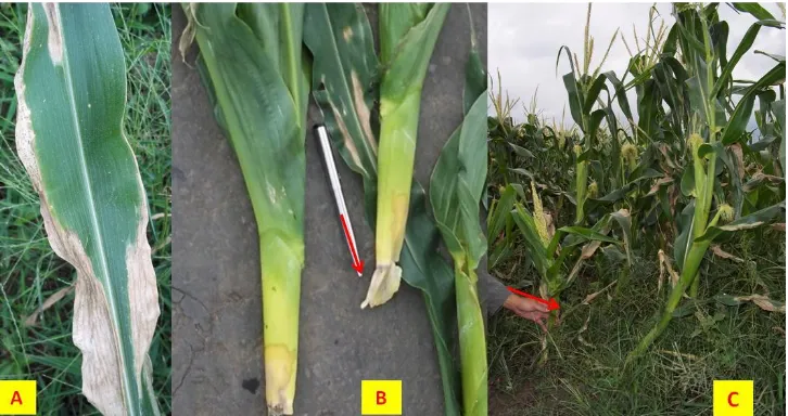
Figure 1. Disease symptoms on corn observed in the field. A, leaf blight; B, stalk rot; C, dwarf Bacterial Pathogen Isolation and Hypersensitive
Assay
Four bacterial strains i.e. BB1, BD1, BB2, BB3 were isolated from leaves and stalks of corn showing disease symptoms. The bacteria were able to grow on NA medium in 24 hours at room temperatures. Colonies were circular, 1-2 mm, somewhat convex with flat edges. All isolates were Gram-negative rod, facultative an-aerobic and showed yellow colonies in YDC medium. All bacterial strains produced HR in tobacco leaves at 48 hours after infiltration.
Pathogenicity Test
When 10 days old seedlings were infiltrated with suspension of all bacterial strains, water soaked emerged in 2 days after inoculation. The symptoms changed into blight with irregular edges two days later. When the bacterial suspensions were injected into the stems of 10 days old corn plants, only BD1 and BB2 strains produced wilt symptoms within a week (Figure 2). These strains induced consistent symptom in corn plants similar to those observed in the field. Control plants inoculated with water did not develop any symptoms. Bacterial pathogens were successfully reisolated from the symptom of plants inoculated with strain BD1 and BB2. Colonies recovered diseased plants were also yellow, Gram negative, and facultative anaerobic.
Physiological and Biochemical Characteristics The results of physiological and bio-chemical tests showed that strain BD1 and BB2 isolated from leaves and stem respectively had nearly similar characteristics, with the exception on the use of carbon from meso inositol assay. Characteristics of BD1 and BB2 strains resembled more to P. ananatis, but they had unique main characters similar to P. stewartii subsp. stewartii i.e. non-motile and were unable to produce indole. These results showed both BD1 and BB2 strains have physiological and biochemical characteristics quite differently from P. stewartii described by Schaad et al. (2000).
290
Lilis Suryani et al.: Characterization of Bacterial Pathogen Causing Wilt and Leaf……….
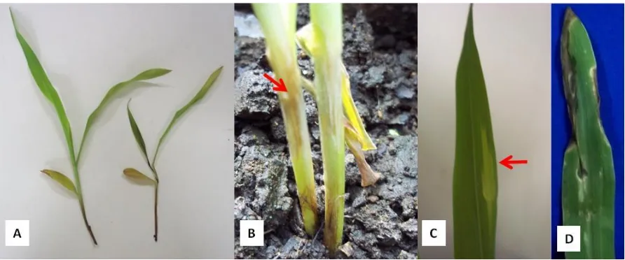
Figure 2. Symptoms on the pathogenicity test, A. wilt symptom (right) compared to control (left), B. watersoak appeared on stalk, C. water soak appeared on leaf, D. leaf blight
Detection of Bacterial Strains using Two Species-Specific Primer Pairs For P. stewartii subsp. stewartii
Primers ES16/ESIG2c were designed for the detection of P. stewartii subsp. stewartii to amplify the unique sequence of 16S to 23S rRNA intergenic transcribed spacer (ITS) region (Coplin et al.,2002). It is known that DNA sequences in the 16S to 23S intergenic spacer region exhibit a great deal of sequence and length variation. The variations in this region have been shown to be useful for differentiating species of prokaryotes (Barry et al., 1991).
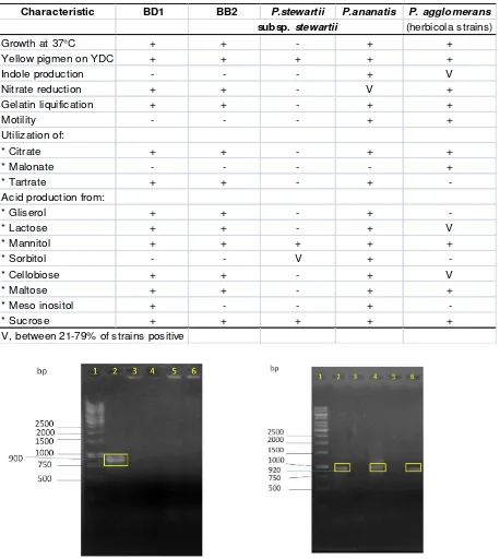
Electrophoresis product using primer ES16/ESIG2c (Figure 3) indicated that 0,92 kb amplicons were detected on the BD1 and BB2 strains, but no amplicons were detected on the BB1 and BB3 strains. Positive control of genomic DNA of P. stewartii subsp. stewartii DM5262 strain also showed the amplicon, suggesting the positive result of both BD1 and BB2 strains.
The PCR assay using HRP1d and HRP3r primers derived from P. stewartii subsp. stewartii hrpS gene region showed that no amplicon was shown in all of 4 bacterial strains. Positive control of genomic DNA of P. stewartii subsp. stewartii DM5262 strain showed the amplicon, suggesting the negative result of the existence of P. stewartii subsp. stewartii hrpS gene in all 4 bacterial strains.
The ability of plant-pathogenic bacteria to elicit the hypersensitive response (HR) in resistant or non-host plants is correlated with their ability to cause disease in susceptible hosts. These capabilities are determined by hypersensitive response and pathogenicity (hrp) genes, which encode components of a protein secretion pathway, regulatory factors, and a number of secreted effector proteins. P. stewartii subsp. stewartii posses hrpS, one of hrp genes which involved in the HR elicitation in non host plants as well as pathogenicity in host plants (Lindgren, 1997)
291
Lilis Suryani et al.: Characterization of Bacterial Pathogen Causing Wilt and Leaf……….
Table 1. Physiological and biochemical characteristics of the bacterial strains isolated from corn, and published strains of Pantoea stewartii subsp. stewartii, Pantoea ananatis and Pantoea agglomerans (Schaad et al., 2000)
Characteristic BD1 BB2 P.stewartii P.ananatis P. agglomerans subsp. stewartii (herbicola strains)
Growth at 37°C + + - + +
Yellow pigmen on YDC + + + + +
Indole production - - - + V
Nitrate reduction + + - V +
Gelatin liquification + + - + +
Motility - - - + +
Utilization of:
* Citrate + + - + +
* Malonate - - - - +
* Tartrate + + - +
-Acid production from:
* Gliserol + + - +
-* Lactose + + - + V
* Mannitol + + + + +
* Sorbitol - - V +
-* Cellobiose + + - + V
* Maltose + + - + +
* Meso inositol + - - +
-* Sucrose + + + + +
V, between 21-79% of strains positive
292
293
Lilis Suryani et al.: Characterization of Bacterial Pathogen Causing Wilt and Leaf……….
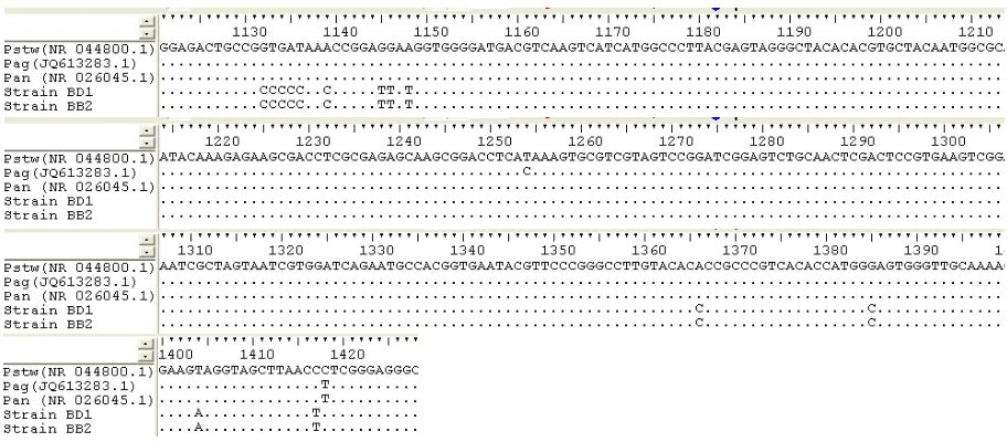
Figure 4. Sequence alignment of the 16S rRNA genes of P. stewartii subsp. stewartii (Pstw), Pantoea ananatis (Pan), Pantoea agglomerans (Pag), strain BD1, and strain BB2. Identical nucleotides are indicated by dotes, and deletions are indicated by dashes.
Multiple alignments of nucleotide sequences were performed using Clustal W program. The sequences were align with the nucleotide sequen-ce of P. stewartii subsp. stewartii strain ATCC 8199 as well as those of P. ananatis and P. agglomerans which has close relationship to P. stewartii subsp. stewartii. The BD1 and BB2 showed high similarity to P. stewartii subsp. stewartii, although several bases have been changed (Figure 4).
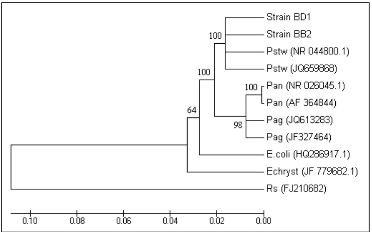
The phylogenetic relationship derived from a neighbor-joining analysis of the pairwise comparison among the partial 16S rRNA gene sequences of both BD1 and BB2 strains with six sequences of well described species of the genus Pantoea is shown in Figure 5. Escherichia coli, Erwinia chrysantemi, and Ralstonia solana-cearum were used as the out group taxons. The sequences of P. ananatis, P. agglomerans, P. stewartii, E. coli, E. chrysantemi, R. solanacea-rum were obtained from the GenBank/EMBL databases. The tree diagram demonstrated that the sequences of BD1 and BB2 are clustered in a group with P. stewartii subsp. stewartii supported by high bootstrap value of 100.
294
Lilis Suryani et al.: Characterization of Bacterial Pathogen Causing Wilt and Leaf……….
Figure 5. Phylogenetic tree showing the relationship among selected partial 16S rRNA gene sequences from Pantoea spp. and strains isolated from corn. The numbers at the nodes indicate the levels of bootstrap support based on data for 1,000 replicates. Accession numbers were showed in brackets. Bar represents genetic distance. Pan = P. ananatis, Pag = P. agglomerans, Pstw = P. stewartii, E. coli = Escherichia coli, Echryst = E. chrysantemi, Rs = R. solanacearum
CONCLUSION
Based on all tests performed, bacterial pathogen causing wilt and leaf blight on corn found in Batu was Pantoea spp. The bacteria had physiological and biochemical characteris-tics quite different from other published strains of P. stewartii subsp. stewartii, indicated that BD1 and BB2 are possibly unique strains of Pantoea spp. Further research on the disease is necessary to develop strategies to manage the disease as well as to prevent greater losses.
ACKNOWLEDGEMENTS
We would like to thank Drs. David L. Coplin and Doris R. Majerczak from Ohio University for their kindness to provide DNA sample of P. stewartii subsp. stewartii DM5262 strain as positive control. This research was funded by Dit.Litabmas Dikti, through Grand Research of Decentralized University received by Dr. Arifin Noor Sugiharto.
REFERENCES
Akil, M. and H.A. Dahlan. 2010. Budidaya jagung dan diseminasi teknologi. Dalam Jagung: teknik produksi dan pengem-bangan. Litbang Deptan. Jakarta. p.192-204. Ausubel F.M., R. Brent, R.E. Kingston, D.D.
Moore, J.G. Seidman, J.A. Smith and K. Struhl. 1996. Current protocols in mole-cular biology. John Wiley and Sons, New York.
Barry, T., G. Colleran, M. Glennon, L. Dunican and F. Gannon. 1991. The 16S/23S ribo-somal spacer as a target for DNA probes to identify eubacteria. PCR Methods Appl. p.151-156.
BPS. 2011. Tanaman pangan. http://www.bps. go.id/tnmn_pgn.php
295
Lilis Suryani et al.: Characterization of Bacterial Pathogen Causing Wilt and Leaf……….
Coplin, D.L., D.R. Majerczak, Y. Zhang, W.S. Kim, S. Jock and K. Geider. 2002. Identi-fication of Pantoea stewartii subsp. stewartii by PCR and strain differentiation by PFGE. Plant Dis. 86: 304-311.
Freeman, N.D. and J.K. Pataky, J.K. 2001. Levels of Stewart’s wilt resistance ne -cessary to prevent reductions in yield of sweet corn hybrids. Plant Dis. 85: 1278-1284.
Kasryno, F., E. Pasandaran, Suyamto, M.O. Adnyana. 2010. Gambaran umum eko-nomi jagung Indonesia. Dalam Jagung: teknik produksi dan pengembangan. Litbang Deptan. Jakarta. p. 474-497. Lindgren P.B. 1997. The role of hrp genes
during plant bacterial interactions. Annual Review of Phytopath. 35: 129-152.
Lipps, P.E., A.E. Dorrance and D.R. Mills. 2003. Stewart's bacterial wilt and leaf blight of corn. Ohio State University Fact Sheet. http://ohioline.osu.edu.
Menelas, B., C.C. Block, P.D. Esker and F.W. Jr. Nutter. 2006. Quantifying the feeding periods required by corn flea beetles to acquire and transmit Pantoea stewartii. Plant Dis. 90: 319-324.
Mergaert, J., L. Verdonck and K. Kersters. 1993. Transfer of Erwinia ananas (synonym, Erwinia uredovora) and Erwinia stewartii to the Genus Pantoea emend. as Pantoea ananas (Serrano 1928) comb. nov. and Pantoea stewartii (Smith 1898) comb, nov., Respectively, and description of Pantoea stewartii subsp. indologenes subsp. nov. Int. J. Sistematic Bacteriol. 43(1): 162-172.
Michener, P. M., J. K. Pataky and D.G. White. 2002. Rates of transmitting Erwinia stewartii from seed to seedlings of a sweet corn hybrid susceptible to Stewart’s wilt. Plant Dis. 86: 1031-1035.
Pataky, J.K. 2003. Stewart’s wilt of corn. APSnet, American Phytopathological So-ciety, St. Paul. MN, U.S.A. Published on-line.
Pepper, E. H. 1967. Stewart’s bacterial wilt of corn. Monograph 4. American Phyto-pathological Society, St. Paul, MN, U.S.A Rahma, H. and Armansyah. 2009. Penyebaran
penyakit Stewart oleh bakteri Pantoea stewartii sebagai penyakit baru pada tanaman jagung (Zea mays) studi kasus di Pasaman Barat. Working Paper. Fakul-tas Pertanian. Univ. Andalas. http://lp. unand.ac.id/?pModule=penelitian&pSub= penelitian&pAct=detail&id=9&bi=2
Schaad, N.W., J.B. Jones and W. Chun . 2000. Laboratory guide for identification of plant pathogenic bacteria. Third Edition. Gail Hoover. Maryland. pp.372.
Tamura, K., D. Peterson, N. Peterson, G. Stecher, M. Nei and S. Kumar. 2011. MEGA5: Molecular evolutionary genetics analysis using maximum likelihood, evolutionary distance, and maximum parsimony methods. molecular biology and evolution (submitted).