Considerations of tooth extraction during pregnancy
M. Hendra Chandha Departement of Oral Surgery
Faculty of Dentistry Hasanuddin University
ABSTRACT
Pregnancy is a common sense. However, when dental care, is likely to perform in particular tooth extraction, a dentist should be aware of any systemic changes occurred during pregnancy that can lead to several problems in dental practice. This paper is aimed to discuss the consideration of taking dental radiography and prescribing medicine for an expectant mother that can't be avoided during oral surgery.
Key words: tooth extraction, pregnancy
ABSTRAK
Kehamilan adalah suatu kondisi yang umum terjadi. Akan tetapi jika pada kondisi demikian membutuhkan perawatan dalam bidang kedokteran gigi, seorang dokter gigi harus waspada. Hal ini disebabkan terdapat beberapa perubahan sistemik yang cenderung dapat menjadi masalah, khususnya jika tindakan pencabutan gigi yang akan dilakukan. Makalah ini mendiskusikan pertimbangan dari praktisi kedokteran gigi pada ibu hamil mengenai pemberian obat-obatan dan radiografi, karena hal ini tidak dapat dipisahkan dari prosedur pembedahan.
Kata kunci: pencabutan gigi, kehamilan
Correspondence: M. Hendra Chandha, Department of Oral Surgery, Faculty of Dentistry Hasanuddin University, Jl. Kandea No.5 Makassar, Indonesia.
INTRODUCTION
Tooth extraction during pregnancy seems to be a common practice. Nevertheless, the opinion found in our society suggests that dental care of an
expectant mother is directed to prevent and restore the teeth while surgical procedure should be delayed until delivery.
problems that are normally absent before pregnancy. This can be marked with the increased sensitivity of gingival area to irritation. When pericoronal infection occurs during pregnancy, tooth extraction may be provided.¹·²
This article is aimed to present detail information of systemic changes during pregnancy, the considerations as well as the safer management for mother and fetus
when extraction or other oral surgery procedure is indicated.
Systemic changes during pregnancy To define rational management guidelines, review of the normal processes of pregnancy and fetal developmental is first necessary. Endocrine changes are the most significant basic alterations that occur with pregnancy. The result as the production of maternal and placental hormones increases and activity of target end organs are modified.³
Increased hormonal secretion and fetal growth induce several systemic, as well as local physiologic and physical changes in a pregnant woman. Fatigue is a common physiologic finding in the first trimester that
has psychologic impact.
A tendency also exists for syncope and postural hypotension. During the second
trimester, patients typically have a sense of well being and relatively few symptoms. Local physical changes occur in different parts of the body, including the oral cavity. During the third trimester, increasing fatigue, discomfort, and mild depression may be seen.³
Cardiovascular change
The main cardiovascular changes are
an increase in the total blood volume and cardiac output, a decrease in blood pressure, and the potential occurrence of the supine hypotension syndrome. Blood volume increases 40%, cardiac output increases 30% to 40% whereas the red blood cell volume increases only about 15% to 20%.3,4
The total blood volume increases by 40% to 50% by the 32nd week of gestation, caused primarily by a 40% to 50% increase in plasma volume. In addition to an increase in the plasma volume there is also a 30% increase in the red cell volume contributing to the increase of the total blood volume. An increase in cardiac output by 30% to 50% occurs between the 25th and the 33rd week of pregnancy secondary to an increase in stroke volume. These changes produce a
The increase in cardiac output also
The respiratory changes occurring during pregnancy accommodate the increasing size of the developing fetus and the maternal-fetal oxygen requirements. The main changes in the respiratory system are dyspnea, hyperventilation, alterations in the oxygen intake and reserve, and an increase in both the tidal volume and minute ventilation rate.4
Anxiety and pain can cause a pregnant patient to increase her respiratory rate to the point of significant hypocapnea, which results in faintness and perioral numbness. These ventilator changes cause increased rate of respiration (tachypnea) and dyspnea that is aggravated by the supine position.3,5
Hematological changes
Significant hematological changes include an increase in red blood cells, white blood cells, erythrocyte sedimentation rate, and all coagulation factors, except factors XI and XIII, and a decrease in the hemoglobin content of blood. The increase in plasma volume is disproportionately greater than the increase in the red cell volume resulting in hemodilution and, hence, a physiological
anemia.4
Anemia occurs because blood volume increase more than red blood cell mass does. As a result a fall in hemoglobin and a marked need for additional folate and iron occurs. White blood cell count also increase because of a neutrophilia. This increased level of neutrophils can complicate interpretation of the complete blood count during infection.3
Although changes in platelets are usually insignificant, several blood clotting factors (especially fibrinogen; factors VII, VIII, IX and X, and fibrin-plit products) are increased. Estrogen increases the hepatic production of coagulation factors, yielding a 30-50% increase in fibrinogen and factors VII, VIII, IX, and X. This hypercoagulation
Gastrointestinal and liver changes
Mechanical changes resulting from an enlarging fetus, in combination with hormonal changes, are responsible for alterations in the gastrointestinal (GI) system. The main GI changes are nausea, vomiting, and heartburn. Nausea and vomiting occur in about 66% of pregnant women beginning approximately 5 weeks after the last menstrual period and peaking
between 8 and 12 weeks. Thereafter, the symptoms decline gradually.
Hypermesis gravidarum (excessive and an uncontrolled vomiting), occurs in less than 1% of all pregnancies. The pathophysiology of nausea and vomiting during pregnancy is poorly understood, but is thought to be due to the hormonal effects of estrogen and progesterone. For pregnant women with hypermesis gravidarum requiring dental treatment, morning appointments should be avoided.4
Physiologic changes in the GI tract during pregnancy increase the risk of aspiration during surgery and anesthesia. During dental procedures, pregnant patients should be seated in semi-supine or comfortable position. In case of vomiting,
the procedure should be stopped immediately and the patient should be repositioned upright. When the vomiting is
over, rinsing the mouth with cold water or a mouthwash is recommended.4,5
Renal and genitourinary changes
The principal renal and genitourinary changes increased glomerular filtration rate (GFR), biochemical changes in the urine and blood, increased frequency of urination, urinary statis, and urinary tract infections. The 50% increase in blood volume during
pregnancy results in an increased renal plasma flow and an increased GFR.4,5
Oral and facial changes
Oral changes seen in pregnancy include gingivitis, gingival hyperplasia, pyogenic granuloma, and salivary changes. Elevated circulating estrogen, which causes increased capillary permeability, predisposes pregnant women to gingivitis and gingival hyperplasia. Increased angiogenesis, due to the sex hormones coupled with gingival irritation by local factors such as plaque, is believed to cause pyogenic granuloma. It occurs mainly on the labial aspect of the interdental papilla. It can happen at any time during pregnancy, but is reported to be most common in first pregnancies, during the first
and second trimesters.
and an increase in potassium, protein, and estrogen levels. Salivary estrogen also increases the proliferation and desquamation of the oral mucosa and an increase in subgingival crevicular fluid levels. The desquamating cells provide a suitable environment for bacterial growth by providing nutrition predisposing the pregnant woman to dental caries.
There is an increase in facial
pigmentation called melasma or “mask of
pregnancy,” appearing as bilateral brown
patches in the mid-face. These facial changes begin during the first trimester and are seen in up to 73% of pregnant women. The etiology of this condition is unknown, but is believed to be related to the increase in serum estrogen and progesterone. Melasma usually resolves after parturition.4
administration. It is virtually impossible to perform an oral surgical procedure properly with neither radiographs nor the
administration of medications; therefore one option is to defer any elective oral surgery until after delivery to avoid fetal risk.
However, if surgery during pregnancy cannot be postponed, efforts should be made to lessen fetal exposure to teratogenic factors.6
The first trimester
The most critical and rapid cell division and active organogenesis occur between the second and the eight week of postconception. Therefore, the greater risk
of susceptibility to stress and teratogens occurs during this time and 50% to 75% of all spontaneous abortions occur during this period. Drugs and infection during this period can interfere with this process and lead to grave congenital anomalies. If an oral surgical procedure is necessary during the first trimester of pregnancy, local anesthetic would be the method of choice, if possible.4,7
The second trimester
Organogenesis is completed and therefore the risk to the fetus is low. This is the safest period for providing dental care during pregnancy.4
The third trimester
of delivery. Although there is no risk to the fetus during this trimester, the pregnant mother may experience an increasing level of discomfort. Short dental appointments should be scheduled with appropriate positioning while the chair to prevent supine hypotension. It is safe to perform routine dental treatment in the early part of the third trimester, but from the middle of the third trimester routine dental treatment should be
avoided.4,7
Dental radiographs
Dental radiography is one of the more controversial areas in the management of a pregnant patient. Irradiation should be avoided during pregnancy, especially during the first trimester, because the developing fetus is particularly susceptible to radiation damage. However, should dental treatment become necessary, radiographs may be required to accurately diagnose and treat the patient. Therefore the dentist must be aware of how to proceed safely in this situation.3
Teratogenecity of radiation depends of fetal age and the dose of radiation. The greatest risk of the fetus for teratogenecity and death is during the first 10 days after
conception. The most critical period of fetal development is between 4 and 18 weeks after conception.4
The safety of dental radiography has been well established, provided features such as fast exposure techniques (e.g. high speed film or digital imaging), filtration, collimation, and lead aprons are used. Of all aids, the most important for the pregnant patient is the protective lead apron.3
The National Commission for Radiation Protection (NCRP) recommends that the cumulative fetal dose should not
exceed 0.005 Gy. Fetal exposure to radiation of more than 0.20 Gy will cause microcephaly and mental retardation. Radiographs employed in dentistry such as the panoramic and full mouth intraoral series are generally safe during pregnancy.4
Despite the negligible risks of dental radiography, the dentist should not be cavalier regarding its use during pregnancy (or at any other time, for that matter). Radiographs should be used selectively and only when necessary and appropriate to aid in diagnosis and treatment.3
Drug administration during pregnancy4 Another controversial area in treating the pregnant dental patient is drug administration. The principal concern is that
cause maternal hypoxia, resulting in fetal hypoxia, injury, or death. Ideally, no drug should be administered during pregnancy, especially the first trimester. However, adhering to this rule is sometimes impossible.
The FDA has categorized the potential for drugs to cause birth defects, providing definitive guidelines for prescribing drugs during pregnancy. They
are as follow: (1) category A-Controlled human studies indicate no apparent risk to the fetus. The possibility of risk to the fetus is remote. (2) Category B-Animal studies do not indicate fetal risk. Well-controlled human studies have failed to demonstrate a risk. (3) Category C-Animal studies show an adverse effect on the fetus but there are no controlled studies in humans. The benefits from use of such drugs may be acceptable. (4) Category D-Evidence of human risk, but in certain circumstances the use of such a drug may be acceptable in pregnant women despite its potential risk. (5) Category X-Risk of use in pregnant women clearly outweights possible benefits.
Local anesthetics
Local anesthetics pass the placental barrier by passive diffusion, but most of
them are considered to be safe and nonteratogenic.4,7
If possible, it would be wise to avoid local anesthetics with vasoconstrictors. The using of epinephrine, a natural hormone, in local anesthesia in the doses used for dental treatment is not associated with fetal abnormality, and is considered to be safe during pregnancy. Although epinephrine is not teratogenic, caution should be taken to
avoid accidental intravenous administration. 4,8
Antibiotics
Selection of an antibiotic for pregnant or nursing women must be made with equal consideration for mother and child. Antibiotics with systemic effects cross the placenta and reach the fetus. When prescribing approved antibiotics to pregnant women, it is important to remember that overall physiological changes that accompany pregnancy, particularly in the third trimester, reduce the serum concentration of antibiotics. Consequently, an adaptation often a doubling-of the therapeutic dose is recommended.9
Analgesics
pregnancy. It can be used in any stage of pregnancy and in nursing mothers. Maternal anemia and fetal renal disease was reported, however, used in high doses.4
Sedatives and hypnotics
Nitrous oxide (N2O) has not been classified into any category by the FDA and its use in pregnancy is controversial due to unproven deleterious effects on the pregnant
women and fetus. Nitrous oxide also causes
vasoconstriction and may reduce uterine blood supply. However, a single exposure of nitrous oxygen (N2O-O2) for less than 35 minutes has not been associated with any human fetal anomalies, including low birth rate. Chronic exposure of pregnant dental health workers to N2O for more than 3 hours without the use of scavengers has resulted in decreased fertility and
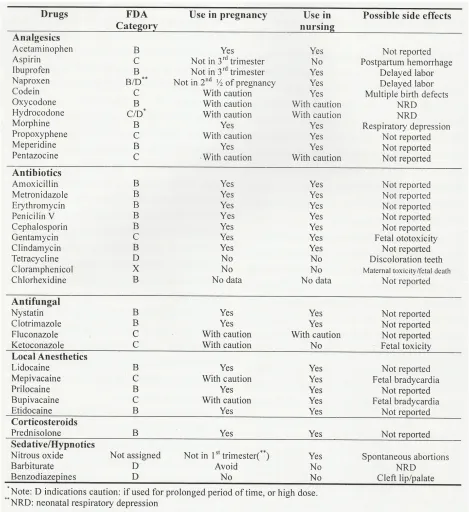
Tabel 1. Common drugs used in dentistry 6
Maternal physiology in pregnancy pre-eclampsia
milimeter of mercury (mmHg) after 20 weeks of pregnancy, usually with higher-than-normal levels of protein found in a urine sample. The condition is characterized by elevated blood pressure, pathologic edema, and proteinuria. Seizures, renal failure, pulmonary edema and thrombocytopenia may be associated with PIH and result in eclampsia.2,10
Pre-eclampsia progressing to
eclampsia if seizures and comma develop. It is essential thatany pregnant patient with hypertension, edema or abnormal weight gain be thoroughly evaluated for this condition before dental care.2,3
Hypotension
Hypotension must be avoided during pregnancy. Compression of the inferior vena cava by the enlarged uterus may induce hypotension and syncope. A sustained episode of this type may result in fetal hypoxia and injury. The supine position may be optimal for many dental procedures but it should be avoided whenever possible and particularly in the third trimester. The supine position may produce maternal and fetal hypoxia and hypotension due to
compression of the inferior vena cava by the enlarged uterus compressing against the spinal column and elevation of the
diaphragm due to the weight of abdominal contents.2
Pregnant dental patients should be placed in a semi-reclined or upright position for dental treatment. If hypotension or syncope occurs during dental treatment of patients in the third trimester, the patient Supplemental oxygen also is recommended during such episodes. If hypotension is associated with sustained bradycardia, administration of atropine may be necessary.2
Pulmonary response
Pulmonary response of the third trimester is in part due to the elevation of the diaphragm, which results in tachypnea especially when reclining. Postural response often makes the patient wish to sit up with hands on knees, leaning forward with legs parted to accommodate the
fetus. While this is not a good position for much dental treatment, sitting in the chair
Hematologic response
The hematologic response of pregnancy is to produce a hypercoagulable state in the average patient due to inhibited fibrinolysis. There is an increased risk of deep vein thrombosis and pulmonary embolism from the first trimester. Compression of the inferior vena cava in the supine position, continuous flexing of the
knees or pressure to the back of the calves in the dental chair should be avoided. These can produce venous stasis which when combined with the hypercoagulability state can result in deep vein thrombosis.2
SUMMARY
In the summary, the dentist who will provide dental care to a pregnant patient must understand about the conditions occured to their patient, such as systemic and oral changes. There are considerations that a dentist should be aware while performing surgical procedure for a pregnant patient. In the first trimester, a higher risk may be occurred because they are more sensitive to stress and teratogenic agents, those can lead to a spontaneous
abortion. Administration of antibiotics to treat infection in this period can cause some congenital anomalies. Second trimester is a
safest period to provide a dental care. Same condition can be found in the early trimester that dentist can arrange the schedule for dental treatment. However in the middle third trimester, dentist must be careful with the routine dental treatment, because in this period the blood volume is highly increased. There are
some physiological problems in an expectant mother, that the dentist must be
aware, include pre-eclampsia, hypotension, pulmonary reaction, and hematologic reactions. emergencies in dentistry. Toronto: W.B Saunders Company; 2002.p.494-7
3. Little JW, Falace DA, Miller CS, Rhodus NL. Dental management of the medically compromised patient. St Louis: Mosby; 2002.p.303-4,306-10.
5. Newton ER. Trauma and pregnancy. Availableat:
http://www.wmedicine.com/med/topic 3268.htm.Accessed:February 26, 2006. 6. Peterson LJ. Oral and maxillofacial Surgery. 4th edition. Ohio: Mosby; 2003.p.20
7. Archer WH. Oral and maxillofacial Surgery. 5th edition. Toronto: W.B. Saunders Company; 1975.p.20-1
8. University of Southern California School of Dentistry. Center for Diagnostic Science bulletin. Available at:http://www.usc.edu/hsc/dental/stude nts/cds_bulletin/2005_may_cd. pdf-. Accessed: 26 Februari 2006
9. Newman MG, Winkelhoff AJ. Antibiotic and antimicrobial use in dental practice.
2nd edition. Prague: Quintessence Publishing Co; 2001.p.236-7