P-ISSN.2503-0817, E-ISSN.2503-0825
CrossMark
Abstract
Objective: Myrmecodia pendens is often used as a traditional medicine to treat various diseases. A previous study showed that ethanol extract of sarang semut plant (hypnophytum furmicarum jack) 4.65 mg was effective in fastening establishment of socket granulation tissue after tooth extraction; likewise, the myrmecodia pendens at 3% also fastens the healing process of soft-tissue wound after tooth extraction. The aim of this study is to assess the effect of ethanol extract of 10% myrmecodia pendens on the expression of TGF-β1 and determine the number of osteoblasts that remain after tooth extraction.
Material and Methods: This study used 24 male marmots divided into 2 groups. All of the groups were extracted in the 1st left-incisor mandibula and 0.1 ml of 0.5% CMC was applied into the socket control group. The treatment group was given 0.1 ml ethanol extract
of myrmecodia pendens 10% dissolved with CMC 0.5%. On day 3, 7, 14 and 21, marmots were decapitated and histopathologic preparations were observed to study the expression of TGF-β1 and the number of osteoblast cells. Data were analyzed using Kruskall–Wallis test with a probability p < 0.05.
Results: Based on the Kruskal–Wallis test, there was a significant difference in the TGF-β1 expression and the number of osteoblast cells after the administration of ethanol extract of myrmecodia pendens between the control group and treatment group treatment (p < 0.05).
Conclusion: The ethanol extract of myrmecodia pendans can fasten the healing of the wound resulting from tooth extraction; it accelerates the healing process by increasing the expression of TGF-β1 and the number of osteoblast cells.
Keywords: Myrmecodia pendens, Osteoblasts, TGF-β1, Tooth extraction
Cite this Article: Ismardianita E, Nasrul E, Yanwirasti, Hemiawati M. 2017. Effect of ethanol extract of myrmecodia pendens on TGF-β1 expression and osteoblast cells after tooth extraction (experimental research on cavia cobaya). Journal of Dentomaxillofacial Science 2(3): 150-154. DOI: 10.15562/jdmfs.v2i3.608
Effect of ethanol extract of myrmecodia pendens on
TGF-β1 expression and osteoblast cells after tooth
extraction (experimental research on cavia cobaya)
Efa Ismardianita1*, Ellyza Nasrul2, Yanwirasti3, Mieke Hemiawati4
I
ntroduction
Tooth extraction often causes wounds. The wound can heal normally, but sometimes it causes some complications that slow down the healing. The rate of likelihood of complications arising from wounds caused by tooth extraction is between 1% and 11.5%1
; research shows that as many as 11 million patients experienced discomfort and complications after tooth extraction, such as pain, swelling, hema-tom and difficulties in mastication and speech.2
To prevent such complications, dentists made some efforts in finding out the right technique to min-imize post-surgical trauma and prescribe medicines appropriately, to treat both systematic or local complications. Antiseptic is the choice for local application in treatment. Antiseptic has several advantages; it kills bacteria and prevents bacter-imia; however, there are advantages it using anti-septics, including the difficulty in finding out the right kind of medicinal plant that provides the best possible remedies . Myrmecodia penden is a medic-inal plant. Based on phytochemical test, we found that myrmecodia pendens contains flavonoids,
triterpenoid, phenols and tanin that can be used for anti-inflamation, antibacterial and antioxidant effects.3
The wound healing process that takes place after tooth extraction involves chemotaxis of various cells, cytokines and growing factor. The growing factor responses to early injury on soft and hard tissues include TGF-β1, BMPs, VEGF, IGFs and PDGF. Transforming growth factor Beta-1 during the inflammation phase initiates and controls chemotaxis, activation and survival of inflamma-tion cells and induces monocytes transformainflamma-tion into macrophages.4
Proliferation phase mediates migration and proliferation of endothelial cells, keratinocytes and fibroblasts composed of granulation tissue stimulates VEGF and bFGF angiogenic growth factor expression. Osteogenesis regulates the establishment of bone by inhibiting fibroblasts; it stimulates migration, proliferation, and differentiates mesenchyme cells, transforming them into osteoblasts and increases synthesis and mineralization of bone matrix.5,6
1Department of Oral and Maxillo-facial Surgery, Faculty of Dentistry, University Baiturrahmah, Padang, Indonesia
2Department of Clinical Pathologic, Faculty of Medicine, Andalas Uni-versity, Padang, Indonesia 3Departement of Biomedic, Faculty of Medicine, Andalas University, Padang, Indonesia
4Departement of Microbiology, Padjajaran University, Bandung, Indonesia
Univer-Proliferation of osteoprogenitor cells occurs on day 4, the differentiation of osteoblasts occurs on day 5 and counting is done until day 14. The final stage of osteoblast differentiation and maturation begins on day 15 and lasts until day 28.7
In the early stages of osteoprogenitor cell differentiation expression, an increase in TGF-β1 was detected; otherwise in the final stages of differentiation, the expression of TGF-β decreased.8
M
aterial and
m
ethods
The extract of ethanol contained the following: Myrmecodia pendens 70%, CMC 0.5%, Ketamin 10% (Ketalar®,Parke Davis) and Xylazine (Bayer), buffer formalin 10%, plank-Rychlo’s solution, alco-hol, ethanol, xylol, nitrat acid, PBS, xylol, H2O2, kit TGF-β1 (Santa Cruz Biotechnology), biotinylated anti-goat antibody (Dako, Glostrup, Denmark), streptavidin-horseradis peroxidase conjugate, diamino benzidine (DAB; Sigma Chemical Co.), and HE.
Myrmecodia pendens was brought from Ayawasi village Sorong district, Papua. It was extracted in Chemical Laboratory Research Center Padjajaran University Bandung. Roots of myrmecodia pendens were peeled and sliced to a length of 3-5 mm and were dried (temp 50°C) and grilled with blender. Then a wet mixture containing 250 grams of powder and ethanol 70% (1:5) was prepared. Maceration was performed (shaking the mixture for the first 6 hours and allowing the mixture to settle for the next 18 hours) repeatedly until the filtrate changed color; the filtrate was evaporated using a rotary evaporator (50°C), then fed into the oven until all the water evaporated and the extract became totally dry.
This research is a pure experimental research using post-test control-group design, using 24 cavia cobaya, aged 6-7 weeks. The animals were anesthetized by intramuscular administration of 80 mg/kg Ketamin and 160 mg/kg of Xylazine (1:1). The extraction of the lower incisors was left with needle holder, control group socket was administered 0.1 cc CMC 0.5% and the treatment group was given 0.1 ml of Myrmecodia pendens extract which was dissolved with CMC 0.5%. On days 3, 7, 14 and 21, 3 animals were decapitated and given histopathologic preparations to conduct immune histochemical test, for determining of the levels of TGF-β1 and HE and osteoblasts; the bodies were observed under light microscope with 40x magnification.
The presence of immunopositive cells according to intensity of staining score was measured based on the ollowing: (I): 0 = no expression;
1 = weak expression; 2 = medium expression; and 3 = strong expression. The presence of immu-nopositive cells based on distribution was scored as follows: (p): 1 = brown color ≤10%; 2 = brown >10–50%; and 3 = brown >50–100%. The final score of TGF-β1 immunopositive cells was calcu-lated by multiplying (p) score and (I) score with the following classification: 0 = no expression; 1: 1–2 = weak positive expression; 2: 3–5 = medium positive expression; and 3: >6 = strong positive expression. The number of osteoblasts was scored as follows: 0=<10: very less; 1 = 11–50: less; 2 = 51-80: medium; 3 =>80: strong.9
Osteoblasts were scored as follows: <10: very less; 11–50: less; 51–80: moderate; and >81: fair.
Re
sults
TGF-β1 expression was detected as brown color on the cytoplasm, osteoblast membrane cells and a new matrix that was formed (collagen and osteoid). The mean of TGF-β1 expression of control group and treatment group is presented in figure 1.
TGF-β1 imunohistochemical expression in control group and treatment group is shown in
figure 2.
Based on the results of statistical analysis (Kruskall–Wallis test), we could see there were significant differences in TGF-β1 expression between the control group and the treatment group; p = 0.0125. It can be concluded that the extract ethanol Myrmecodia pendens can increase the expression of TGF-β1.
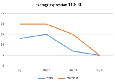
The mean number of osteoblasts in control group and treatment group is presented in figure 3.
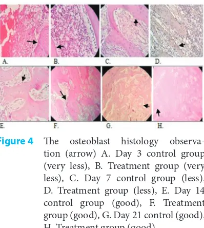
The number of osteoblast cells in the control group and the treatment group is presented in
figure 4.
Results of Kruskall–Wallis test showed there is a significant difference in the number of osteoblasts between the control group and the treatment group;
p value = 0.0034. Thus, results proe that that the extract ethanol myrmecodia pendens can increase the number of osteoblast cells.
Di
scussion
TGF-β1 expression in both groups shows a different pattern. On day 3, the TGF-β1 expression in the treatment group increased and reached its peak. The present study shows results which vary from the previous studies. Expression of TGF-B1 increased post-trauma and lasted until day 4.10,11
The highest level of TGF-β1 expression occurred
day 3 in the treatment group, which could have been caused by myrmecodia pendens’ active substances. Phytochemical test result showed that myrmecodia pendens contained flavonoid, saponin, triterpenoid, tannin and phenolics, all of which could have aided the healing process through different mechanisms.
As an anti-inflammatory agent, flavonoid decreases the permeability of capiler and substracted oedem.12 Inhibitory functions of arachidonate
acid through lipoxygenase and cyclooxygenase pathway cause the inhibition of prostaglandin and leukotriene biosynthesis; as a result, production of prostaglandin, prostacyclic, thromboxane and leukotriene is inhibited so that inflammation is contained.10Inflammation phase becomes shorter
and comes to the proliferation phase rapidly.12
Flavonoid inhibites neutrophils degranulation so that it subtracts the extrication of arachidonate acid. Flavonoid could also inhibit leukocytes accu-mulation in inflammation area, decrease leukocytes adhesion into endotel and reduce the inflammation response.13,14
As antimicrobial agents flavonoid and tannins disturb nucleic acid synthesis and ATPase enzyme activities inhibit metabolism stripe so that bacteria cannot cause further infection and if infection could not be contained the patient’s condition will become lethal.15,16Triterpenoid inhibits the growth
of bacteria by disturbing the process of forma-tion of bacteria cell membrane.16Tannins inhibits
transcriptase reverse enzyme and topoisomerase DNA, so that bacteria cannot be formed.15Saponin
prevents infection by preventing bacteria from lengthening the pro-inflammation cycle.17,18
In the control group, expression increased on day 3, the peak was observed on day 7, on day 14
Figure 2 Immunohistochemical view for pos-itive identification of expression of TGF-β1 on day 3 in collagen mesh (arrow). A. Control group (medium positive), B. Treatment group (strong positive). Day 7 in fibrous tissue, C. control group (strong positive) D. Treatment group (strong positive). Day 14 in osteoblasts. E. Control group (weak positive), F. Treatment group (medium positive). Day 21 in trabecular bone, G. Control group (weak positive), H. Treatment group (weak positive)
Figure 3 The mean of osteoblast cell number in control group and treatment group
and day 21 the expression decreased (following a normal healing pattern). The TGF-β1 distribution and intensity increased after trauma and lasted until day 4, due to the absence of macrophages migra-tion, endothelial cells, keratinocytes, fibroblasts and osteoclasts on the wound area in which the cell secreted TGF-β1; as the result, TGF-β1 increased.8,9
TGF-β1 expression on day 7 in the treatment group is different from Lalani’s research10
; on day pendens extract indeed helps fasten the inflammation phase and pushes the inflammation process into proliferation phase. Flavonoid can manage cell function by stimulating the production of TGF-β1, fibroblasts, keratinocytes, endothelial cells and osteoblasts. This moment is the early stage of osteoblast differentiation.5,6,12
During this process of differentiation, TGF-β1 expression reaches its peak,8
which in turn leads to a considerable increase in the proliferation of osteoblasts.12
On day 14, TGF-β1 expression decreases; the said decrease is caused not only by a decrease in the number of chemotactic and mitogenic endotel cells, macrophages, fibroblasts and keratinocytes, but also by various processes such as angiogenesis, epithelization, and fibroplasia; fibroplasia is the last step, that is, it is stage of suppression of inflam-mation. In the stage of suppression, angiogenesis, epithelization and collagen fibers reach normal levels, and thus proliferation is no longer needed. Some fibroblasts turn to be fibrocytes and others differentiate into osteoblasts.12
On day 21, in both groups, TGF-β1 expression decreased; it could have been caused by the last step of differentiation. In the last step of differentiation and osteoblast maturation (from day 15 to day 28) TGF-β expression begins to decrease because in the last step of differentiation, TGF-β1 inhibits the proliferation of osteoblasts; as result proliferation of osteoblasts is also reduced.8,12,19,20
Bone tissue regeneration is formed equally between bone resorption by osteoclasts and synthesis by osteoblasts. It correlates with various growth factors. Transforming growth factor induces osteo-blast activities, modulating the bone growth.4
In this research, the number of osteoblasts in both groups on day 3, 7 and 14 showed the same pattern. The mean number of osteoblasts for the treatment group is higher than that of the control group. On day 21, the number of osteoblasts observed in the treatment group is stable, whereas the number in the control group was still increasing.
On day 3, the mean number of osteoblasts in both groups was very low; this agrees well with the result of osteoprogenitor cell proliferation that happens on day 4; thus, it indicated that the osteoblasts that were found were from the rest of the old bone. On day 4, the number of osteoblasts increased; the mean number of treatment group was higher than that of control group, higher but still low in terms of research criteria. On day 14, the number of osteo-blasts increased in both groups. The rate of increase in the number of osteoblasts was very high in the treatment group. It was caused by active substances of myrmecodia pendens. Flavonoid can manage cell function by stimulating TGF-β1 production, whereas TGF-β induces migration and prolifer-ation of osteoblasts.13
Triterpenoid can stimulate osteoblast cells to form bone and flavonoid can cause osteoblast differentiation. Accordingly on day 14, the number of osteoblasts was increasing to induce osteogenesis process because osteo-blast proliferation is still needed to fill the bone defect caused by the wound resulting from tooth extraction.
C
onclusion
The application of the ethanol extract myrmecodia pendans can assist the wound healing process after tooth extraction by increasing the expression of TGF-β1. The increase in TGF-β1 stimulates migration, proliferation, and differentiation of mesenchyme cells into osteoblasts, leading to an increase in the number of osteoblast cells.
C
onflict of Interest
The authors report no conflict of interest.
Re
ferences
1. Simon E, Matee M. Post-extraction complications seen at a referral dental clinic in Dar Es Salaam. N Dent J 2001; 51: 273-276.
2. Friedman JW. The prophylactic extraction of third molars: a public health hazard. Am J Public Health 2007; 97: 1554-1559.
3. Kareem JJ. Post operative complications associated with non-surgical tooth extraction. MDJ 2008; 5: 104-113. 4. Zhao GQ. Consequences of knocking out BMP signaling
in the mouse. Genesis 2003;35: 43-56.
the large latent complex from the extracellular matrix of growth plate chondrocytes by matrix vesicle stromelysin-1 (MMP-3). Calcif Tissue Int 2002;70: 54-65.
7. Dimitriou R, Tsiridis E, Giannoudis PV. Current concepts of molecular aspects of bone healing. Injury 2005;36: 1392–1404.
8. Lalani ZW, Brey EM, Mikos AG, et al. Spatial and temporal locazation of transformasing growth factor-beta 1, bone morphogenetic protein-2 and platelet-derived growth factor-A in healing tooth extraction sockets in rabbit model. J Oral Maxillofac Surg 2003;61: 1061-1072. 9. Mohamed MAH, Younis WH, Yaseen NY. The effect
of autologous bone marrow-derived stem cells with estimation of molecular events on tooth socket healing in diabetic rabbits (immunohistochemical study). J Bagh College Dentistry 2013;25.
10. Sabir. Pemanfaatan flavonoid di bidang kedokteran gigi. Majalah kedokteran Gigi Edisi Khusus Temu Ilmiah Nasional III 2003: 81-83.
11. Nijveldt RJ, Nood E, Hoorn DEC, et al. Flavonoids: a review of probable mechanism of action and potential application. Am J Clin Nutr 2001;74: 418-425.
12. Lakhanpal P, Rai DK. Quercetin: a versatile flavonoid. IJMU 2007;2: 22-37.
13. Akiyama H, Kazuyasu F, Osamu Y, et al. Antibacterial action of several tannins against staphylococcus aureus. J Antimicrob Chemot 2001;48: 487-491.
14. Ajizah, A. Sensitifitas salmonella typhimurium terhadap ekstrak daun psidium guajava l. Journal Bioscient 2004; 1: 31-38.
15. Gautam GK, Mishra SH, Kumar A, et al. Phytochemical evaluation of the etanolit extract of bauhinia tomentosa Linn. (Root). IJPLS 2012;3: 1675-1676.
16. Robbinson T. ”The basic of higher plants 6th ed”. In Disadur PK. Kandungan organik tumbuhan tinggi. Bandung: ITB; 1995.
17. Huang Z, Nelson ER, Smith RL, et al. The sequential expression profiles of growth factors. J Tissue Eng 2007;13: 2311-2320.
18. Iwata J, Hosokawa R, Sanchez-Lara PA, et al. Transforming growth factor-beta regulates basal transcriptional regulatory machinery to control cell proliferation and differentiation in cranial neural crest-derived osteoprogenitor cells. J Biol Chem 2010;285: 4975-4982.
19. Spinella-Jaegle S, Roman-Roman S, Faucheu C, et al. Opposite effects of bone morphogenetic protein-2 and transforming growth factor-beta1 on osteoblast differentiation. Bone 2001;29: 323.
20. Zhang Z, Nakashima K, Zhou X, et al. The novel zinc finger-containing transcription factor osterix is required for osteoblast differentiation and bone formation. Cell 2008;108: 17-29.