© 2010 Psychology Press, an imprint of the Taylor & Francis Group, an Informa business
www.psypress.com/socialneuroscience DOI: 10.1080/17470911003708032
PSNS
Dysfunctions in brain networks supporting empathy:
An fMRI study in adults with autism spectrum disorders
Brain Correlates of Empathy in Autism
Martin Schulte-Rüther and Ellen Greimel
University Hospital Aachen, Aachen, and Research Center Jülich, Jülich, Germany
Hans J. Markowitsch
Bielefeld University, Bielefeld, Germany
Inge Kamp-Becker and Helmut Remschmidt
Philipps University Marburg, Marburg, Germany
Gereon R. Fink
Research Center Jülich, Jülich, and University Hospital Cologne, Cologne, Germany
Martina Piefke
Research Center Jülich, Jülich, and Bielefeld University, Bielefeld, Germany
The present study aimed at identifying dysfunctions in brain networks that may underlie disturbed empathic behavior in autism spectrum disorders (ASD). During functional magnetic resonance imaging, subjects were asked to identify the emotional state observed in a facial stimulus (other-task) or to evaluate their own emotional response (self-task). Behaviorally, ASD subjects performed equally to the control group during the other-task, but showed less emotionally congruent responses in the self-task. Activations in brain regions related to theory of mind were observed in both groups. Activations of the medial prefrontal cortex (MPFC) were located in dorsal subregions in ASD subjects and in ventral areas in control subjects. During the self-task, ASD subjects activated an additional network of frontal and inferior temporal areas. Frontal areas previously associated with the human mirror system were activated in both tasks in control subjects, while ASD subjects recruited these areas during the self-task only. Activations in the ventral MPFC may provide the basis for one’s “emotional bond” with other persons’ emotions. Such atypical patterns of activation may underlie disturbed empathy in individuals with ASD. Subjects with ASD may use an atypical cognitive strategy to gain access to their own emotional state in response to other people’s emotions.
Keywords: Emotion; Medial prefrontal cortex; Neuroimaging; Self; Theory of mind; Autism.
Correspondence should be addressed to: Martin Schulte-Rüther, Institute of Neuroscience and Medicine (INM-3), Research Center Jülich, 52425 Jülich, Germany. E-mail: [email protected]
2 SCHULTE-RÜTHER ET AL.
INTRODUCTION
Empathy can be defined as the result of psychological inferences about other persons’ mental and emotional states allowing for socially appropriate emotional responses. The ability to empathize entails both emotional and cognitive components. On the emo-tional side, empathy allows for emoemo-tional “contagion” (Singer, 2006), that is, our ability to share other people’s emotions. However, a crucial aspect of empathy is that it includes self-referential emotional cognition in order to evaluate the relationship between other people’s emotional states and one’s own emotions (Decety & Jackson, 2004; Schulte-Rüther, Markowitsch, Fink, & Piefke, 2007). Cogni-tive components of empathy are closely related to the concepts of “theory of mind” (ToM) and “mentaliz-ing” (Frith & Frith, 2003). ToM refers to the aware-ness that mental states of other people may differ from one’s own mental state. The ability to adopt others’ mental states and evaluate them from one’s own men-tal perspective drives the ability to infer and predict the intentions, beliefs, and feelings of other people and allows for successful social interaction.
Difficulties in social interaction and social cogni-tion are hallmarks of autism spectrum disorders (ASD). It has been suggested that many aspects of the observed problems in social interaction can be explained by an ASD-specific deficit in ToM (Baron-Cohen, Tager-Flusberg, & Cohen, 2000) and empathy (Gillberg, 1992). It has repeatedly been reported that individuals with ASD have profound difficulties in gaining access to intentions, beliefs, and emotions of other people (see Baron-Cohen et al., 2000 for review). Even ASD subjects with high cog-nitive abilities show impairments in diverse tasks with ToM demands (Happé, 1994). Most previous studies of ASD focused solely on the ability to infer other
people’s thoughts and intentions (for example using false-belief tasks or tasks requiring the inference of emotional states from faces), although the representa-tion of both other and self may be altered in autism (Rogers & Pennington, 1991). This idea is supported by several behavioral findings. For example, ASD subjects use fewer descriptions of their own mental or emotional states when talking about personal everyday experiences and daydreams (Hurlburt, Happe, & Frith, 1994). Furthermore, ASD subjects do not show the commonly observed memory advantage for self-related materials or self-experienced events (Lombardo, Barnes, Wheelwright, & Baron-Cohen, 2007; Millward, Powell, Messer, & Jordan, 2000; Toichi et al., 2002) and this effect might be intrinsically linked with mea-sures of empathy (Lombardo et al., 2007). Finally,
many studies report the atypical use of first-person pronouns in autistic children (e.g., Lee, Hobson, & Chiat, 1994), suggesting an ASD-related delay of the development of a self-concept.
Little is known about the neural bases of atypical self-reference and empathy in individuals with ASD. In control subjects, tasks requiring self-reference typi-cally activate the medial prefrontal cortex (MPFC), precuneus, and posterior cingulate cortex (PCC) (Amodio & Frith, 2006; Cavanna & Trimble, 2006; Mitchell, Macrae, & Banaji, 2006; Schulte-Rüther et al., 2007). Interestingly, mentalizing about other persons and self-referential cognition activate overlapping regions in the MPFC (Amodio & Frith, 2006). These findings have led to the suggestion that ToM may involve “simulation” strategies, i.e., the understanding of another person’s mind may be mirrored in first-person experiences. In particular, affective mentaliz-ing may strongly draw on self-reference (Mitchell et al., 2006). In the context of emotional face-to-face situa-tions, mirror mechanisms (in particular, mirror neurons in the inferior frontal cortex; IFC, BA44/45) have also been proposed as a neural basis of simula-tion strategies (Carr, Iacoboni, Dubeau, Mazziotta, & Lenzi, 2003; Dapretto et al., 2006; Schulte-Rüther et al., 2007). Recent neuroimaging work revealed dysfunc-tions of mirror mechanisms in children with ASD (Dapretto et al., 2006), which may give rise to the per-sisting social and empathic deficts in ASD (Williams, Whiten, Suddendorf, & Perrett, 2001). However, pre-vious approaches (Dapretto et al., 2006) used tasks requiring the imitation and observation of emotional faces which lacked the demand of explicit self-reference and empathizing. Therefore, important behavioral and neurofunctional aspects of self-related emotional pro-cessing and its possible disturbance in ASD may have been overlooked.
other-perspective and thus allowed for the construction of an interpersonal context in which self- and other-related empathic social cognition could emerge. On the behavioral side, we expected ASD subjects to show fewer contagious emotional responses. On the neural level, we hypothesized to find decreased activation in adults with ASD (compared to control subjects) in the networks supporting ToM (MPFC, temporoparietal regions, temporal poles), self-ref-erential emotional cognition (MPFC, precuneus/ PCC), and frontal components (IFC, BA44/45) of the human mirror system (hMS) during both self and other conditions (in comparison to a control-task). Aberrant neural activation of ASD subjects during the self-task (in comparison to a control-task) was expected to reflect atypical strategies of assessing one’s own emotions in the absence of contagious emotional responses.
METHODS
Participants
Eighteen male adults (mean age ±SD = 27.40 ± 9.34) with a diagnosis of ASD and 18 male control subjects (mean age ±SD = 25.05 ± 6.69) without a history of neurological or psychiatric disease and matched for age and IQ took part in this study; 14 participants in each group were included in the final fMRI data analysis. Only participants with a general IQ of at least 85 (as assessed with the German version of the WAIS-III) were included. ASD subjects were diag-nosed by experienced clinicians for Asperger’s syn-drome (n = 7) or high-functioning autism (n = 7) according to the criteria of ICD-10 and DSM-IV using a semi-structured diagnostic interview. For all
participants, diagnosis was confirmed with the Autism Diagnostic Observation Schedule (ADOS) conducted by trained examiners (EG, IK-B). Further-more, all participants completed the Autism Spectrum Questionnaire (AQ) and the Empathy Questionnaire (EQ) (Baron-Cohen, Wheelwright, Robinson, & Woodbury-Smith, 2005). Demographic and clinical data are summarized in Table 1. To exclude con-founding psychiatric and neurological disorders, ASD and control subjects completed the Brief Symptom Inventory (Derogatis, 1993), and a brief demographic and medical anamnesis was performed. Subjects who scored above threshold for clinically relevant psychiatric problems were excluded from the study. At the time of examination, two subjects of the ASD group were medi-cated (Subject 1: Venlafaxin® 150 mg, Mirtazapin® 15 mg; Subject 2: Risperdal® 4 mg, Eunerpan® 50 mg). The study was approved by the local ethics committee (according to the Declaration of Helsinki), and all subjects gave written informed consent prior to participation.
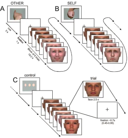
Experimental paradigm
Subjects were asked to empathize with emotional facial expressions presented on a computer screen by “feeling into” the depicted person and either to judge the emotional state of each face (other-task), or to report the emotions elicited in themselves by the emotional faces (self-task). The instructions were as follows. Other-task: “Try to empathize with the depicted person. For each face that appears on the screen you should decide how this person feels.” Self-task: “Try to empathize with the depicted person. For each face that appears on the screen you should decide how you feel yourself when you look at that
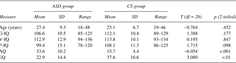
TABLE 1
Demographic and clinical characteristics of the ASD and control sample
ASD group CS group
T (df = 26) p (2-tailed)
Measure Mean SD Range Mean SD Range
Age (years) 27.4 9.3 18–48 25.1 6.7 19–46 −0.764 .452
G-IQ 106.6 10.5 85–125 112.1 10.4 89–129 1.388 .177
V-IQ 112.9 12.9 94–136 113.8 10.1 93–134 0.195 .847
P-IQ 99.4 15.1 78–120 108.1 11.3 86–125 1.715 .098
AQ 33.6 10.2 15.7 4.4 −6.054 <.001
EQ 22.9 14.4 37.6 10.6 3.080 <.01
4 SCHULTE-RÜTHER ET AL.
face.” To reduce potential social desirability bias, subjects were explicitly told that there were no correct or wrong answers in the self-task. Response options were “sad,” “neutral,” or “happy.” For the self- and the other-task, stimulus faces had either a happy or a sad emotional expression with either high or low intensity. A perceptual decision on the width of neu-tral faces was included as a control condition using “thin,” “normal,” or “wide” as response options. Only neutral faces were used in the control condition to avoid the elicitation of implicit emotional responses during the control-task. We did not include a further low-level baseline (e.g. resting condition) because comparison of experimental tasks against this kind of baseline may yield ambiguous results (Morcom & Fletcher, 2007). Importantly, brain activity during resting conditions may be associated with self-referential processing (Gusnard, Akbudak, Shulman, & Raichle, 2001) and therefore provides a non-optimal control condition for social cognitive processes such as empa-thy. The three experimental tasks (self-task, other-task, and control-task) alternated blockwise in a pseu-dorandomized counterbalanced order. Twelve blocks of each task were presented, resulting in 36 blocks. Each block contained 6 trials, resulting in a total of 192 trials (64 trials per task; see Figure 1 for the exact time course of stimulus presentation). Across self-and other-blocks, intensity (high, low) self-and quality of emotion (happy, sad) were counterbalanced. A blocked design was chosen to maximize design effi-ciency for the detection of differences between tasks. Furthermore, as initial pilot testing indicated that switching between tasks on a trial-by trial basis was very difficult even for control subjects, a blocked presentation of tasks was considered as the best choice for our paradigm. A block contained stimuli of either low or high intensity. Low-intensity stimuli were included in the stimulus set to avoid potential ceiling effects. Using only high-intensity stimuli might have rendered the task too easy for controls as well as for ASD subjects. Emotion categories were mixed within blocks, that is, stimulus faces of the same emotion category did not appear more then three times in a row and each emotion appeared at least twice within a block. This procedure was chosen (i) in order to avoid habituation effects related to empathizing with persons displaying the same emotion category; (ii) to prevent subjects from adopting response strategies related to predictable stimulus sequences. Subjects responded with button-presses using three fingers of their right hand while the stimuli were on the screen. Responses were collected for each presented lus face and were counted from 150 ms post stimu-lus-onset until the onset of the next stimulus face. The
software Presentation 9 (Neurobehavioral Systems, Albany, CA; www.neurobs.com) was used for stimu-lus presentation and response collection. Prior to scan-ning, subjects were trained on the experimental tasks to ensure that they were able to respond within the required time window. After the fMRI experiment, subjects were questioned about their strategies used to perform the tasks and other performance-related aspects. Of the 18 ASD and 18 control subjects (CS) who initially participated in the study, 4 participants in each group were not able to describe the difference between the self- and the other-task and indicated that they had always responded “according to how the other person felt” without any reference to their own feelings. These were not included in any analysis, resulting in a final sample of 28 participants (14 ASD, 14 CS) which entered in the fMRI analyses.
Stimuli
Stimulus faces were constructed using FaceGen 3.1 (Singular Inversions, Vancouver, Canada). Photos of volunteers showing a neutral facial expression were transformed into three-dimensional representations that were subsequently morphed for quality and inten-sity of emotional expressions. According to estab-lished conventions of the Facial Action Coding System (FACS; Ekman & Friesen, 1978), each face was morphed to a male adult with a happy and a sad expression (with either high or low intensity), and a neutral expression. Only male stimulus pictures were used because all participants were male and empathiz-ing is facilitated with perceived similarity to the observed person (see, e.g., Preston & de Waal, 2002). Furthermore, possible confounds related to differ-ences in empathizing with men or women could be excluded. Validity of emotional expressions was corroborated in a behavioral pilot study. Ten male volunteers rated faces (i) for emotion category (happy, sad, neutral) and intensity (high, low). A total of 72 faces were included in the final stimulus set. These faces had a mean ratio of correct responses for the identification of emotion categories (88.5% ± 17.5
SD) and for the sorting into emotion intensity categories (75.1% ± 19.8 SD).
Eye movement data
at a sampling rate of 50 Hz and a resolution of 600 × 800 pixels. Due to technical problems, eye movement data were not available for seven participants. Eye movement data from the remaining 21 participants (9 of 14 ASD, 12 of 14 CS) were further processed with eye-movement data analysis software (ILAB 3.6.0, Gitelman, 2002). Eye blinks were filtered out and fixations during the presentation of facial stimuli were determined (minimum duration of 50 ms and no consecutive dispersion of more than 20 pixels).
Mean durations of fixation for each experimental con-dition within predefined regions of interest (eyes, mouth, and whole face region) were determined for each participant.
MR technical parameters
MR imaging was accomplished on a 1.5-T Avanto MR scanner (Siemens, Erlangen, Germany) using a
6 SCHULTE-RÜTHER ET AL.
standard head coil. For functional imaging, gradient-echo, echoplanar T2*-weighted images (EPI) were acquired (TE = 60 ms, TR = 3000ms, α = 90°, FOV = 200mm, voxel size = 3.1 × 3.1 × 4 mm3, matrix size = 64 × 64, 30 transversal slices, slice acquisition: ascending) in one session (∼14 min). Anatomical images were acquired using a T1-weighted 3D mag-netization-prepared, rapid acquisition gradient echo (MP-RAGE) pulse sequence (TE = 3.93 ms, TR = 2200 ms, α = 15°, FOV = 256 mm, voxel size = 1 × 1 × 1 mm3, matrix size = 256 × 256, 160 sagittal slices, slice thickness = 1 mm).
Image processing and data analysis
Twenty-eight subjects (14 CS, 14 ASD) were included in the final sample for the analysis of fMRI data. Functional volumes were analyzed with SPM5 (Wellcome Department of Imaging Neuroscience, London; www.fil.ion.ucl.ac.uk/spm) implemented in MATLAB 7 (The Mathworks, Inc., Natick, MA). The first four volumes of each functional time-series were discarded to allow the MR signal to reach a steady state. The remaining 285 images were realigned using rigid body transformation, normalized into the Mon-treal Neurological Institute (MNI) coordinate space and resampled at 2 × 2 × 2 mm3. Normalization parameters were determined by applying the “unified segmentation” routine (Ashburner & Friston, 2005) to each individual subject’s mean EPI image. This rou-tine gives normalization parameters that are at least as precise as the standard normalization routine in SPM5, but may even provide more precision due to the parallel and recursive segmentation and normali-zation procedure. Anatomical scans were normalized into MNI space using the same method. Prior to stat-istical analysis, functional volumes were smoothed with an 8 × 8 × 8 mm3 Gaussian kernel (full width half maximum) to compensate for residual variations in individual anatomy and to meet the requirements of the Gaussian random fields theory.
Boxcar functions of 19.2 s duration (corresponding to the onset of each experimental block, starting with the first presentation of a face) were convolved with a model of the hemodynamic response (canonical HRF implemented in SPM) and its first-order temporal derivative (to compensate for timing differences in slice acquisition). Movement parameters were included as additional regressors of no interest. A high-pass cut-off filter of 128 s was used to account for low-frequency drifts in the imaging data. To handle within-subject autocorrelations an approximate AR(1) model was estimated at omnibus F-significant voxels
(p < .001), used globally over the whole brain. Param-eter estimates of the resulting general linear model were calculated for each voxel and each regressor.
For population inference, the contrast estimates for the simple effect of each experimental condition were taken to the second level (using the first regressor of the first-level HRF model as an estimate of response height) and a random effects analysis was performed (mixed ANOVA, factors: condition × group × sub-ject). Departures from sphericity assumptions were accommodated using the non-sphericity correction in SPM5 (modeling of variance components). For this procedure, unequal variance was assumed for all factors; non-independence was assumed for the factor condi-tion (repeated measures). Specific effects at each voxel were tested by applying appropriate linear con-trasts to the parameter estimates. Experimental condi-tions containing high- and low-intensity stimuli were modeled separately. However, since initial assess-ment of results related to stimulus intensity did not reveal differential effects, high- and low-intensity trials were collapsed for subsequent data analysis. Further analysis related to group differences in empa-thizing focused on the separate comparison of both empathizing tasks with the control-task.
To constrict the analysis of group differences to brain regions that play a role in empathizing, we used the respective within-group contrasts as functional regions of interest for the assessment of interactions with the factor group (i.e., group differences of the self- or other-task relative to the control-task). These analyses were performed using the SPMs of a within-group contrast as an inclusive mask (threshold used for masking: p < .01) and as a functional region of interest (ROI) for the interaction contrast. The statist-ical threshold for both within- and between-group comparisons (interactions) was set to p < .05, cor-rected for multiple comparisons at the cluster level (for cluster-level inference, SPMs were thresholded at
precuneus/PCC (precuneus, posterior cingulate cortex). A ROI of the TPJ was constructed using the coordi-nates of rTPJ given in a recent meta-analysis on empathy, ToM, and attention (Decety and Lamm, 2007) and the corresponding mirrored coordinate of lTPJ, each surrounded by a 10 mm sphere. These ROIs were used for small volume corrections in SPM. Peak activated voxels resulting from these analyses were further inspected in a whole brain analysis to ensure that these voxels represented peak activations within the respective ROI and not merely an overlap-ping activation cluster from neighboring regions.
To assess correlations between brain activation and individual empathic abilities (as measured by the EQ), whole brain regression models were constructed using individual EQ values and first-level contrast estimates of either the other-control or the self-control compari-son. Areas showing positive correlations between brain activation and EQ values across all subjects were identified within brain regions that were reliably activated in the respective contrast of the ANOVA analysis (see above), either for ASD or for CS subjects. These analyses were performed using the combined SPMs (logical OR) of both within group contrasts of the ANOVA analysis as an inclusive mask (threshold used for masking p < .01) and as a ROI for the sion analyses. The statistical threshold for the regres-sion analyses was set to p < .05, corrected for multiple comparisons at the cluster level (for cluster-level inference, SPMs were thresholded at p < .005, voxel level).
Localization of activations
SPMT maps resulting from the group analysis were superimposed onto a group mean MR image calcu-lated from the normalized anatomical T1-images of each subject (see above). MNI coordinates of the local maxima within areas of significant relative changes in neural activity were determined and anatomically localized by comparing activation maps superim-posed on the anatomical group mean brain with a standard atlas of brain anatomy (Duvernoy, 1999). In addition, an SPM toolbox (Eickhoff et al., 2005) was applied which allows for the integration of probabilis-tic cytoarchitectonic maps of the brain and functional neuroimaging data.
Analysis of behavioral data
Behavioral data were analyzed with the software package SPSS 15 (SPSS Inc., Chicago, IL). For each experimental condition and each subject, percentage of correct (i.e., correct attribution of the emotional
state of a stimulus face in the other-task) and congruent responses (i.e., responses during the self-task mirroring the emotional state of a stimulus face), as well as mean reaction times (RTs) were calculated. Since Kolmogorov-Smirnoff tests indicated normal distri-bution of all variables of interest, parametric analyses (mixed ANOVAs and t-tests) were employed to test for statistically significant differences between groups and experimental conditions. For all behavioral analy-ses, significance was determined using two-tailed testing.
RESULTS
Behavioral data
RTs were analyzed with a 2 × 2 × 2 mixed ANOVA (task × intensity × group) and a 3 × 2 mixed ANOVA (task × group). RTs were faster for the other- than for the self-task, F(1, 26) = 6.94, MSE = 21.95, p < .05, and faster for the high than the low emotion intensity stimuli, F(1, 26) = 22.65, MSE = 31.06, p < .001. Interactions and the main effect of group were nonsig-nificant. RTs of the three tasks differed, F(2, 52) = 3.339, MSE = 59.10, p < .05, but self- and other-task did not differ significantly from the control-task, respectively (post-hoc pairwise comparisons, p > .262). Interactions and the main effect of group were also nonsignificant.
For the analyses of correct/congruent responses, 2 × 2 × 2 mixed ANOVAS were calculated (task × intensity × group).The number of correct responses for the other-task was higher than the number of con-gruent responses for the self-task, F(1, 26) = 12.83,
MSE = 0.085, p < .001, and higher for the high emo-tional intensity than the low emoemo-tional intensity stimuli, F(1, 26) = 104.73, MSE = 0.037, p < .001. There was also a main effect of group, F(1, 26) = 5.936, MSE = 0.125, p < .05, and a significant task × group interaction, F(1, 26) = 7.78, MSE = 0.085, p < .010. These effects were due to a group difference of congruent responses in the self-task (t-test for inde-pendent samples, t = 2.906, df = 15.47, p < .05) but no differences in the other-task (t = 0.159, df = 22.931,
8 SCHULTE-RÜTHER ET AL.
experimental groups, mean congruent responses during the self-task were correlated with EQ values (Spearman’s rho = .414, p < .028), confirming the interrelationship between empathic abilities and performance in the experimental task. To relate
performance in the self-task (as measured by the per-centage of congruent responses) to performance in the other task (as measured by the percentage of correct responses) a correlation analysis was performed. Behavioral data for the self- and the other-task were significantly correlated for control subjects (r = .610, p
< .01), but not for ASD subjects (r = .163, p < .289).
Eye movement data
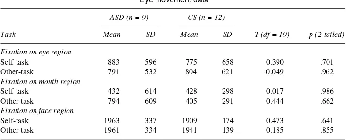
Unlike previous studies reporting atypical visual scanning patterns in ASD for emotional faces (Dalton, Nacewicz, Alexander, & Davidson, 2007), we did not find differences between groups in the time spent on fixating the face, the eye, or the mouth region during any experimental condition (t-tests for independent samples; t(19) < 0.707, p > .489). Neuroimaging data are thus not confounded by aberrant fixation patterns that may occur in ASD subjects (see also Dapretto et al, 2006). Eye movement data are summarized in Table 2.
FMRI data
In the following paragraphs, we report fMRI results for each group separately, as well as results of the dir-ect statistical comparisons between groups (interac-tion analyses). Note that differences in the activa(interac-tion patterns of the respective group results are not indica-tive of a difference between groups, unless significant in the direct comparison, and that post-hoc exclusion of medicated subjects from the fMRI data analysis did not change the pattern of results reported here.
Other-task vs. control-task
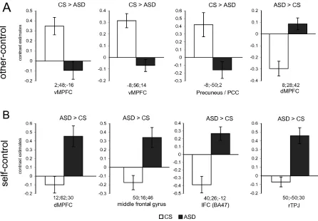
In control subjects, significant increases in neural activity were observed in bilateral medial cortical structures (ventral portions of the MPFC (vMPFC), precuneus/PCC), the left lingual gyrus, bilateral mid-dle temporal gyrus/superior temporal sulcus (STS), left temporoparietal junction (TPJ), and right IFC (BA44/45). ASD subjects showed increased neural activation in the dorsal part of the left MPFC (dMPFC), bilateral precuneus, right middle temporal gyrus/STS and left TPJ. There was no significant acti-vation in ASD subjects in the right IFC even at an uncorrected threshold (p < .001, voxel level). Signifi-cant differences in brain activation between groups could be revealed in the direct comparison (interac-tion analysis). Control subjects showed differential activation in the vMPFC and precuneus/PCC while
there was differential activation in the dMPFC in ASD subjects (see Table 3).
Self-task versus control-task
In the control group, increased neural activity was evident in areas similar to those observed for the other-task. However, additional activations were located in the dMPFC, left IFC, left TPJ, and right cerebellum. In contrast, ASD subjects showed increases in neural activity which extended into wide-spread frontal areas (left superior frontal gyrus, bilat-eral middle frontal gyrus, bilatbilat-eral IFC), bilatbilat-eral TPJ, inferior temporal gyrus (ITG), and temporal pole. Sig-nificant differences in brain activation between groups could be revealed in the direct comparison (interaction analysis). ASD subjects showed differen-tial activation in the right IFC (pars orbitalis, BA47), right dMPFC, right middle frontal gyrus, and the right TPJ (see Table 3). No significant differential activations were observed for the control group at the pre-defined statistical threshold. At a more liberal, exploratory threshold (p < .05, uncorrected) the biggest cluster of activation emerged in the vMPFC.
Conjunction of other-task vs. control-task and self-task vs. control-self-task
The conjunction of the two experimental tasks (compared to the control-task) revealed activations bilaterally in the vMPFC and precuneus/PCC, right STS, and left TPJ in control subjects. In ASD sub-jects, conjoint activation could be observed in bilat-eral precuneus/PCC and left dMPFC. Using a ROI analysis, a trend towards significance could be observed for a cluster in right STS (p < .0689, FWE-corrected).
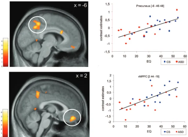
Correlations of brain activation and empathy
Whole brain regression analyses yielded signific-ant correlations between empathic abilities and brain activation during the other task in the vMPFC (MNI coordinates of peak activated voxel: [2, 44, −18]) and precuneus bilaterally (MNI coordinates of peak acti-vated voxel: [−6, −46, 48], see Figure 3). Whole brain analyses were performed across the whole group. However, to elucidate potential group differences in correlations, individual contrast estimates were extracted at peak activated voxels and tested for a positive correlation with EQ values separately for ASD and CS subjects. These analyses revealed signi-ficant correlations for each group in the vMPFC (ASD: R = 0.575, p < .05; CS: R = 0.538, p < .05) and precuneus (ASD: R = 0.725, p < .01; CS: R = 0.598, p
< .05). The whole brain regression analysis of EQ and the self-task did not reveal foci of activation at the selected threshold. However, using the same pre-cuneus and vMPFC coordinates reported above, we found significant correlations between activation in the self-task and EQ values in both vMPFC (R = 0.449, p < .01) and precuneus (R = 0.387, p < .05) across all participants. Separate correlation analyses for each group yielded marginally significant results for the precuneus in both groups (CS: R = 0.428, p = .0633; ASD: R = 0.452, p = .0522) and for the vMPFC only in control subjects (CS: R = 0.398, p = .0801; ASD: R = 0.289, p = .1578).
DISCUSSION
The paradigm of the present study is unique in that it enabled us to assess emotional self- and other-related
TABLE 2
Eye movement data
ASD (n = 9) CS (n = 12)
T (df = 19) p (2-tailed)
Task Mean SD Mean SD
Fixation on eye region
Self-task 883 596 775 658 0.390 .701
Other-task 791 532 804 621 −0.049 .962
Fixation on mouth region
Self-task 432 614 428 298 0.017 .986
Other-task 794 609 405 291 0.444 .662
Fixation on face region
Self-task 1963 337 1909 174 0.473 .641
Other-task 1961 334 1941 139 0.185 .855
10
TABLE 3
Peaks of activation in the experimental tasks
H BA
CS ASD CS > ASD ASD > CS
Anatomical region x y z t x y z t x y z t x y z t
Other-task (vs. control-task)
vMPFC R 11 8 56 −10 5.72 2 48 −16 3.54
vMPFC L 11 −8 58 −18 4.81 −8 56 −14 4.21
dMPFC L 8 −8 58 36 4.58*a
dMPFC R 32 8 28 42 4.44*a
IFC pars opercularis/triangularis R 44/45 54 26 12 3.91*e
Precuneus/PCC R 23 12 −50 28 5.44
Precuneus/PCC M 23 0 −58 34 4.38
Precuneus/PCC M 23 0 −48 26 5.18 −6 −48 26 4.45 −8 −50 2 4.33*d
Lingual gyrus L 18/19 −16 −58 −2 4.97
Middle temporal gyrus/STS R 21/22 54 −38 −4 6.01 58 −38 −4 4.04*b
Middle temporal gyrus/STS/TPJ L 21/22/42 −60 −60 16 4.23 −50 −58 30 3.57*c
Middle temporal gyrus L 21/22 −58 −40 −4 3.41*b
Self-task (vs. control-task)
dMPFC L 10 −2 56 8 5.81
dMPFC R 10 4 62 18 6.99
dMPFC L 9/10 −4 56 26 5.39 −8 56 34 7.73
dMPFC R 9/10 8 56 28 3.86 8 58 30 5.73*a 12 62 30 4.50
vMPFC L 11 −4 54 −16 5.20
vMPFC R 11 8 58 −6 4.70
Superior frontal gyrus L 8/9 −16 34 52 7.64
Middle frontal gyrus L 9/46 −38 20 44 6.52
Middle frontal gyrus R 9 50 16 46 4.32
IFC (p. orbitalis) L 47 −40 30 −14 4.57 −46 36 −8 5.58
IFC (p. orbitalis/triangularis) L 45/47 −52 30 −4 5.66
IFC (p. triangularis) L 45 −50 26 8 5.56*e
IFC (p. opercularis) L 44 −46 12 10 4.10*e −48 22 6 5.68
IFC (p. triangularis) R 45 56 28 −4 4.33*e
11
Precuneus/PCC L 23 −6 −52 28 5.03 −2 −54 34 7.23
Precuneus L 7 −4 −64 32 5.14
TPJ L 22/21/39 −52 −58 24 5.61 −50 −62 34 7.89
TPJ R 22/39 50 −54 30 5.63 50 −50 30 4.65
Middle temporal gyrus/STS R 21/22 54 −36 −4 5.36 60 −36 −4 5.08
Middle temporal gyrus/STS L 21/22 −58 −40 −6 5.33
Middle temporal gyrus L 20/21 −56 −14 −18 4.97
Middle temporal gyrus/temporal pole R 20/38 50 6 −30 5.03
Inferior temporal gyrus R 20 48 4 −40 4.90
Inferior temporal gyrus L 20 −54 −30 −18 4.76
Temporal pole L 21/38 −52 8 −22 3.88*
Cerebellum R 34 −76 −36 5.41 28 −80 −36 7.39
Cerebellum L −24 −86 −42 5.35
Conjunction: self-task (vs. control-task) and other-task (vs. control-task)
dMPFC L 9/10 −8 58 36 4.58*a
vMPFC L 11 −6 56 −14 4.79
vMPFC R 11 8 58 −6 4.70
Precuneus/PCC R 23 12 −50 30 5.35 0 −58 34 4.38
Precuneus/PCC L 23 −2 −48 28 4.98 −6 −48 26 4.45
Precuneus L 7 −2 −66 32 4.32*d
TPJ L 22/21/37 −58 −62 14 5.36
Middle temporal gyrus/STS R 21/22 54 −36 −4 5.36 58 −38 −4 4.04b
(trend, p < .069)
12 SCHULTE-RÜTHER ET AL.
processing in ASD subjects during an interactive empathic situation. Other studies have examined emotional self- or other-related social cognition in ASD using a paradigm that required the recognition of one’s own face (Uddin et al., 2008) or abstract evaluation of trait adjectives (Kennedy & Courchesne, 2008), or have examined resting-state conditions that are considered to be associated with self-referential processing (Kennedy, Redcay, & Courchesne, 2006). To our knowledge, this is the first fMRI study that examines subjects with ASD in an explicit empathizing task. Previous imaging studies on empathy in ASD either focused on brain activation related to imitation and observation of facial expressions as indirect measures of empathy (Dapretto et al., 2006) or used affective pictures to induce emotion and correlate respective brain acti-vation with empathy questionnaires (Silani et al., 2008).
The present study aimed at identifying brain dysfunctions underlying atypical self- and other-related emotional processing in adults with ASD in the context of facial expressions of emotions. With respect to behavioral performance, there were no significant differences in RTs between ASD sub-jects and control subsub-jects for any experimental condition. It is thus unlikely that differences in neural activations are related to domain-general performance deficits (such as differences in per-ceptual processing speed). Moreover, the percent-age of correct responses in the other-task did not differ between groups, suggesting that ASD sub-jects were able to infer other persons’ emotions from facial displays. In contrast to previous studies (Dalton et al., 2007), we did not observe differences in eye-movements and visual scanning patterns between groups. One can thus exclude that our neuroimaging results are confounded by aber-rant fixation patterns in ASD subjects. Consistent with previous findings of reduced emotional conta-gion (Scambler, Hepburn, Rutherford, Wehner, & Rogers, 2007) and empathy (Baron-Cohen & Wheelwright, 2004), individuals with ASD reported fewer contagious emotional responses during the self task. Note that incongruent responses were mostly “neutral” responses. Choos-ing an opposite emotion was rare, suggestChoos-ing an absence of emotional contagion rather than the emergence of inappropriate incongruent emotional responses. The finding that in control subjects the amount of emotional contagion was correlated with the correct identification of emotional expres-sions suggests that these two processes are closely interrelated components of empathic processing.
Brain networks involved in ToM and
empathy
one’s own emotions and the observation of emotional faces may thus account for the activation of the TPJ in both groups. It might be speculated that for ASD sub-jects, such switching processes need more attentional resources than in control subjects (as indicated by stronger activation of the TPJ and the ventral inferior frontal cortex) because of their lack of emotional conta-gion (i.e., their internal emotional state was more often incongruent with the observed emotional faces).
Using a similar paradigm, Schulte-Rüther et al. (2008) showed that males (in comparison to females) also show enhanced recruitment of the TPJ during the assessment of their own emotions in an empathic situ-ation. Furthermore, emotional reactions to the observed faces were less pronounced in males than in females. Taken together, our results demonstrate that gender dif-ferences in these brain functions and behavior show sim-ilarities with ASD-related neurofunctional and behavioral deviations. The data thus provide preliminary support for theories that relate ASD to an extreme variant of a typical “male brain” (Baron-Cohen, Knickmeyer, & Belmonte, 2005).
Medial prefrontal cortex
The MPFC has been implicated in diverse emotional and non-emotional social tasks. Recently, it has been proposed that the MPFC can be segregated into neu-rofunctional submodules along a caudal–rostral axis (Amodio & Frith, 2006). According to the model, the most ventral parts of the MPFC (approximately defined by z < 2) are involved in autonomic and vis-ceral aspects of emotional responses (Koski & Paus, 2000), which are typically associated with the moni-toring of the value of future outcomes. In contrast, dorsal (posterior rostral) MPFC areas are considered to be primarily engaged in action monitoring and the evaluation of observed actions. Moreover, Amodio and Frith (2006) argue that processing within the MPFC proceeds from the most dorsal and most ven-tral parts towards an anterior rosven-tral transition zone. In this transition zone, more abstract metacognitive rep-resentations supporting self-reference and mentaliz-ing are supposed to be implemented. Overall, this model is in accordance with the idea that social cogni-tive judgments rely primarily on the dMPFC while the vMPFC is more related to self-referential emotional cognition (D’Argembeau et al., 2007; Mitchell et al., 2006; Schulte-Rüther et al., 2007). Consistently, a large number of neuroimaging studies have impli-cated the vMPFC in self-referential thinking (e.g., Macrae, Moran, Heatherton, Banfield, & Kelley, 2004; Schmitz, Kawahara-Baccus, & Johnson, 2004),
especially in the context of emotions (Moran, Macrae, Heatherton, Wyland, & Kelley, 2006). Since the vMPFC is strongly interconnected with emotion pro-cessing areas including the amygdala, ventral striatum, and orbitofrontal cortex (Ongur, Ferry, & Price, 2003), it is conceivable that self-related cognition, emotion processing, and external socially significant cues are integrated in this region. Such integration may allow for one’s “emotional bonding” with other persons in empathic situations. This view is corrobo-rated by our finding of a positive correlation between empathic abilities and activation in the vMPFC during empathizing (see Figure 3).
Note, however, that recent meta-analyses of brain imaging studies investigating theory of mind (Spreng, Mar, & Kim, 2009) and experience of emotion (Kober et al., 2008) demonstrated similar activations of ventral and dorsal portions of the MPFC during tasks that require emotional social cognition. These find-ings speak against the view of a clear-cut ventral/ dorsal neurofunctional segregation within the MPFC. However, one needs to consider that many studies included in the meta-analysis (Kober et al., 2008) used facial expressions or pictures showing complex social scenes (e.g. the International Affective Picture System, IAPS) as stimulus materials which may evoke both empathic reactions and ToM reasoning. Furthermore, ToM paradigms typically contain not only cognitive, but also affective components. Lesion studies are better suited than meta-analyses to differentiate between effects of emotional process-ing and ToM within subregions of the MPFC. It has recently been demonstrated that lesions of the vMPFC selectively affect performance in ToM tasks that require the empathic understanding of other people’s feelings (e.g., detecting “faux-pas” situations), but do not impair cognitive aspects of ToM (e.g., under-standing of second-order false belief; Shamay-Tsoory, Tomer, Berger, Goldsher, & Sharon-Peretz, 2005; Stone, Baron-Cohen, & Knight, 1998). Moreover, patients with vMPFC lesions rate themselves as having less empathic ability than other people (Shamay-Tsoory, Sharon-Peretz, & Perry, 2009).
14 SCHULTE-RÜTHER ET AL.
activation was located more dorsally in subjects with ASD and more ventrally in control subjects. In the direct comparison between ASD subjects and control subjects, the activation patterns show a clear
dissociation, with differential dMPFC recruitment in ASD subjects and differential vMPFC recruitment in control subjects. This brain activation pattern is paralleled by a reduction of contagious emotional
Figure 3. Covariation of brain activation and empathic abilities. Brain activity during the other task (vs. high-level baseline) correlated with individual EQ values (Baron-Cohen and Wheelwright, 2004) across all participants. SPM is thresholded at p < .005 (voxel-level). Circled clus-ters are significant at p < .05, corrected for multiple comparisons at the cluster level. Scatterplots illustrate the correlation in the peak activated voxels of both significant clusters in vMPFC and precuneus, respectively. Solid lines represent the linear best fit.
responses in subjects with ASD. In controls, empa-thizing with other persons is thus likely to be trig-gered by emotional self-referential cognition instantiated in vMPFC regions, whereas in ASD subjects cognitive components of ToM (e.g., detection of intentions) and action monitoring (relying on dMPFC regions) may predominate. As the vMPFC plays an important role for monitoring the value of future outcomes (Amodio and Frith, 2006), it is con-ceivable that ASD subjects lack the direct link between metacognitive representations and the emo-tional value of social interactions. Clinical observa-tions indicating that individuals with ASD can develop ToM abilities at an abstract level, but lack intuitive ToM abilities in dyadic social interactions (Bowler, 1992; Happé, 1994), are consistent with this suggestion. Note that the differential activation of
dMPFC in ASD subjects for the other-task was at least in part also driven by a deactivation in control subjects for this task (in comparison to the control task) (see Figure 5). Enhanced emotional self-referen-tial cognition as evidenced by vMPFC activation may perhaps have suppressed processing in the dMPFC. However, further studies investigating functional con-nectivity patterns of dMPFC and vMPFC are needed to substantiate such a claim.
Self-related emotional cognition in ASD
Besides the MPFC, ASD and control subjects also recruited the precuneus and the adjacent PCC during both the self- and other-tasks, and precuneus activation was also positively correlated with empathic abilities,
16 SCHULTE-RÜTHER ET AL.
as assessed with the EQ. These regions have been implicated in a broader range of self-referential cogni-tive and emotional processes such as first-person per-spective taking (Vogeley et al., 2001), representation of the mental self (Lou et al., 2004), and autobio-graphical memory (Piefke et al., 2008; Piefke, Weiss, Zilles, Markowitsch, & Fink, 2003). Interestingly, activation in these areas could be observed during
both self- and other-conditions. This pattern of results is in accordance with other studies indicating that overlapping brain areas are implicated in judging other people’s and one’s own mental states, especially in cases where the other person is perceived as similar to oneself (Mitchell, Banaji, & Macrae, 2005; Mitch-ell et al., 2006). MitchMitch-ell et al. (2006) conclude that the judgment of other persons may be built on the simulation of judging oneself. Together with these data, our results suggest that empathizing with other people may draw on simulation mechanisms by acti-vating the neural networks underlying self-referential cognitive and emotional processing. In the present study, this idea is supported by the results of the con-junction analysis (self- and other-tasks vs. control-task): During both tasks, conjoint neural activation could be observed in PCC/precuneus and MPFC (dMPFC in ASD and vMPFC in healthy controls). This simulation mechanism may be disturbed in indi-viduals with ASD. In support of this conclusion, our behavioral data demonstrate for control subjects that the tendency for emotionally congruent responses (self-task) was positively correlated with the ability to correctly infer an emotion in the other person (other-task). This was not the case in ASD subjects. Thus, in typical adults the capacity to identify the emotions of other people may benefit from the capacity of emo-tional contagion. These behavioral data are also con-sistent with the view that self-reflection may facilitate sensitive judgments about the mental states of other persons (Dimaggio, Lysaker, Carcione, Nicolo, & Semerari, 2008).
We observed significantly less activation of the vMPFC and precuneus/PCC in ASD subjects relative to the control group. Similar aberrant activation pat-terns in subjects with ASD have been reported for resting state conditions (Kennedy et al., 2006), which may be linked to automatic processes of self-referen-tial cognition (Gusnard et al., 2001). Further evidence for altered activation patterns in ASD in these medial cortical areas has been observed during a task that required subjects to make judgments about the rela-tionship of trait adjectives to oneself or a well-known other person (Kennedy & Courchesne, 2008). In com-bination with these data, our results point to a defi-ciency in the neural networks subserving
self-referential processing in ASD as one reason for reduced empathic abilities. This may in particular be based on dysfunctions of MPFC regions and the precuneus/PCC. Our findings thus show a neurofunc-tional mechanism for the frequently observed impair-ments of self-referential cognition in ASD (Hurlburt et al., 1994; Lombardo et al., 2007; Toichi et al., 2002) and the related deficits in empathic behavior.
Due to a lack of self-referential emotional process-ing (possibly resultprocess-ing in diminished emotional conta-gion), individuals with ASD may recruit different strategies to infer emotional states of other persons. ASD subjects showed activation in a widespread frontal network (including mid-dorsolateral and ventrolateral areas) and inferior temporal regions (including the temporal poles) during the self-task (see Figure 6). Mid-dorsolateral and ventrolateral areas of the pre-frontal cortex have been implicated in diverse execut-ive demands (e.g., monitoring cognitexecut-ive processes and problem solving (Duncan & Owen, 2000). These acti-vations may reflect the need for additional cognitive resources for resolving the cognitive–emotional requirements of the self-task.
conclusions regarding the involvement of ITG and temporal poles in compensatory strategies will require further investigation.
Mirror mechanisms
The human mirror system (hMS) may play an import-ant role in social cognition, especially in the context of emotional face-to-face interactions (Carr et al., 2003; Dapretto et al., 2006; Schulte-Rüther et al., 2007; but also see Hickok, 2009, for a critique). In support of this notion, we demonstrate that areas pre-viously associated with the hMS (e.g., BA44/45 in the IFC) are activated when empathy is elicited by facial expressions of emotions. It is currently a matter of debate whether an early deficiency of the hMS in individuals with ASD may lead to their typical social and emotional deficits (Williams et al., 2001). Dapretto et al. (2006) demonstrated that children with ASD show less activation in frontal components of
the hMS during the observation and imitation of emo-tional facial expressions. However, children with ASD are not necessarily impaired in the understand-ing and imitation of non-emotional actions (Hamilton, Brindley, & Frith, 2007). Furthermore, it remains unclear at which level of imitative process problems may arise in individuals with ASD (Southgate & Hamilton, 2008). It has thus been argued that the claim of a direct link between imitation deficits and a core dysfunction of the mirror system in autism is speculative, to date. In the present study, we did not observe a significant difference between the two groups in the IFC (BA44/45) at the selected statistical threshold, in either the self- or the other-task. There was right-hemispheric activation in the IFC (BA44/ 45) during the other-task in the control group, but no activation above threshold in ASD subjects. In the self-task, however, left-hemispheric activation in frontal parts of the hMS was evident in ASD subjects as well. In adults with ASD, components of the hMS may thus become engaged in emotional face-to-face
18 SCHULTE-RÜTHER ET AL.
interactions especially when subjects are explicitly instructed to attend to their own emotional reaction to other people’s emotions. The data suggest that ASD does not necessarily affect basic functions of the hMS. Rather, hMS recruitment during social interac-tion appears to be modulated by a combinainterac-tion of task, context, and instruction in individuals with ASD. Further studies are needed to clarify under which cir-cumstances aberrant activations in the hMS may occur in ASD subjects and under which circum-stances hMS function in this patient group is compa-rable to that of control subjects.
Limitations
Several potential limitations of the paradigm should be kept in mind. Our choice of computerized faces warranted high naturalism and optimum standardiza-tion of stimuli. More natural stimuli (e.g., videoclips of emotional faces in a naturalistic context) may perhaps trigger stronger empathic reactions; however, they are not well controlled experimentally. Potential differences between computerized facial stimuli and real faces have not been investigated in ASD yet, and should be explored in future studies. For example, ASD subjects might find it harder to empathize with a computerized person, and perceive such persons as more dissimilar to themselves. Such effects could contribute to our observed dorsal/ventral dissociation in MPFC (Mitchell et al., 2005, 2006).
An important aspect of the paradigm is that the behavioral responses during the self-task can be interpreted as an index of emotional contagion. Though this self-report response might be biased (e.g. by social desirability), our interpretation of reduced emotional contagion in ASD is in line with several previous studies (Scambler et al., 2007; Baron-Cohen & Wheelwright, 2004). Furthermore, a recent study systematically investigated social desir-ability bias in subjects with Asperger syndrome and control subjects, and found no group differences (Dziobek et al., 2008). However, more objective mea-sures of emotional contagion (e.g. skin conductance, video recordings of facial reactions) should be employed in future studies to rule out such potential biases. Another issue related to the self-task refers to conditions of alexi-thymia. Subjects suffering from alexithymia have pro-found difficulties in verbalizing and identifying their own emotional states. It has been suggested that there may be an overlap between ASD and alexithymia with respect to social difficulties, affective interaction, and emotional awareness. (Fitzgerald & Bellgrove, 2006).
However, fMRI studies did not reveal a direct relation-ship between difficulties in emotional awareness and self-reflection and mentalizing (Silani et al., 2008). Future studies should therefore investigate the role of alexithymia in ASD in more detail.
CONCLUSION
The present data provide novel insights into the brain networks involved in explicit emotional self-reference and emotion identification, two processes closely related to empathy. Furthermore, we demonstrate atypical neural activation associated with these proc-esses in individuals with ASD. Importantly, our find-ings support the idea of a ventral–dorsal neurofunctional segregation in the MPFC. Atypical MPFC function during emotional face-to-face interac-tions in individuals with ASD may at least in part con-tribute to the impairment in self-referential emotional processing associated with the disease.
Manuscript received 24 August 2009 Manuscript accepted 12 February 2010 First published online 13 October 2010
REFERENCES
Amodio, D. M., & Frith, C. D. (2006). Meeting of minds: The medial frontal cortex and social cognition. Nature Reviews Neuroscience, 7, 268–277.
Ashburner, J., & Friston, K. J. (2005). Unified segmenta-tion. NeuroImage, 26, 839–851.
Baron-Cohen, S., Knickmeyer, R. C., & Belmonte, M. K. (2005). Sex differences in the brain: Implications for explaining autism. Science, 310, 819–823.
Baron-Cohen, S., Ring, H. A., Wheelwright, S., Bullmore, E. T., Brammer, M. J., Simmons, A., et al. (1999). Social intelligence in the normal and autistic brain: An fMRI study. European Journal of Neuroscience, 11, 1891–1898. Baron-Cohen, S., Tager-Flusberg, H., & Cohen, D. (2000).
Understanding other minds: Perspectives from autism and developmental cognitive neuroscience. Oxford, UK: Oxford University Press.
Baron-Cohen, S., & Wheelwright, S. (2004). The empathy quotient: An investigation of adults with Asperger syn-drome or high functioning autism, and normal sex differ-ences. Journal of Autism and Developmental Disorders, 34, 163–175.
Baron-Cohen, S., Wheelwright, S., Robinson, J., & Wood-bury-Smith, M. (2005). The Adult Asperger Assessment (AAA): A diagnostic method. Journal of Autism and Developmental Disorders, 35, 807–819.
Blakemore, S. J., & Frith, C. (2003). Self-awareness and action. Current Opinion in Neurobiology, 13, 219–224. Bowler, D. M. (1992). “Theory of Mind” in Asperger’s
Carr, L., Iacoboni, M., Dubeau, M. C., Mazziotta, J. C., & Lenzi, G. L. (2003). Neural mechanisms of empathy in humans: A relay from neural systems for imitation to limbic areas. Proceedings of the National Academy of Sciences of the United States of America, 100, 5497–5502. Castelli, F., Frith, C., Happé, F., & Frith, U. (2002). Autism, Asperger syndrome and brain mechanisms for the attri-bution of mental states to animated shapes. Brain, 125, 1839–1849.
Cavanna, A. E., & Trimble, M. R. (2006). The precuneus: A review of its functional anatomy and behavioural corre-lates. Brain, 129, 564–583.
Corbetta, M., Patel, G., & Shulman, G. L. (2008). The reori-enting system of the human brain: From environment to theory of mind. Neuron, 58, 306–324.
Dalton, K. M., Nacewicz, B. M., Alexander, A. L., & Davidson, R. J. (2007). Gaze-fixation, brain activation, and amygdala volume in unaffected siblings of individu-als with autism. Biological Psychiatry, 61, 512–520. Damasio, H., Tranel, D., Grabowski, T., Adolphs, R., &
Damasio, A. (2004). Neural systems behind word and concept retrieval. Cognition, 92, 179–229.
Dapretto, M., Davies, M. S., Pfeifer, J. H., Scott, A. A., Sigman, M., Bookheimer, S. Y. et al. (2006). Under-standing emotions in others: Mirror neuron dysfunc-tion in children with autism spectrum disorders. Nature Neuroscience, 9, 28–30.
D’Argembeau, A., Ruby, P., Collette, F., Degueldre, C., Balteau, E., Luxen, A., et al. (2007). Distinct regions of the medial prefrontal cortex are associated with self-ref-erential processing and perspective taking. Journal of Cognitive Neuroscience, 19, 935–944.
Decety, J., & Grezes, J. (2006). The power of simulation: Imagining one’s own and other’s behavior. Brain Research, 1079, 4–14.
Decety, J., & Jackson, P. L. (2004). The functional architec-ture of human empathy. Behavioral and Cognitive Neuroscience Reviews, 3, 71–100.
Decety, J., & Lamm, C. (2007). The role of the right tempo-roparietal junction in social interaction: How low-level computational processes contribute to meta-cognition. The Neuroscientist, 13(6), 580–593.
Derogatis, L. R. (1993). Brief Symptom Inventory (BSI) administration, scoring, and procedures manual (3rd ed.). Minneapolis, MN: National Computer Services. Dimaggio, G., Lysaker, P. H., Carcione, A., Nicolo, G., &
Semerari, A. (2008). Know yourself and you shall know the other . . . to a certain extent: Multiple paths of influ-ence of self-reflection on mindreading. Consciousness and Cognition, 17, 778–789.
Duncan, J., & Owen, A. M. (2000). Common regions of the human frontal lobe recruited by diverse cognitive demands. Trends in Neurosciences, 23, 475–483. Duvernoy, H. M. (1999). The human brain: Surface,
three-dimensional sectional anatomy with MRI, and blood supply (Vol. 2). New York: Springer.
Dziobek, I., Rogers, K., Fleck, S., Bahnemann, M., Heek-eren, H., Wolf, O. T., et al. (2008). Dissociation of cog-nitive and emotional empathy in adults with Asperger syndrome using the Multifaceted Empathy Test (MET). Journal of Autism and Developmental Disordors, 38, 464–473.
Eickhoff, S. B., Stephan, K. E., Mohlberg, H., Grefkes, C., Fink, G. R., Amunts, K., et al. (2005). A new SPM toolbox
for combining probabilistic cytoarchitectonic maps and functional imaging data. NeuroImage, 25(4), 1325–1335. Ekman, P. & Friesen, W. V. (1978). Facial Action Coding
System: Manual. Palo Alto, CA: Consulting Psycholo-gists Press.
Farrer, C., Franck, N., Georgieff, N., Frith, C. D., Decety, J., & Jeannerod, M. (2003). Modulating the experience of agency: A positron emission tomography study. NeuroImage, 18, 324–333.
Farrer, C., & Frith, C. D. (2002). Experiencing oneself vs another person as being the cause of an action: The neural correlates of the experience of agency. NeuroImage, 15, 596–603.
Fitzgerald, M., & Bellgrove, M. A. (2006). The overlap between alexithymia and Asperger’s syndrome. Jour-nal of Autism and Developmental Disorders, 36(4), 573–576.
Frith, U., & Frith, C. D. (2003). Development and neuro-physiology of mentalizing. Philosophical Transactions of the Royal Society of London, Series B: Biological Sciences, 358, 459–473.
Gauthier, I., Anderson, A. W., Tarr, M. J., Skudlarski, P., & Gore, J. C. (1997). Levels of categorization in visual rec-ognition studied using functional magnetic resonance imaging. Current Biology, 7, 645–651.
Gauthier, I., Tarr, M. J., Anderson, A. W., Skudlarski, P., & Gore, J. C. (1999). Activation of the middle fusiform ‘face area’ increases with expertise in recognizing novel objects. Nature Neuroscience, 2, 568–573.
Gillberg, C. L. (1992). The Emanuel Miller Memorial Lecture 1991. Autism and autistic-like conditions: subclasses among disorders of empathy. Journal of Child Psychology and Psychiatry, and Allied Disci-plines, 33, 813–842.
Gitelman, D. R. (2002). ILAB: A program for postexperi-mental eye movement analysis. Behavior Research Methods, Instruments, & Computers, 34, 605–612. Gusnard, D. A., Akbudak, E., Shulman, G. L., & Raichle,
M. E. (2001). Medial prefrontal cortex and self-referen-tial mental activity: Relation to a default mode of brain function. Proceedings of the National Academy of Sci-ences of the United States of America, 98, 4259–4264. Hamilton, A. F., Brindley, R. M., & Frith, U. (2007).
Imi-tation and action understanding in autistic spectrum disorders: How valid is the hypothesis of a deficit in the mirror neuron system? Neuropsychologia, 45, 1859–1868.
Happé, F. G. E. (1994). An advanced test of theory of mind: Understanding of story characters’ thoughts and feelings by able autistic, mentally-handicapped, and normal chil-dren and adults. Journal of Autism and Developmental Disorders, 24, 129–154.
Happé, F., Ehlers, S., Fletcher, P., Frith, U., Johansson, M., Gillberg, C. et al. (1996). ‘Theory of mind’ in the brain: Evidence from a PET scan study of Asperger syndrome. NeuroReport, 8, 197–201.
Hickok, G. (2009). Eight problems for the mirror neuron theory of action understanding in monkeys and humans. Journal of Cognitive Neuroscience, 21(7), 1229–1243. Hurlburt, R. T., Happé, F., & Frith, U. (1994). Sampling the
form of inner experience in three adults with Asperger syndrome. Psychological Medicine, 24, 385–395. Kennedy, D. P., & Courchesne, E. (2008). Functional
20 SCHULTE-RÜTHER ET AL.
other-reflection in autism. Social Cognitive and Affective Neuroscience, 3, 177–190.
Kennedy, D. P., Redcay, E., & Courchesne, E. (2006). Fail-ing to deactivate: RestFail-ing functional abnormalities in autism. Proceedings of the National Academy of Sciences of the United States of America, 103, 8275–8280. Kober, H., Barrett, L. F., Joseph, J., Bliss-Moreau, E.,
Lindquist, K., Wager, T. D., et al. (2008). Functional grouping and cortical–subcortical interactions in emo-tion: A meta-analysis of neuroimaging studies. Neu-roImage, 42(2), 998–1031.
Koski, L. & Paus, T. (2000). Functional connectivity of the anterior cingulate cortex within the human frontal lobe: A brain-mapping meta-analysis. Experimental Brain Research, 133, 55–65.
Lee, A., Hobson, R. P., & Chiat, S. (1994). I, you, me, and autism: An experimental study. Journal of Autism and Developmental Disorders, 24, 155–176.
Lombardo, M. V., Barnes, J. L., Wheelwright, S. J., & Baron-Cohen, S. (2007). Self-referential cognition and empathy in autism. PLoS ONE, 2, e883.
Lou, H. C., Luber, B., Crupain, M., Keenan, J. P., Nowak, M., Kjaer, T. W., et al. (2004). Parietal cortex and represen-tation of the mental self. Proceedings of the National Academy of Sciences of the United States of America, 101, 6827–6832.
Macrae, C. N., Moran, J. M., Heatherton, T. F., Banfield, J. F., & Kelley, W. M. (2004). Medial prefrontal activity pre-dicts memory for self. Cerebral Cortex, 14, 647–654. Maldjian, J. A., Laurienti, P. J., Burdette, J. B., & Kraft, R. A.
(2003). An automated method for neuroanatomic and cytoarchitectonic atlas-based interrogation of fMRI data sets. NeuroImage, 19, 1233–1239.
Manjaly, Z. M., Bruning, N., Neufang, S., Stephan, K. E., Brieber, S., Marshall, J. C., et al. (2007). Neurophysio-logical correlates of relatively enhanced local visual search in autistic adolescents. NeuroImage, 35, 283–291. Millward, C., Powell, S., Messer, D., & Jordan, R. (2000). Recall for self and other in autism: Children’s memory for events experienced by themselves and their peers. Journal of Autism and Developmental Disorders, 30, 15–28.
Mitchell, J. P., Banaji, M. R., & Macrae, C. N. (2005). The link between social cognition and self-referential thought in the medial prefrontal cortex. Journal of Cognitive Neuroscience, 17, 1306–1315.
Mitchell, J. P., Macrae, C. N., & Banaji, M. R. (2006). Dis-sociable medial prefrontal contributions to judgments of similar and dissimilar others. Neuron, 50, 655–663. Moran, J. M., Macrae, C. N., Heatherton, T. F., Wyland, C. L.,
& Kelley, W. M. (2006). Neuroanatomical evidence for distinct cognitive and affective components of self. Journal of Cognitive Neuroscience, 18, 1586–1594. Morcom, A. M. & Fletcher, P. C. (2007). Does the brain
have a baseline? Why we should be resisting a rest. NeuroImage, 37, 1073–1082.
Ongur, D., Ferry, A. T., & Price, J. L. (2003). Architectonic subdivision of the human orbital and medial prefrontal cortex. Journal of Comparative Neurology, 460, 425–449. Piefke, M., Prestinger, M., Arin, T., Kohl, B., Kastrau, F., Schnitker, R., et al. (2008). The neurofunctional mecha-nisms of traumatic and non-traumatic memory in patients with acute PTSD following accident trauma. Neurocase, 13, 342–357.
Piefke, M., Weiss, P. H., Zilles, K., Markowitsch, H. J., & Fink, G. R. (2003). Differential remoteness and emotional tone modulate the neural correlates of auto-biographical memory. Brain, 126, 650–668.
Preston, S. D. & de Waal, F. B. M. (2002). Empathy: Its ultimate and proximate bases. Behavioral and Brain Sciences, 25, 1–20.
Rogers, S. J. & Pennington, B. F. (1991). A theoretical approach to the deficits in infantile autism. Development and Psychopathology, 3, 137–162.
Scambler, D. J., Hepburn, S., Rutherford, M. D., Wehner, E. A., & Rogers, S. J. (2007). Emotional responsivity in children with autism, children with other developmental disabilities, and children with typical development. Jour-nal of Autism and Developmental Disorders, 37, 553–563. Schmitz, T. W., Kawahara-Baccus, T. N., & Johnson, S. C. (2004). Metacognitive evaluation, self-relevance, and the right prefrontal cortex. NeuroImage, 22, 941–947. Schulte-Rüther, M., Markowitsch, H. J., Fink, G. R., &
Piefke, M. (2007). Mirror neuron and theory of mind mechanisms involved in face-to-face interactions: A func-tional magnetic resonance imaging approach to empathy. Journal of Cognitive Neuroscience, 19, 1354–1372. Schulte-Rüther, M., Markowitsch, H. J., Shah, N. J., Fink,
G. R., & Piefke, M. (2008). Gender differences in brain networks supporting empathy. NeuroImage, 42, 393–403. Schultz, R. T., Gauthier, I., Klin, A., Fulbright, R. K., Anderson, A. W., Volkmar, F., et al. (2000). Abnormal ventral temporal cortical activity during face discrimina-tion among individuals with autism and Asperger syndrome. Archives of General Psychiatry, 57, 331–340. Shamay-Tsoory, S. G., Tomer, R., Berger, B. D., Goldsher, D., & Sharon-Peretz, J. (2005). Impaired “affective theory of mind” is associated with right ventromedial prefrontal damage. Cognitive and Behavioral Neurology, 18, 55–67. Shamay-Tsoory, S. G., Sharon-Peretz, J., & Perry, D.
(2009). Two systems for empathy: A double dissociation between emotional and cognitive empathy in inferior frontal gyrus versus ventromedial prefrontal lesions. Brain, 132, 617–627.
Silani, G., Bird, G., Brindley, R., Singer, T., Frith, C., & Frith, U. (2008). Levels of emotional awareness and autism: An fMRI study. Social Neuroscience, 3, 97–112. Singer, T. (2006). The neuronal basis and ontogeny of empathy and mind reading: Review of literature and implications for future research. Neuroscience and Biobehhavioral Reviews, 30, 855–863.
Southgate, V. & Hamilton, A. F. (2008). Unbroken mirrors: Challenging a theory of autism. Trends in Cognitive Sciences, 12, 225–229.
Spreng, R. N., Mar, R. A., & Kim, A. S. N. (2009). The common neural basis of autobiographical memory, prospection, navigation, theory of mind, and the default mode: A quantitative meta-analysis. Journal of Cognitive Neuroscience, 21, 489.
Stone, V. E., Baron-Cohen, S., & Knight, R. T. (1998). Frontal lobe contributions to theory of mind. Journal of Cognitive Neuroscience, 10, 640–656.
Toichi, M., Kamio, Y., Okada, T., Sakihama, M., Young-strom, E. A., Findling, R. L., et al. (2002). A lack of self-consciousness in autism. American Journal of Psychia-try, 159, 1422–1424.
Automated anatomical labeling of activations in SPM using a macroscopic anatomical parcellation of the MNI MRI single-subject brain. NeuroImage, 15(1), 273–89. Uddin, L. Q., Davies, M. S., Scott, A. A., Zaidel, E.,
Bookhe-imer, S. Y., Iacoboni, M., et al. (2008). Neural basis of self and other representation in autism: An FMRI study of self-face recognition. PLoS ONE, 3, e3526.
Vogeley, K., Bussfeld, P., Newen, A., Herrmann, S., Happé, F., Falkai, P., et al. (2001). Mind reading: Neural mecha-nisms of theory of mind and self-perspective. NeuroImage, 14, 170–181.
Vogeley, K., & Fink, G. R. (2003). Neural correlates of the first-person-perspective. Trends in Cognitive Sciences, 7, 38–42.
Wang, A. T., Lee, S. S., Sigman, M., & Dapretto, M. (2007). Reading affect in the face and voice: Neural cor-relates of interpreting communicative intent in children and adolescents with autism spectrum disorders. Archives of General Psychiatry, 64, 698–708.