R E V I E W
Bioactive polymer scaffold for fabrication of vascularized
engineering tissue
Irza Sukmana
Received: 1 December 2011 / Accepted: 2 April 2012 / Published online: 21 April 2012 The Japanese Society for Artificial Organs 2012
Abstract Tissue engineering seeks strategies to design polymeric scaffolds that allow high-cell-density cultures with signaling molecules and suitable vascular supply. One major obstacle in tissue engineering is the inability to create thick engineered-tissue constructs. A pre-vascular-ized tissue scaffold appears to be the most favorable approach to avoid nutrient and oxygen supply limitations as well as to allow waste removal, factors that are often hurdles in developing thick engineered tissues. Vasculari-zation can be achieved using strategies in which cells are cultured in bioactive polymer scaffolds that can mimic extracellular matrix environments. This review addresses recent advances and future challenges in developing and using bioactive polymer scaffolds to promote tissue con-struct vascularization.
Keywords Tissues engineeringBioactive polymer
Scaffold vascularizationAngiogenesis
Introduction
The development of a functional vascular network within an engineered human tissue construct constitutes a promising
hope in tissue engineering and regenerative medicine [1]. Although there have been some successes in tissue-engi-neering approaches, these have been limited to thin and avascular tissues (e.g., cartilage, skin and bladder) [2]. However, the development of thick tissues (i.e., pancreas, liver, heart, and kidneys) is problematic because of the lack of construct vascularization, resulting in cell and tissue death [1,3]. The focus of current research efforts in tissue engi-neering has mainly been on developing strategies to promote microvascularization within tissue constructs [3,4].
One possible strategy for creating thick engineered-tis-sue substitutes in vitro is to use a bioactive polymer scaf-fold that allows the development of microvessel formation in order to provide a vascularized tissue construct [5]. The idea of pre-vascularizing engineering tissue substitutes was initiated by Mikos [6] when comparing the performance of pre-vascularized tissues to non-vascularized ones in vitro. The pre-vascularization strategy was also developed to improve the performance of skeletal muscle tissue con-structs in vivo [7].
Further advances in tissue engineering have also brought significant knowledge about the mechanisms and parame-ters related to the development of vascularization and angiogenesis [7,8]. The tissue engineering scientific soci-ety relies on the increasing knowledge about vasculo- and angiogenesis within the polymer scaffolds. This review will report the current status and developments related to bioactive polymer scaffolds and strategies to promote vascular networks inside engineered thick tissue constructs.
Polymer scaffolds in tissue engineering
The need for engineered tissue substitutes is important. Currently, the demand for organ transplants is higher than I. Sukmana (&)
Medical Devices and Implant Technology (Mediteg) Research Group, Department of Biomechanics and Biomedical Materials, Universiti Teknologi Malaysia, Block P23 UTM Skudai, 81310 Johor Bahru, Johore, Malaysia
e-mail: [email protected]
I. Sukmana
Department of Mechanical Engineering, University of Lampung, Gedung H lantai 2, Jl. Prof. Soemantri Brojonegoro No. 1, Bandar Lampung 35143, Indonesia
the supply. In the United States alone, 79,512 patients were on the transplantation waiting list in 2002, and only 24,422 received organs; 6,297 died while waiting [8]. In addition, although organ transplantation is one of the less expensive therapies in regenerative medicine, tissue engineering offers hope for more consistent and rapid treatment of those in need [1,8,9].
As an interdisciplinary approach between engineering and life science, tissue engineering seeks the opportunity to develop suitable biomaterial-cell hybrid constructs to sup-port the regeneration and restoration of tissue structure and function. Facing the critical challenges in tissue engineer-ing today relies on our knowledge and ability to fabricate tissue and organ replacements that can carry out physio-logical functions [10]. Also, the success of tissue-engi-neering methods relies on the ability of the construct to integrate with the native tissue at the implantation site. Tissue engineering is facing important clinical and prac-tical problems, such as cell sourcing, rejection, healing, and cell/tissue death [11,12].
Various key concepts in tissue engineering and regen-erative medicine have been pursued to overcome those problems; these concepts include injection of tissue-spe-cific viable cells directly into damaged tissue (for example, brain cells in the case of Parkinson’s or Alzheimer’s dis-ease), encapsulation of specific cell types within synthetic permeable matrices that allow release of therapeutics (e.g., the release of insulin or dopamine from pancreatic islets in the treatment of diabetes), and seeding scaffolds with living cells in vitro, allowing their maturation before being implanted [9, 13]. This article is mainly interested in the last concept.
If an isolated cell population can be expanded in vitro using cell culture and bioreactor techniques, in theory, only a very small number of cells from donors would be nec-essary to prepare such biological implants. Since the iso-lated cells cannot form new tissue by themselves, a (temporary) template is needed, which we refer to here as a scaffold. Scaffolds are expected to provide control over tissue architecture and mechanical properties. They can allow cells to adhere, proliferate, and migrate [14,15] in order to form a required structure and to synthesize their own extracellular matrix (ECM) molecules [15,16], thus hopefully allowing tissue regeneration or repair.
It is believed that successfully developing tissue con-structs depends on many factors, such as cell sourcing, the type of biomaterials used to make scaffolds, and tissue culture methods, to name only a few. For example, the use of cells from other species, such as pigs, remains in debate because of the risk of transferring diseases from animals to humans [17]. Using cells from the same genotype or close relatives of a patient could avoid problems associated with immune rejection, which can result in tissue death [18].
The behavior of individual cells and the dynamic state of multicellular tissues are regulated by the interaction between cells and their surrounding matrix. Therefore, the design of scaffolds from the macroscopic scale (e.g., pore structure) to the nanoscopic level (e.g., surface properties) is very important. Firstly, the decision to use either natural or synthetic scaffolds should be based on their ability to provide a specific microenvironment that mimics the nat-ural environment of the targeted anatomical site [19]. Secondly, the three-dimensional scaffold should fulfill some requirements with respect to: biocompatibility, deg-radation rate, porosity, mechanical properties (e.g., stiff-ness), and chemistry (e.g., surface chemical/protein composition) [20].
In the in vivo environment, cells interact with their ECM in a dynamic manner. The concept of dynamic ‘‘commu-nication’’ between cells and their matrix has opened wide exploration of the ECM molecules and scaffold materials that can be used in tissue engineering. Scaffolds can be made from synthetic polymers, naturally occurring mate-rials, or a combination of both.
Synthetic polymers
Synthetic polymers have been investigated and used to make scaffolds in tissue engineering for a variety of possible applications. The principal advantage of using synthetic polymers is that their properties (e.g., biodegradation, physicochemistry, and mechanical stiffness) can be con-trolled by manipulating their molecular weight and compo-sitions, for example [20]. Among them, synthetic degradable polymers from the poly(a-esters) family, such as poly(lactic acid) (PLA), poly(glycolic acid) (PGA), and their co-poly-mers, have been extensively investigated in biomaterials and tissue engineering [20,21].
Synthetic polymers from the poly(a-esters) group are degraded mainly through chemical hydrolysis and are mostly insensitive to enzymatic attack [21]; often, the degradation profile does not vary between patients [22]. For example, it was recognized that the constituting monomers of PLA and PGA are nontoxic and metabolized in the body [23]. Therefore, PLA, PGA, and their copolymers [e.g., poly(lactic-co-glycolic acid) (PLGA)] are FDA-approved, and they can be produced to form a variety of implants ranging from screws, meshes, and sutures to porous scaffolds [23,24].
more popular, since their properties can be tailored [26]. For example, the degradation time of PLLA has been reported to be very slow, while PDLLA hydrolyzed in a matter of weeks [24,26]. Even a small amount ofD,L-LA in the polymer chain of PLA can accelerate the degradation time dramatically [26,27].
Several investigators have explored the potential use of polymers from the PLA and PGA family to fabricate scaffolds for the promotion of microvascularization and angiogenesis. For example, Grizzi et al. [28] have produced poly(D,L-lactic-co-glycolic acid) scaffolds to engineer tubular tissues. Furthermore, when endothelial cells were seeded in these constructs, they were able to generate a capillary network inside the scaffold. Also, in a more recent study, Levenberg et al. [29] successfully pre-vas-cularized PLLA/PLGA sponges with pore size ranging from 225 to 500lm. Then, when they implanted the
pre-vascularized scaffold in skeletal muscles of mice, they found that the method could promote angiogenesis in the implant.
In another application, D,L-lactic acid (DLLA) was combined with 1,3-trimethylene carbonate (TMC) at a specific molecular weight ratio of 81:19 (DLLA:TMC) in order to produce a TMC-DLLA copolymer [30]. This copolymer was processed to make a scaffold with 100-lm
average pore size having high interconnectivity. This scaffold was tested in vitro to support cardiomyocytes. Further subcutaneously implanted study in rats showed it elicited an acute inflammatory reaction [31].
Natural polymers
Compared to synthetic polymers, natural polymers have longer histories and have been broadly used in many applications in the biomedical, pharmaceutical, and tis-sue-engineering fields. While synthetic scaffolds offer good mechanical properties and less product variability with a high level of control, natural scaffolds provide a better environment for cell attachment and signalling, resulting in more efficient regulation of cell structures and functions [32]. Natural polymers can be made from proteins (e.g., collagens, gelatin, albumin, and fibrino-gen), polysaccharides (e.g., chitosan, hyaluronic acid, alginate, cellulose and dextran), and their chemical derivatives.
Extracellular matrix-derived polymers are attractive materials for making bioactive scaffolds, since they can provide cells with an environment more similar to the cell native ECM [18,21]. The ECM can be defined as a com-plex protein structure outside the cells, which mainly consists of collagens and proteoglycans. The primary function of the ECM is to support the cellular structure. Some ECM components regulate cellular processes, such
as cell proliferation, motility, differentiation, migration, and adhesion [32].
Each tissue has a unique ECM composition and envi-ronment. Therefore, the design of the ECM-derived scaf-fold should mimic certain features and functions of the ECM for the targeted end use. For example, in the case of scaffold vascularization, the matrix should provide an environment such as in the connective tissue for the endothelial cells to adhere and proliferate as well as to form and remodel vascular structures [32, 33]. Further-more, as endothelial cells line the innermost layer of blood vessels and capillary microvessels, their interaction with the underlying ECM is essential to maintain cellular integrity and functional activity for the development of functional and mature blood vessels [33]. To date, natural polymers such as hyaluronic acid, chitosan, alginate, col-lagens, and fibrin are the most important biodegradable materials for fabricating scaffolds.
Hyaluronic acid (HA), also known as hyaluronan, is a glycosaminoglycan (GAG) that has a linear polysaccharide branch (glucuronic acid N-acetyl D-glucosamine). Hyalu-ronic acid is the embryo’s first ECM material and is present in nearly all adult mammalian tissues [34]. Hyaluronic acid, with high molecular mass (ranging between 10 to 1,000 kDa), has unique characteristics [35]. Indeed, HA shows poor cell adherence and inhibits endothelial cell proliferation, while its degradation products (e.g., oligo-saccharides of HA, o-HA) are pro-angiogenic and, through chemotaxis, can stimulate cell migration, differentiation, and the overall angiogenesis process [35,36]. For example, Toole [35] demonstrated that o-HA (molecular mass
\10 kDa) induced angiogenesis with human umbilical
endothelial cells (HUVEC) in the chorioallantoic mem-brane (CAM) assay. Furthermore, the CD44 receptors in endothelial cells were found to bind o-HA and to initiate the expression of early response genes (ERG), resulting in cell proliferation and migration [37]. More recently, o-HA was reported to stimulate angiogenesis, either in vitro or in vivo, with vascular endothelial cells through both CD44 and RHAMM (receptors for HA-mediated motility) [38, 39]. In addition, added fibroblast growth factor (FGF) in the HA matrix improved the neovascularization of the construct [40].
cross-linked with HA [39,41–43]. This scaffold improved endothelial cell proliferation, and induced capillary net-work and angiogenesis development inside the construct [39,43].
Alginate gel is a hydrogel that is not affected by tem-perature changes [44]. It has been tested for drug delivery and cell transplantation [45]. Furthermore, incorporating vascular endothelial growth factor (VEGF) in alginate gels promoted neovascularization in the matrix [45, 46]. This system has been suggested as a promising approach for clinical applications [46].
Collagens are the most abundant proteins, being found in nearly all tissues in mammals [47]. Type I, II, III, and IV are the most abundant forms and make up approximately 90 % of the collagens in the human body [47,48]. To date, over 25 types of collagens have been identified and have been processed into various forms, including films, spon-ges, fibers, and gels [48]. Collagen type I, II, III, V and XI can self-assemble into fibrils. Other collagens (e.g., type IV, VIII, and X) form networks and are found in the basement membrane.
In tissue engineering, to increase the mechanical strength and to avoid rapid degradation of collagens, often physical or chemical cross-linking is used [49]. Photo-oxidation, de-hydrothermal treatment, and ultraviolet irra-diation are examples of physical cross-linking methods, while chemical methods include treatment with carbodii-mides, glutaraldehydes, and poly(glycidyl ether) [50]. Often, chemical methods result in a higher degree of cross-linking; they are therefore more common than physical methods. Also, chemical treatment parameters (e.g., time and temperature) as well as catalyst concentration can be adapted to vary mechanical and degradation properties of collagen scaffolds [51]. On the other hand, chemical methods could leave some potentially toxic chemical res-idues [50].
Collagens can be purified from animal and human sources, but concern about immunological and disease transmissions, especially for animal collagens, still remains. To avoid these risks, Toman et al. [51] have suggested a method to produce recombinant collagens. Recombinant human collagen types I and II are commer-cially available now (e.g., FibroGen Inc., San Francisco, CA, USA). An engineered tissue substitute for skin replacement called ApligraftTM(Organogenesis Inc., Can-ton, MA, USA) is made from collagen type I and was the first commercialized man-made tissue substitute. Collagen can also be extracted from tilapia (Oreochromis niloticus). Indeed, Sugiura et al. [52] have produced collagen sponge from tilapia. In vivo implantation of the scaffold into rabbit muscle revealed that tilapia collagen caused fewer inflammatory responses when compared to porcine colla-gen [52].
Other studies have reported the use of type I and IV collagens to carry out angiogenesis and vasculogenesis assays [53]. In vitro culture of endothelial cells in a 3D matrix made of type I collagen resulted in an increased number of tube-like structures and supported angiogenesis development [54]. Also, with FGF, a collagen type IV scaffold supported endothelial cell growth and differenti-ation, thus regulating capillary development [54,55]. Xu et al. [56] concluded that denaturation of collagen type IV can promote a specific angiogenic cryptic epitope (i.e., HUIV26), which can bind to the cellular integrinavb3. To date, at least four different collagen-binding integrins on endothelial cells are known, includinga1b1,a2b1,a10b1, anda11b1 [56,57].
Human fibrinogen is a large, complex, and fibrous gly-coprotein with a molecular weight of 340 kDa. It is 45 nm long and composed of two symmetric ‘‘D’’ domain mole-cules and a central ‘‘E’’ domain. Each domain consists of one set of three different polypeptide chains termed the Aa, Bb, and cchain [58]. In the body, fibrinogen is present in human blood plasma at a concentration of approximately 2.5 g/l. The protein is essential for hemostasis, wound healing, inflammation, angiogenesis, and other biological events. Fibrinogen is a soluble macromolecule that can be converted to an insoluble gel (i.e., fibrin) to stabilize the hemostatic plug and to provide a temporary matrix for subsequent cellular responses involved in wound healing [58,59]. The role of fibrin in this process is not passive, but the protein rather actively directs cellular responses through specific receptor-mediated interactions with blood cells (e.g., leucocytes) as well as endothelial cells of the vessel wall [60]. Therefore, the use of fibrin as a bioactive scaffold to support tissue vascularization is of interest.
The formation of fibrin clots during wound healing is initiated by the release of thrombin, a serine protease enzyme, which subsequently activates the coagulation cas-cade [58,60]. After the release, thrombin cleaves peptide fragments from fibrinogen to generate the fibrin monomer by the clotting cascade into protofibrils. Afterward, in the presence of the chloride ion and transglutaminase factor XIII or factor XIIIa, protofibrils undergo intermolecular cross-linking to form a stable fibrin gel [60,61].
Changing the fibrinogen or thrombin concentration can change the resulting fibrin material, affecting both bio-chemical and mechanical properties [62]. For example, Vailhe´ et al. [63] have shown that capillary-like structures made from HUVEC seeded on fibrin depended on the mechanical factor of the gel. Harder gel, made using a higher concentration of fibrinogen, led to a decreased number of capillary-like structures. No capillary-like structures were found in a softer matrix (\0.5 mg/ml of
fibrinogen) or in a too rigid one ([4 mg/ml of fibrinogen)
Increasing fibrinogen concentration can also reduce the matrix pore size, thus hindering endothelial cell migration and capillary formation [64]. In addition, Rowe et al. [61] found that decreasing thrombin concentration resulted in both an increased gel compaction and micro-fiber size, thus causing a different cellular morphology and alignment of vascular smooth muscle cells. These examples illustrate that fibrin gel properties and subsequent cell responses can be modulated to some extent, opening the door to more applications [64,65].
To date, fibrin is commercially available as fibrin sealant or fibrin glue (e.g., TisseelTM, Baxter AG, Vienna, Austria) for surgical applications [65]. Also, fibrin scaffolds have been used in many tissue-engineering applications, including as a matrix to treat bone and skin defects [66], for drug delivery in neurological and cardiovascular disorders [66, 67], and for three-dimensional angiogenesis assays [63,68,69]. Fibrinogen has induced adhesion, spreading, and microfilament organization of human endothelial cells in 2D and 3D in vitro culture systems [68,69].
Also, culturing endothelial cells on microcarrier beads and then embedding these beads in fibrin have been proposed by Nehls and Drenckhahn [70]. This system resulted in the formation of capillary structures and sprouting [70]. However, the system failed to model sprouting angiogenesis containing a multi-cellular lumen surrounded by polarized endothelial cells, which is important during blood microvessel development [71–73]. More recent vascularization studies using fibrin gels have been presented by [74] and [75]. In another study [76], endothelial cells proliferated and migrated along pat-terned polymer fibers. In longer culture time, fibrin was degraded along with the formation of cell-cell interac-tions, leading to the formation of tube-like structures, and eventually to sprouting and lumen formation with adja-cent vessels [77].
Unlike synthetic hydrogels, fibrin is an active matrix for cells. It can bind many growth factors and bioactive cloth
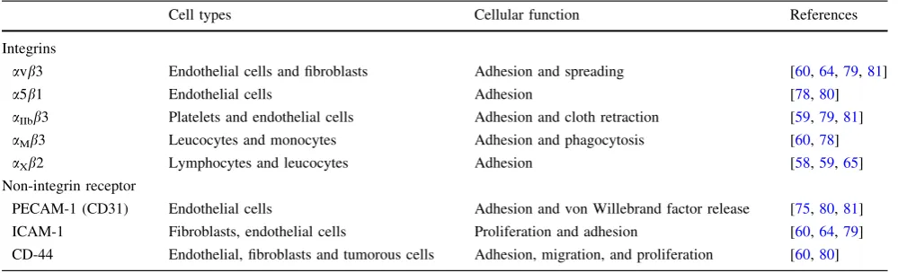
components including fibronectin, hyaluronic acid, and von Willebrand factor [60]. Human fibrinogen can bind to endothelial cells through either integrin or non-integrin binding sites (Table1). For example, fibrin has two pairs of RGD binding sites and a non-RGD site at the c chain, which can interact with endothelial cell integrins (i.e., a5b1,avb3, andaIIbb3) as well as with leucocyte integrins (i.e., aMb3 and aXb2) [65, 77, 78]. Other non-integrin receptors that can bind to endothelial cells include ICAM-1, CD-44 surface receptor, and platelet endothelial cell adhesion molecule-1 (PECAM-1, also known as CD-31) [60,79–81].
In other tissue-engineering applications, fibrin was combined with collagen for the development of blood vessel substitutes [82,83]. Collagen type I is the predom-inant structural component of the media as well as the adventia layers of blood vessels, while the inner layer of natural blood vessels, called theintimallayer, is lined with endothelial cells [27, 81]. Therefore, the combination of collagen and fibrin can be used to make scaffolds with good mechanical and biochemical properties [82]. For example, Isenberg et al. [83] have investigated a tubular scaffold made of type I collagen and fibrinogen to engineer small-diameter artificial arteries.
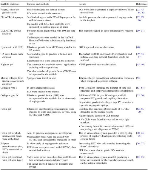
A summary of the uses of synthetic and natural poly-mers that have been applied to support tissue vasculariza-tion is presented in Table2.
Material properties
Scaffold and ECM materials are selected based on bulk and surface properties, which can be tuned with the aim to modulate cell adhesion and proliferation as well as phe-notypic cell expression [54,84]. Among the properties of importance, scaffold porosity and matrix stiffness play significant roles in cell and tissue responses [85, 86], and these will be briefly discussed below.
Table 1 Integrins and non-integrin receptors that can bind to fibrin(ogen)
Cell types Cellular function References
Integrins
avb3 Endothelial cells and fibroblasts Adhesion and spreading [60,64,79,81]
a5b1 Endothelial cells Adhesion [78,80]
aIIbb3 Platelets and endothelial cells Adhesion and cloth retraction [59,79,81]
aMb3 Leucocytes and monocytes Adhesion and phagocytosis [60,78]
aXb2 Lymphocytes and leucocytes Adhesion [58,59,65]
Non-integrin receptor
PECAM-1 (CD31) Endothelial cells Adhesion and von Willebrand factor release [75,80,81]
ICAM-1 Fibroblasts, endothelial cells Proliferation and adhesion [60,64,79]
Table 2 The use of bioactive polymers to support engineering-tissue construct vascularization
Scaffold materials Purpose and methods Results References
Poly(D,L -lactic-co-glycolic acid)
Scaffold designed for tubular tissues
ECs were seeded in the construct
ECs were able to generate a capillary network inside the scaffolds
[22,45, 84,85]
PLLA/PLGA sponges Scaffolds designed with 225–500-lm pores for skeletal muscle tissue
Pre-seeded with MC, then scaffolds were implanted in skeletal muscles of mice
Scaffold pre-vascularization promoted angiogenesis in the implant
[27,29, 86]
DLLA/TMC porous scaffold
For heart tissue engineering with 100lm pore size
Cardiomyocytes were seeded in the scaffold. Then scaffolds were subcutaneously implanted in rats
This method elicited an acute inflammatory reaction [30,31]
Hyaluronic acid (HA) Fibroblast growth factor (FGF) was added in the HA matrix
FGF improved neovascularization [40]
HA cross-linked with chitosan
Scaffold designed to produce a human skin equivalent
Endothelial cells were seeded in the construct
The hybrid scaffold improved EC proliferation and induced capillary network formation inside the scaffold
[39,43, 87]
Alginate gel The construct was made for several applications including cell encapsulation
Vascular endothelial growth factor (VEGF) was incorporated in the scaffold
VEGF promoted neovascularization. [45,46]
Marine collagen from tilapia (Oreochromis niloticus)
Sponges were tested in vivo Tilapia collagen caused fewer inflammatory responses when compared to porcine collagen
[52]
Collagen type I In vitro angiogenesis assay
ECs were seeded in the matrix
Type I collagen increased the number of tube-like structures and supported angiogenesis development
[53,54]
Collagen type IV Fibroblast growth factor (FGF) was
incorporated in the scaffold for in vitro study of angiogenesis
Addition of FGF in type IV collagen scaffold supported EC growth and capillary formation
Degradation product of collagen type IV promoted a specific angiogenic epitope
[55,56]
Fibrin gel Fibrinogen and thrombin concentrations were changed to study angiogenesis, in vitro, using HUVEC and VSMC
Capillary-like structures (CLS) made of HUVEC depended on the matrix rigidity
Higher rigidity decreased CLS number
•No CLSs were found in very soft or very rigid matrices.
•Decreasing thrombin concentration caused different morphology and alignment of VSMC
[62–64, 70]
Fibrin gel in which microcarrier beads were embedded
Aim: to generate angiogenesis development
Microcarrier beads were pre-coated with HUVEC and subsequently embedded in fibrin
This in vitro culture system provided a step-by-step process of capillary development containing multi-cellular lumen
[70,74, 75]
Polymer
monofilaments (i.e., PET) embedded in fibrin
In vitro study of angiogenesis guidance
PET fibers were pre-coated with HUVEC, then sandwiched in fibrin
Pre-coating PET with cells enabled increasing the fibers’ bioactivity
PET fibers were able to guide ECs to orient microvessels
[76,77]
Fibrin gel combined with collagen type I
SMCs were grown on a sheet-like scaffold, and then wrapped around a tubular vessel
The vessel allowed transfer of nutrients and oxygen
This in vitro culture system enabled producing a better environment for the vascularization of small-caliber arterial substitutes
[82,83]
Scaffold porosity
Porosity is defined as the fraction of the void space over the total volume of a scaffold. Pore size corresponds to the distance between two solid sections of the porous matrix [87, 88]. In tissue engineering, a highly porous scaffold (about 90 % porosity) is more desirable, since it should increase mass and nutrient transport [88]. Higher porosity can increase cell adhesion and provide a sufficient area for cell-matrix interactions and new ECM production by cells [89]. However, bulk and mechanical properties should also be considered. At higher porosity, the total solid volume of the solid part of the scaffold is lower when compared to scaffolds with lower porosity, thus resulting in weaker mechanical support [88,89].
The effect of pore size on cell behavior has been investigated in culturing bone tissue substitutes. Pores in the range of 300–400lm have been found to be optimal
for osteoblast attachment, growth, and proliferation [90]. The important role of such porous structures in endothelial cell organization and angiogenesis development was pio-neered by Clowes et al. [91] more than 20 years ago. In a more recent study, pore size was found to have a significant effect on cell binding, morphology, and phenotype, thus inducing endothelial cell migration and capillary formation inside the scaffold [92,93].
In the scientific literature, there are some suggestions concerning the optimum pore size to support vasculariza-tion. For example, many mature cell types, including fibroblasts and endothelial cells, have been found to be unable to spread and completely colonize the bulk of scaffolds with pore sizes higher than 300lm because of
the difficulty in bridging the distance [93]. In an in vitro angiogenesis study, it was shown that cell’s ability to bridge the distance in 3D scaffolds is important for sup-porting the vascularization process [76]. Using HUVEC-covered poly(ethylene terephthalate) (PET) monofilaments as contact guidance in HUVEC-seeded fibrin, it was sug-gested that the optimum fiber-fiber distance to support microvessel development was 100lm [76,77].
Furthermore, hepatocytes were also reported to spread well on a gelatin-chitosan scaffold (3:1) with a pore size of 20lm, while fibroblasts and endothelial cells spread better
on the same matrix but with pores ranging between 100–150lm compared to pores from 20–80 lm [93,94].
The role of pore size to promote the endothelial cell lining has been investigated in vascular grafts. For example, Zhang et al. [95] found that an external pore size of 30lm was
preferable compared to 20lm or smaller pores in terms of
promoting rapid tissue ingrowth and endothelial cell growth in the expanded poly(tetrafluoroethylene) (ePTFE) graft.
In addition, Marshall et al. [96] found that fibrin with 35lm pores significantly supported angiogenesis
development when compared to fibrin with either 20 or 70lm pores. Fibrin with pore sizes of approximately
30lm promotes the ingrowth of vascularized fibrous tissue
in engineered blood vessels [97]. However, the optimum porosity and pore size of the scaffold are still open to question and are based on the application as well as the cell type [88,93,95].
Matrix stiffness
Substrate mechanical properties, such as stiffness, are known to be important parameters affecting cell responses. In cell biology, matrix stiffness is sensed by cell receptors, and integrins transmit mechanical stress across the cell surface to the cell cytoskeleton, converting mechanical signals into biochemical ones [47,55,98].
Therefore, the ECM stiffness will influence cellular functions, including cell adhesion, proliferation, migration, and phenotype differentiation [99]. For example, Pelham and Wang [100] examined the effect of a collagen-coated poly(acrylamide) scaffold on the behavior of rat epithelial and fibroblast cells. They found that on more rigid (higher stiffness) surfaces, cells were more spread, and showed increased motility and focal adhesion contacts [99, 100]. Furthermore, increasing surface stiffness was found to result in increased cell contractility [101], more organized cells cytoskeleton and actin stress fibers, and higher adhesion strength [102]. The phenomenon related to the effect of environmental stiffness on cell behavior is known to as durotaxis [103].
Substrate stiffness and cell contractility also play sig-nificant roles in microvascular development. For example, while endothelial cells proliferate more on rigid surfaces, they form tube-like structures on softer substrates [102, 104]. Vasculogenesis decreased with an increase in matrix stiffness, which was a result of an increase in collagen [48, 51,105] or fibrinogen [63,65,106,107] concentrations.
Concluding remarks
Acknowledgments The author would like to acknowledge Prof. Patrick Vermette (Universite´ de Sherbrooke, QC, Canada) for his critical review. The author is supported by the FRGS-Malaysian Ministry of Higher Education (MOHE), the Universiti Teknologi Malaysia ST-Grant (vote # 4D045) and UTM Tier-1 Grant.
References
1. Atala A. Tissue engineering and regenerative medicine: con-cepts for clinical application. Rejuvenation Res. 2004;7:15–31. 2. Orlando G, Wood KJ, Stratta RJ, Atala A, Soker S. Regenerative medicine and organ transplantation: past, present, and future. Transplantation. 2011;91:1310–7.
3. Jain RK. Molecular regulation of vessel maturation. Nat Med. 2003;9:685–93.
4. Neumann T, Nicholson BS, Sanders JE. Tissue engineering of perfused microvessels. Microvasc Res. 2003;66:59–67. 5. Godbey WT, Atala A. In vitro systems for tissue engineering.
Ann N Y Acad Sci. 2002;961:10–26.
6. Mikos AG, Sarakinos G, Lyman MD, Ingber DE, Vacanti JP, Langer R. Prevascularization of porous biodegradable polymers. Biotechnol Bioeng. 1993;42:716–23.
7. Levenberg S, Langer R. Advances in tissue engineering. Curr Top Dev Biol. 2004;61:113–34.
8. Nerem RM. Cell-based therapies: from basic biology to replace-ment, repair, and regeneration. Biomaterials. 2007;28:5074–7. 9. He W, Ye L, Li S, Liu H, Wu B, Wang Q, et al. Construction of
vascularized cardiac tissue from genetically modified mouse embryonic stem cells. J Heart Lung Transplant. 2012;31:204–12. 10. Koh CJ, Delo DM, Lee JW, Siddiqui MM, Lanza RP, Soker S, Yoo JJ, Atala A. Parthenogenesis-derived multipotent stem cells adap-ted for tissue engineering applications. Methods. 2009;47:90–7. 11. Conway EM, Carmeliet P. The diversity of endothelial cells: a
challenge for therapeutic angiogenesis. Genome Biol. 2004;5:207. 12. Gimble J, Guilak F. Adipose-derived adult stem cells: isolation, characterization, and differentiation potential. Cytotherapy. 2003;5:362–9.
13. Langer R, Vacanti JP. Tissue engineering. Science. 2003;260: 920–6.
14. Howe A, Aplin AE, Alahari SK, Juliano RL. Integrin signaling and cell growth control. Curr Opin Cell Biol. 1998;10:220–3. 15. Frescaline G, Bouderlique T, Huynh MB, Papy-Garcia D,
Courty J, Albanese P. Glycosaminoglycans mimetics potentiate the clonogenicity, proliferation, migration and differentiation properties of rat mesenchymal stem cells. Stem Cell Res. 2012;8:180–92.
16. Chang C, Werb Z. The many faces of metalloproteases: cell growth, invasion, angiogenesis and metastasis. Trends Cell Biol. 2001;11:S37–43.
17. Bach FH. Xenotransplantation: problems and prospects. Annu Rev Med. 1998;49:301–10.
18. Lee CH, Huang GS, Chao KH, Wu SS, Chen Q. Differential pretensions of a flexor tendon graft for anterior cruciate ligament reconstruction: a biomechanical comparison in a porcine knee model. Arthroscopy. 2005;21:540–6.
19. Fuchs JR, Nasseri BA, Vacanti JP. Tissue engineering: a 21st century solution to surgical reconstruction. Ann Thorac Surg. 2001;72:577–91.
20. Chaikof EL, Matthew H, Kohn J, Mikos AG, Prestwich GD, Yip CM. Biomaterials and scaffolds in reparative medicine. Ann N Y Acad Sci. 2002;961:96–105.
21. Larson JW III, Chu CR. Tissue engineering of articular carti-lage. In: Guelcher SA, Hollinger JO, editors. An introduction to biomaterials. 2nd ed. Boca Raton: CRC Press; 2006. p. 525–36.
22. Mooney DJ, Sano K, Kaufmann PM, Majahod K, Schloo B, Vacanti JP, Langer R. Long-term engraftment of hepatocytes transplanted on biodegradable polymer sponges. J Biomed Mater Res. 1997;37:413–20.
23. Tabata Y. Biomaterial technology for tissue engineering appli-cations. J R Soc Interface. 2009;6:S311–24.
24. Lee JW, Lan PX, Kim B, Lim G, Cho DW. Fabrication and characteristic analysis of a poly(propylene fumarate) scaffold using micro-stereolithography technology. J Biomed Mater Res B Appl Biomater. 2008;87:1–9.
25. Kikuchi A, Okamoto S, Takahashi S, Asano S, Nishikawa T. Linear chronic cutaneous graft-versus-host disease. J Am Acad Dermatol. 1997;37:1004–6.
26. Vert M, Mauduit J, Li S. Biodegradation of PLA/GA polymers: increasing complexity. Biomaterials. 1994;15:1209–13. 27. Bramfeldt H, Sarazin P, Vermette P. Characterization,
degra-dation, and mechanical strength of poly(D,L -lactide-co-epsilon-caprolactone)-poly(ethylene glycol)-poly(D,L -lactide-co-epsilon-caprolactone). J Biomed Mater Res A. 2007;83:503–11. 28. Grizzi I, Garreau H, Li S, Vert M. Hydrolytic degradation of
devices based on poly(DL-lactic acid) size-dependence. Bioma-terials. 1995;16:305–11.
29. Levenberg S, Rouwkema J, Macdonald M, Garfein ES, Kohane DS, Darland DC, Marini R, van Blitterswijk CA, Mulligan RC, D’Amore PA, Langer R. Engineering vascularized skeletal muscle tissue. Nat Biotechnol. 2005;23:879–84.
30. Pego AP, Siebum B, Van Luyn MJ. Preparation of degradable porous structures based on 1,3-trimethylene carbonate andD,L -lactide (co)polymers for heart tissue engineering. Tissue Eng. 2003;9:981–94.
31. Pego AP, Van Luyn MJ, Brouwer LA, van Wachem PB, Poot AA, Grijpma DW, Feijen J. In vivo behavior of poly(1,3-tri-methylene carbonate) and copolymers of 1,3-tripoly(1,3-tri-methylene car-bonate withD,L-lactide or epsilon-caprolactone: degradation and tissue response. J Biomed Mater Res A. 2003;67:1044–54. 32. Aird WC. Phenotypic heterogeneity of the endothelium: II.
Representative vascular beds. Circ Res. 2007;100:158–73. 33. Hutchings H, Ortega N, Plouet J. Extracellular matrix-bound
vascular endothelial growth factor promotes endothelial cell adhesion, migration, and survival through integrin ligation. FASEB J. 2003;17:1520–2.
34. Brekke JH, Goldman SM, Ieska K, Issack P, Bong MR, Tian H. Hyaluronan as a biomaterials. In: Guelcher SA, Hollinger JO, editors. An introduction to biomaterials. 2nd ed. Boca Raton: CRC Press; 2006. p. 219–48.
35. Toole BP. Hyaluronan: from extracellular glue to pericellular cue. Nat Rev Cancer. 2004;4:528–39.
36. Rao CM, Deb TB, Gupta S, Datta K. Regulation of cellular phosphorylation of hyaluronan binding protein and its role in the formation of second messenger. Biochim Biophys Acta. 1997;1336:387–93.
37. Lesley J, Hascall VC, Tammi M, Hyman R. Hyaluronan binding by cell surface CD44. J Biol Chem. 2000;275:26967–75. 38. Slevin M, Kumar S, Gaffney J. Angiogenic oligosaccharides of
hyaluronan induce multiple signaling pathways affecting vas-cular endothelial cell mitogenic and wound healing responses. J Biol Chem. 2002;277:41046–59.
39. Miletti-Gonza´lez KE, Chen S, Muthukumaran N, Saglimbeni GN, Wu X, Yang J, Apolito K, Shih WJ, Hait WN, Rodrı´guez-Rodrı´guez L. The CD44 receptor interacts with P-glycoprotein to promote cell migration and invasion in cancer. Cancer Res. 2005;65:6660–7.
41. Park YD, Tirelli N, Hubbell JA. Photopolymerized hyaluronic acid-based hydrogels and interpenetrating networks. Biomate-rials. 2003;24:893–900.
42. Zielinski BA, Aebischer P. Chitosan as a matrix for mammalian cell encapsulation. Biomaterials. 1994;15:1049–56.
43. Black AF, Hudon V, Damour O, Germain L, Auger FA. A novel approach for studying angiogenesis: a human skin equivalent with a capillary-like network. Cell Biol Toxicol. 1999;15:81–90. 44. Draget KI, Skjak-Braek G, Smidsrod O. Alginate based new
materials. Int J Biol Macromol. 1997;21:47–55.
45. Drury JL, Mooney DJ. Hydrogels for tissue engineering: scaf-fold design variables and applications. Biomaterials. 2003;24: 4337–51.
46. Smidsrod O, Skjak-Braek G. Alginate as immobilization matrix for cells. Trends Biotechnol. 1990;8:71–8.
47. Becker WM, Kleinsmith LJ, Hardin J. The world of the cell. 7th ed. San Francisco: Pearson/Benjamin Cummings; 2009. p. 229–328.
48. Haarer JC, Dee KC. Proteins and amino acid-derived polymers. In: Guelcher SA, Hollinger JO, editors. An introduction to biomaterials. 2nd ed. Boca Raton: CRC Press; 2006. p. 121–38. 49. Chen CS, Yannas IV, Spector M. Pore strain behaviour of col-lagen–glycosaminoglycan analogues of extracellular matrix. Biomaterials. 1995;16:777–83.
50. Ma L, Gao C, Mao Z, Zhou J, Shen J. Biodegradability and cell-mediated contraction of porous collagen scaffolds: the effect of lysine as a novel crosslinking bridge. J Biomed Mater Res A. 2004;71:334–42.
51. Toman PD, Pieper F, Sakai N, Karatzas C, Platenburg E, de Wit I, Samuel C, Dekker A, Daniels GA, Berg RA, Platenburg GJ. Production of recombinant human type I procollagen homotri-mer in the mammary gland of transgenic mice. Transgenic Res. 1999;8:415–27.
52. Sugiura H, Yunoki S, Kondo E, Ikoma T, Tanaka J, Yasuda K. In vivo biological responses and bioresorption of tilapia scale collagen as a potential biomaterial. J Biomater Sci Polym Ed. 2009;20:1353–68.
53. Soker S, Machado M, Atala A. Systems for therapeutic angio-genesis in tissue engineering. World J Urol. 2000;18:10–8. 54. Francis ME, Uriel S, Brey EM. Endothelial cell–matrix
inter-actions in neovascularization. Tissue Eng Part B Rev. 2008;14:19–32.
55. Ingber DE, Folkman J. Mechanochemical switching between growth and differentiation during fibroblast growth factor-stimulated angiogenesis in vitro: role of extracellular matrix. J Cell Biol. 1989;109:317–30.
56. Xu J, Rodriguez D, Petitclerc E, Kim JJ, Hangai M, Moon YS, Davis GE, Brooks PC. Proteolytic exposure of a cryptic site within collagen type IV is required for angiogenesis and tumor growth in vivo. J Cell Biol. 2001;154:1069–79.
57. Senger DR, Claffey KP, Benes JE, Perruzzi CA, Sergiou AP, Detmar M. Angiogenesis promoted by vascular endothelial growth factor: regulation through alpha1beta1 and alpha2beta1 integrins. Proc Natl Acad Sci USA. 1997;94:13612–7. 58. Litvinov RI, Gorkun OV, Owen SF, Shuman H, Weisel JW.
Polymerization of fibrin: specificity, strength, and stability of knob-hole interactions studied at the single-molecule level. Blood. 2005;106:2944–51.
59. Janmey PA, Winer JP, Weisel JW. Fibrin gels and their clinical and bioengineering applications. Interface. 2009;6:1–10. 60. Herrick S. Fibrinogen. Int J Biochem Cell Biol. 1999;31:741–6. 61. Rowe SL, Lee S, Stegemann JP. Influence of thrombin con-centration on the mechanical and morphological properties of cell-seeded fibrin hydrogels. Acta Biomater. 2007;3:59–67. 62. Ferrenq I, Tranqui L, Vailhe´ B, Gumery PY, Tracqui P.
Mod-elling biological gel contraction by cells: mechanocellular
formulation and cell traction force quantification. Acta Biotheor. 1997;45:267–93.
63. Vailhe´ B, Lecomte M, Wiernsperger N, Tranqui L. The for-mation of tubular structures by endothelial cells is under the control of fibrinolysis and mechanical factors. Angiogenesis. 1998;2:331–44.
64. Nehls V, Herrmann R. The configuration of fibrin clots deter-mines capillary morphogenesis and endothelial cell migration. Microvasc Res. 1996;51:347–64.
65. Cox S, Cole M, Tawil B. Behavior of human dermal fibroblasts in three-dimensional fibrin clots: dependence on fibrinogen and thrombin concentration. Tissue Eng. 2004;10:942–54. 66. Catelas I, Sese N, Wu BM, Dunn JC, Helgerson S, Tawil B.
Human mesenchymal stem cell proliferation and osteogenic differentiation in fibrin gels in vitro. Tissue Eng. 2006;12: 2385–96.
67. Ferguson WD, Collins LM, Smith DW. Psychophysical thresh-old variability in cochlear implant subjects. Hear Res. 2003;180:101–13.
68. Tassiopoulos AK, Golts E, Oh DS, Labropoulos N. Current concepts in chronic venous ulceration. Eur J Vasc Endovasc Surg. 2000;20:227–32.
69. Nakatsu MN, Hughes CCW. An optimized three-dimensional in vitro model for the analysis of angiogenesis. Methods Enzymol. 2008;443:65–82.
70. Nehls V, Drenckhahn D. A novel, microcarrier-based in vitro assay for rapid and reliable quantification of three-dimensional cell migration and angiogenesis. Microvasc Res. 1995;50:311–22. 71. Montano I, Schiestl C, Schneider J, Pontiggia L, Luginbu¨hl J, Biedermann T, Bo¨ttcher-Haberzeth S, Braziulis E, Meuli M, Reichmann E. Formation of human capillaries in vitro: the engineering of prevascularized matrices. Tissue Eng Part A. 2009;15:1–15.
72. Carmeliet P, Conway EM. Growing better blood vessels. Nat Biotechnol. 2001;19:1019–20.
73. Yancopoulos GD, Davis S, Gale NW, Rudge JS, Wiegand SJ, Holash J. Vascular-specific growth factors and blood vessel formation. Nature. 2000;407:242–8.
74. Chen Z, Htay A, Dos Santos W, Gillies GT, Fillmore HL, Sholley MM, Broaddus WC. In vitro angiogenesis by human umbilical vein cells (HUVEC) induced by three-dimensional co-culture with glioblastoma cells. J Neurooncol. 2009;92:121–8. 75. Nakatsu MN, Sainson RC, Aoto JN, Taylor KL, Aitkenhead M,
Pe´rez-del-Pulgar S, Carpenter PM, Hughes CC. Angiogenic sprouting and capillary lumen formation modeled by human umbilical vein endothelial cells (HUVEC) in fibrin gels: the role of fibroblasts and angiopoietin-1. Microvasc Res. 2003;66:102–12. 76. Sukmana I, Vermette P. Polymer fibers as contact guidance to orient microvascularization in a 3D environment. J Biomed Mater Res A. 2010;92:1587–97.
77. Sukmana I, Vermette P. The effect of co-culture with fibroblast and angiogenic growth factors on microvascular maturation and multi-cellular lumen formation in HUVEC-oriented polymer fibre constructs. Biomaterials. 2010;31:5100–9.
78. Lishko VK, Kudryk B, Yakubenko VP, Yee VC, Ugarova TP. Regulated unmasking of the cryptic binding site for integrin alpha M beta 2 in the gamma C-domain of fibrinogen. Bio-chemistry. 2002;41:12942–51.
79. Cheresh DA, Berliner SA, Vicente V, Ruggeri ZM. Recognition of distinct adhesive sites on fibrinogen by related integrins on platelets and endothelial cells. Cell. 1989;58:945–53.
80. Weisel JW. Fibrinogen and fibrin. Adv Protein Chem. 2005;70:247–99.
82. Bayless KJ, Davis GE. Sphingosine-1-phosphate markedly induces matrix metalloproteinase and integrin-dependent human endothelial cell invasion and lumen formation in three-dimen-sional collagen and fibrin matrices. Biochem Biophys Res Commun. 2003;312:903–13.
83. Isenberg BC, Williams C, Tranquillo RT. Small-diameter arti-ficial arteries engineered in vitro. Circ Res. 2006;98:25–35. 84. Mooney DJ, Organ G, Vacanti JP, Langer R. Design and
fab-rication of biodegradable polymer devices to engineer tubular tissues. Cell Transplant. 1994;3:203–10.
85. Mooney DJ, Mazzoni CL, Breuer C, McNamara K, Hern D, Vacanti JP, Langer R. Stabilized polyglycolic acid fibre-based tubes for tissue engineering. Biomaterials. 1996;17:115–24. 86. Lesman A, Blinder Y, Levenberg Y. Modeling of flow-induced
shear stress applied on 3D cellular scaffolds: implications for vascular tissue engineering. Biotechnol Bioeng. 2010;105: 645–54.
87. Davis GE, Black SM, Bayless KJ. Capillary morphogenesis during human endothelial cell invasion of three-dimensional collagen matrices. In Vitro Cell Dev Biol Anim. 2000;36:513–9. 88. Kannan RY, Salacinski HJ, Sales K, Butler P, Seifalian AM. The roles of tissue engineering and vascularisation in the development of micro-vascular networks: a review. Biomateri-als. 2005;26:1857–75.
89. Agrawal CM, Ray RB. Biodegradable polymeric scaffolds for musculoskeletal tissue engineering. J Biomed Mater Res. 2001;55:141–50.
90. Linnes MP, Ratner BD, Giachelli CM. A fibrinogen-based precision microporous scaffold for tissue engineering. Bioma-terials. 2007;28:5298–306.
91. Clowes AW, Kirkman TR, Clowes MM. Mechanisms of arterial graft failure. II. Chronic endothelial and smooth muscle cell proliferation in healing polytetrafluoroethylene prostheses. J Vasc Surg. 1986;3:877–84.
92. O’Brien FJ, Taylor D, Clive LT. The effect of pore size on cell adhesion in collagen–GAG scaffolds. Biomaterials. 2005;26: 433–41.
93. Zeltinger J, Sherwood JK, Graham DA, Mu¨eller R, Griffith LG. Effect of pore size and void fraction on cellular adhesion, pro-liferation, and matrix deposition. Tissue Eng. 2001;7:557–72. 94. Huang Y, Onyeri S, Siewe M, Moshfeghian A, Madihally SV.
In vitro characterization of chitosan–gelatin scaffolds for tissue engineering. Biomaterials. 2005;26:7616–27.
95. Zhang Z, Wang Z, Liu S, Kodama M. Pore size, tissue ingrowth, and endothelialization of small-diameter microporous polyure-thane vascular prostheses. Biomaterials. 2004;25:177–87. 96. Marshall A, Barker T, Sage E, Hauch K, Ratner B. Pore size
controls angiogenesis in subcutaneously implanted porous matrices. In: 7th World Biomaterials Congress, Sydney; 2004. 97. Yao L, Swartz DD, Gugino SF, Russell JA, Andreadis ST.
Fibrin-based tissue-engineered blood vessels: differential effects of biomaterial and culture parameters on mechanical strength and vascular reactivity. Tissue Eng. 2005;11:991–1003. 98. Geiger B, Bershadsky A, Pankov R, Yamada KM.
Transmem-brane crosstalk between the extracellular matrix-cytoskeleton crosstalk. Nat Rev Mol Cell Biol. 2001;2:793–805.
99. Ingber DE, Prusty D, Sun Z, Betensky H, Wang N. Cell shape, cytoskeletal mechanics and cell cycle control in angiogenesis. J Biomech. 1995;28:1471–84.
100. Pelham RJ, Wang Y. Cell locomotion and focal adhesions are regulated by substrate flexibility. Proc Natl Acad Sci USA. 1997;94:13661–5.
101. Tseng Q, Wang I, Duchemin-Pelletier E, Azioune A, Capri N, Gao J, et al. A new micropatterning method of soft substrates reveals that different tumorigenic signals can promote or reduce cell contraction levels. Lab Chip. 2011;11:2231–40.
102. Discher DE, Janmey P, Wang YL. Tissue cells feel and respond to the stiffness of their substrate. Science. 2005;310:1139–43. 103. Lo CM, Wang HB, Dembo M, Wang YL. Cell movement is
guided by the rigidity of the substrate. Biophys J. 2000;79: 144–52.
104. Deroanne CF, Lapiere CM, Nusgens BV. In vitro tubulogenesis of endothelial cells by relaxation of the coupling extracellular matrix-cytoskeleton. Cardiovasc Res. 2001;49:647–58. 105. Yeung T, Georges PC, Flanagan LA, Marg B, Ortiz M, Funaki
M, Zahir N, Ming W, Weaver V, Janmey PA. Effects of sub-strate stiffness on cell morphology, cytoskeletal structure, and adhesion. Cell Motil Cytoskelet. 2005;60:24–34.