CHAPTER IV
RESULTS
The goal of this study was to identify the underlying genetic defect in patients with MR
and epilepsy. Mutation analysis from the syndromic patients were performed, from the non
syndromic patients then do further screening to confirm the mutation to the parents of the
suspected patient.
IV.1 Obtaining MR with Epilepsy patients
First step of this study was obtaining MR with Epilepsy patients list. Starting total 527
MR patients that already collected from previous study 16, screening of the clinical data from all
patients performed. Patients that positively diagnosed with epilepsy by medical doctor from
Bandung and Semarang Indonesia are all included in this study.
The patients clinical features were collected, possible dysmorphology data from the
pictures, results of measurements, and cytogenetic analysis. Unfortunately those patients have
had no previous additional brain imaging examination, or blood metabolite analysis yet. History
and pedigree was already taken from the parents and or the teacher in the special school and was
written in the medical record, although not completely captured or reliable.
The clinical features with pictures already presented to the clinical geneticist in charge,
Dr. Tjitske Kleefstra, MD, PhD and Bregje van Bon, MD, PhD in power point format.
Differential diagnosis gained based on LMD (London Medical Database) software and
1 MR M 7y Absence
epilepsy were excluded.One patient with marker at chromosome 15 was excluded from patients
list, so in total there were 31 subjects left.
IV.2 Mutation Screening from The Syndromic Patients List
For the syndromic patients list, there are four patients that were suspected have
dysmorphology that fit to some features in some syndromes. For patient with the possible mosaic
22 (named RGP, male), cytogenetic analysis for mosaicism were recheck, but did not find
anything. Need more specific cytogenetic tools to help the diagnosis for this patient (example:
whole genome array). Explanationof the details can be seen below.
1. PTEN gene: Cowden syndrome patient
Dysmorphology and physical examination of this patient found macrocephaly,
hamartomas on chest and inside mouth and severe MR.
Figure 3. Picture of suspected Cowden syndrome patient.
The pictures of the girl showed narrow palate, and hamarthomas inside the mouth. She also has
hamarthomas on her chest.
2. UPF3B and MED12 gene: Lujan syndrome
Dysmorphology and physical examination of this patient characterized male, had severe
Figure 4. Picture of suspected Lujan syndrome patient.
The pictures of the boy showed arachnoid fingers, typically for marfanoid phenotype.
3. UBE3A gene: Angelman syndrome
Dysmorphology and physical examination of this patient foundsevere MR, speech
impairment, seizures and attention disorder.
Figure5. Picture of suspected Angelman syndrome patient.
The picture of the girl does not show much dysmorphological appearances, but she has severe
MR and atenntion disorder on her behaviour.
4. TCF4 gene: Pitt-Hopkins syndrome
Dysmorphology and physical examination of this patient foundspecific facial features &
Figure 6. Picture of the suspect Pitt-Hopkins syndrome patient.
The picture of the girl showed crowded teeth on her open mouth, facial express of severed MR
patients.
No mutation found in UBE3A, UPF3B and MED12, PTEN, and TCF4 genes sequencing.
IV.3 Mutation Screening from the non syndromic patients list
From the LMD, differential diagnosis for suspected syndromic patients were made. But
for the rest of them, which had no specific dysmorphology, screening with known genes that
responsible for MR and epilepsy were the best option. Those gene are SCN1A, ARX, STXBP1,
and LGI1 genes. Only ARX testing is already performed as routine diagnostic gene in DNA
Diagnostic Human Genetic Department RUN MC Nijmegen.
The rest of them are chosen based on similarity of some clinical features in the patients
list. The tests need to be designed and tested first before applied to the patients list. SCN1A, ARX
and STXBP1 genes were chosen based on supported journals and characteristic of patients list
that matched with characteristics of those genes.
Table 5. List of the patients with the screened genes.
No Name Sex Tested genes for MR/epilepsy
SCN1A ARX STXBP1 LGI1
2 DY M tested tested tested tested
pathogenesis of epilepsy. It is tested for all patients (males and females), it was chosen to be first
tested due to its high prevalence that in the society. It is also a well known gene for FS, GEFS+,
First step was designed the primers of SCN1A. PCR product were run on the gel and got
the results. If there are product on the gel, then the primer worked. Here is the example of the
PCR product of the tested SCN1A primers. After purified the DNA then sequencing were
performed.
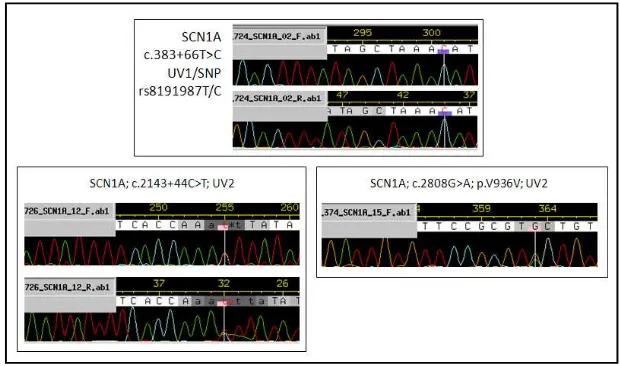
Figure 7. Results of SCN1A primers with three controls and 26 exons tested.
Figure 8. Some of the results of SCN1A sequencing.
Analyzing whether this results are real mutation or just SNPs were performed using
Alamut software. Some of the results are new SNPs, it might be not recorded yet because mostly
Javanese population were sequenced. After comparation from Alamut software that mostly came
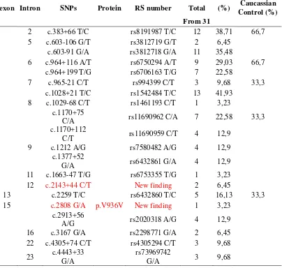
Table 6. SNPs Results from SCN1A screening examination
believed to be rare nonpathogenic variants, since they did not lead to a change in the amino acid
sequence of the protein. Therefore, they are believed not to explain MR and epilepsy from the
patients list.
Splice site prediction programs already performed to those SNPs. Yet, no changes were
found and proven influencing on gene splicing process.
Located in Xp.22.13 makes only 19 male patients tested for this gene. It is chosen
because of the characteristic of ARX that have the widest phenotypic spectrum 79. The patients
list was also not really specific in terms of dysmorphology. No mutation or SNPswere found in
the ARX gene with sequencing.
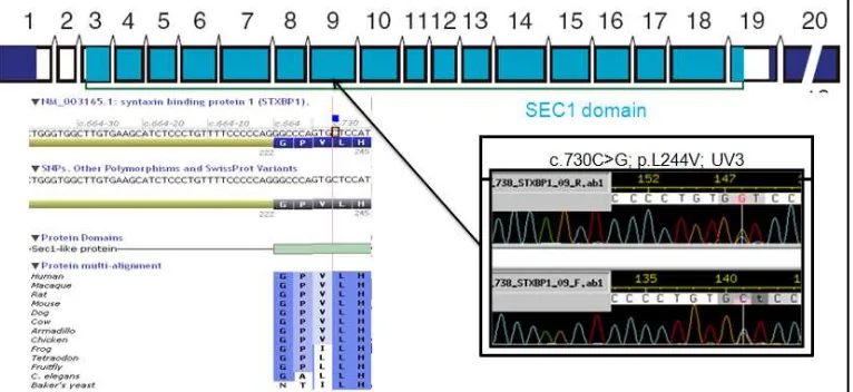
IV.3.3 STXBP1
The third one is the STXBP1 gene. The gene is tested for all patients. It is located in
9q34.1, and it is a good candidate gene for the patients list since Hamdan FF et al.2009 described
a screening of this gene in idiopathic MR with epilepsy, and had positive results in 2 out of 95
patients14. After sequencing, one possible mutation were found in exon nine that can be seen in
the picture below.
Figure 9. Results of STXBP1 sequencing compared with Alamut software and pointed to the
location of UV in exon nine.
From the mutation screening result, decission to predict pathogenicity of that mutation
using prediction programs that based on amino acid changes were made. By using alamut
conservation program, Leucine as highly conserved amino acid, also located in the sec1 domain,
if the comparation were resulted from the range of zero until 215. The conclussion was that it has
no effect on splicing processin Alamut Splicing Predictions program.
In PolyPhen Prediction, this variant is predicted to be benign, probably because Valine
and Leucine has the same type which is non polar. On the other hand, the SIFT prediction
softwareshows that Valine is predicted to be not tolerated. The prediction is mainly based on the
fact that Valine on this position in the protein is fully conserved in evolution. In Poly Phen, black
colour means that protein is non polar, green: uncharged polar, red: basic, and blue: acidic. Seq
rep is the fraction of sequences that contain one of the basic amino acid. Low seq rep means it
has low fraction, it means also that the position severely gapped or unalignable or has a little
information. Since the prediction software shows contradictory results, and the mutation
concerns a highly conserved amino acid in an important domain of the protein. The hypothesis
was that this might be the causative mutation in the the patient.
The clinical features of the patient that has this possible mutation is described as follows.
He was examined when he was 11 years old and he originated from Bandung special school. His
family history is normal, also prenatal, natal and perinatal history. No congenital anomalies. His
developmental milestones was normal. No behavioral problems in this patient. From the
measurement he has microcephaly and has prominent ears. He is severely mentally retarded, and
has also clinodactyly on the fourth right toe. From the neurological examination,he had epilepsy
with general partial type of the seizures since he was 6 months old.
Comparation ofthe clinical features of the patients with previous reported cases by Saitsu
et al in 2008, who described four patients with a mutation in STXBP1 were done 80, 81. All of
them are de novo mutation, and most likely causing loss of function. Possible pathogenic
mutation in exon 9 that haven’t reported before was confirmed by testing the parents.
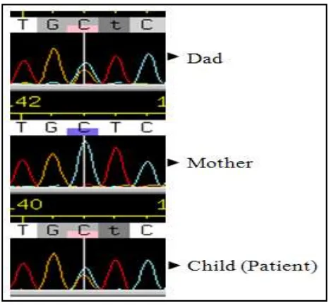
Further mutation screening to confirm the mutation
The result in STXBP1 gene (c.730C>G/ p.Leu244Val) need to be confirmed. This is
where genetic counseling take the most important role.
Figure 10. Result of verification
Thisvariant was also present in the healthy father of patient. That result proved that this UV is
not the cause of the MR and epilepsy in this patient, because the healthy father also had the
exactly same mutation.
No pathogenic mutation were found in STXBP1 gene sequencing in thepatients list.
IV.3.4 LGI1
LGI1 or known as leucine-rich gene, gliomainactivated-1 gene is studied due to its
relationship with Autosomal dominant lateral temporal lobe epilepsy (ADLTE). LGI1 mutations
generalized seizures.Anypathogenic mutation in LGI1 gene were did not found in