Indones. J. Chem., 2016, 16 (2), 222 - 228 222
Molecular Dynamics Simulations and Empirical Observations
on Soy Lecithin Liposome Preparation
Rini Dwiastuti1,2, Muhammad Radifar3,5, Marchaban4,*, Sri Noegrohati1,4, and Enade Perdana Istyastono1,5
1
Faculty of Pharmacy, Sanata Dharma University, Yogyakarta 55282, Indonesia
2Pharmaceutical Sciences Doctoral Program, Universitas Gadjah Mada, Sekip Utara, Yogyakarta 55281, Indonesia
3
Graduate School, Universitas Gadjah Mada, Sekip Utara, Yogyakarta 55281, Indonesia
4Faculty of Pharmacy, Universitas Gadjah Mada, Sekip Utara, Yogyakarta 55281, Indonesia
5
Molecular Modeling Center “MOLMOD.ORG” Yogyakarta, Indonesia
Received July 23, 2015; Accepted January 6, 2016
ABSTRACT
Soy lecithin is a phospholipid often used in liposome formulations. Determination of water and phospholipid composition is one of the problems in the liposome formulation. This study is using molecular dynamics simulation and empirical observation in producing liposome preparations. Phospholipids 1,2-dilauroyl-sn-glycero-3-phosphoethanolamine (DLPE) were objected in molecular dynamics simulations using Coarse Grained Molecular Dynamics (CGMD) approaches. The result showed that the molecular dynamic simulations could be employed to predict the liposome size. The molecular dynamic simulations resulted in liposome size of 71.22 ± 2.54 nm, which was located within the range of the liposome size resulted from the empirical observations (95.99 ± 43.02 nm). Moreover, similar liposome forms were observed on both results of molecular dynamics simulations and empirical approaches.
Keywords: soy lecithin; liposome; 1,2-dilauroyl-sn-glycero-3-phosphoethanolamine (DLPE); Coarse Grained Molecular Dynamics (CGMD)
ABSTRAK
Lesitin dari kedelai merupakan fosfolipid yang sering digunakan dalam formulasi liposom. Penentuan komposisi air dan fosfolipid merupakan salah satu kendala dalam formulasi liposom. Penelitian ini menggunakan pendekatan simulasi dinamika molekuler dan dibuktikan secara empiris untuk menghasilkan sediaan liposom. Simulasi dinamika molekuler menggunakan komposisi air : fosfolipid sesuai dengan perhitungan teoretis dan selanjutnya dibuktikan secara empiris. Fosfolipid yang dimodelkan adalah 1,2-dilauroyl-sn-glycero-3-phosphoethanolamine (DLPE) dengan metode Coarse Grained Molecular Dynamics (CGMD). Hasil penelitian ini menunjukkan bahwa pemodelan komputasi DLPE dapat memprediksi pembuatan liposom menggunakan lesitin. Diperoleh ukuran liposom dan gambaran liposom yang sama antara hasil simulasi dinamika molekuler dan hasil observasi empiris. Ukuran liposom hasil simulasi dinamika molekuler (71,22 ± 2,54 nm) berada pada jangkauan sebaran ukuran liposom hasil observasi empiris (95,99 ± 43,02 nm).
Kata Kunci: lesitin dari kedelai; liposom; 1,2-dilauroyl-sn-glycero-3-phosphoethanolamine (DLPE); Coarse Grained Molecular Dynamics (CGMD)
INTRODUCTION
Liposomes are pharmaceutical preparation with many of advantages for drug delivery system such as high solubility and increasing drug penetration. Liposomes are spherical vesicles composed of phospholipid bilayers or lamellae. Liposome formulation research and development as cosmeceuticals are still
being developed [1]. Liposomes in topical drug delivery preparation is developed because having a good penetration through the skin [2-3]. Liposomes are spherical vesicles composed of single or multiple lipid bilayer which have a high biocompatibility, high transfer efficiency, as well as having a good pharmacokinetic profile [4-6]. Liposomes drug delivery is also suitable for hydrophobic and hydrophilic drugs with the ability to
* Corresponding author.
Email address : [email protected]
Rini Dwiastuti et al.
reduce toxicity and improve stability [7]. The lecithin increases transdermal drug delivery by decreasing of the skin barrier permeability. The lecithin which is often used in liposome formulations is soy lecithin. Permeability liposomes have an important role in improving the efficiency of drug encapsulation of hydrophilic compounds in the liposome structure [8].
Phospholipids from soy lecithin consist of phosphatidylcholine (PC), phosphatidylethanolamine (PE), and phosphatidylinositol (PI). Phospholipids are directly affecting the membrane permeability of the stratum corneum [9]. Formulation has several aspects including formula, process, tool, method and packaging. This study will be related to the determination of water and phospholipids composition in the formula. The approaches used in this study are molecular dynamics simulations and empirical observations in liposome preparations.
Modeling and simulation play an important role in the development of new technologies in the production and design of materials/products. Molecular dynamics simulations could be employed to investigate the interaction of the drug molecule and the membrane phospholipid liposomes. Molecular dynamics simulations provide a comprehensive picture of molecular interactions that occur during the process of the formation of liposomes. Risselada and Marink [10] have conducted research on the effects of temperature and composition of the membrane to the structure and dynamic properties of the liposome membrane with a diameter of 15-20 nm using Coarse Grained Molecular Dynamics (CGMD) simulations. The attempts to reduce the size of the particles can be done by sonication [11]. Ramalho et al. [12] investigated how the presence of a nanoparticle affects fluid-gel transformations in 1,2-dipalmitoyl-sn-glycero-3-phosphocholine (DPPC) lipid.
Pickholz and Giupponi [13] has developed the MARTINI coarse grained force field to model water and lipids as well as study the encapsulation of local anesthetics prilocaine in a liposome structure with a small size by using CGMD. Molecular dynamics simulations using CGMD can be used as an initial step to reduce trial and error in a liposome formulation technology.
The aim of the study was to compare the results of liposome preparation between molecular dynamics simulations and empirical observations. Molecular dynamics simulations could provide qualitative results related to the formed liposomes. Molecular dynamics simulations used in this study were unconstrained molecular dynamics simulations to mimicking the empirical observation. The composition determination of phospholipids and water used in the empirical observation is similar to the composition used in molecular dynamics simulations. This approach is expected to assist the efforts to predict the parameters
of both empirical and theoretical required in the forming of the membrane phospholipid liposomes structures. In this article, the liposome sizes and shapes resulted from the molecular dynamics simulations and the empirical observations are presented.
EXPERIMENTAL SECTION
Computation Details
Molecular dynamics simulations were performed using the CGMD approaches, employing MARTINI 2.0 force field [14]. Prior to the system preparation, the coordinate of 1,2-dilauroyl-sn-glycero-3-phosphoethanolamine (DLPE) molecule was coarse-grained and turned into beads, where each bead is corresponding to 3 to 5 heavy atom of the original molecule. The CGMD in this study used 4 groups bead: uncharged, polar, nonpolar (dipole moment close to molecule used in this study was retrieved by converting the number of DLPE from Hudiyanti et al. [18]. The number of phospholipid in liposome with diameter of 32 nm was calculated by multiplying the number of phospholipid in 20 nm diameter liposome [18] with the square of diameter ratio (32/20). This yielded 8192 DLPE molecules and was rounded up to 8200. All molecular dynamics simulations were performed on a Linux (Ubuntu 12.04 LTS) machine with Intel(R) Xeon(R) CPU E31220 (@ 3.10 GHz) as the processors and 8.00 GB of RAM [19].
Indones. J. Chem., 2016, 16 (2), 222 - 228 224
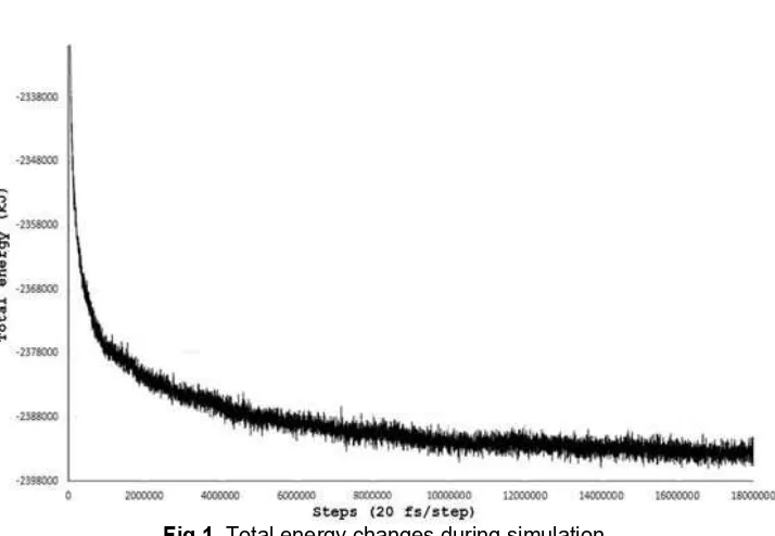
Fig 1. Total energy changes during simulation
Liposomes Preparation
Materials used in this study were: soybean lecithin (Nacalai Tesque Inc., Japan), and distilled water. The instruments used in this study were blender, ultra turrax, sonicator, Particle Size Analyzer (PSA (HORIBA scientific) at Department of Pharmacy, Indonesian Islamic University Yogyakarta), Transmission Electron Microscope (TEM (TEM JEOL JEM 1400) at Department of Chemistry Universitas Gadjah Mada). Preparation of liposomes referred to some previously publish methods [21-22] with some modifications. Liposomes were prepared by dispersing 8.70 g phospholipid in 100 mL distilled water at 60 °C. Phospholipids and water were mixed in a blender for 60 sec. These mixtures further were subjected to sonication for about 30 min at 60 °C. The resulting liposomes subsequently were analyzed using PSA to examine the particle sizes and TEM to see the shape of the liposomes [23].
Comparison of the Liposomes Results
The experimental liposome self-assembly aimed to form liposome with the size of 72 nm. Therefore, the liposome sizes resulted in the molecular dynamics simulations were converted from 32 nm in to 72 nm by employing Eq. 1.
Liposome self-assembly process was observed by inspecting the total energy change and trajectory visualization along the simulation using VMD 1.9.2b. Systems were in equilibrium when both the total energy and the liposome are stable enough. Then the time needed for liposome assembly and the change of total energy were calculated. The sizes of the liposomes were calculated by averaging the maximum distance between PO4 bead in x, y, and z axis in the last 4 ns. The final liposome snapshot was then compared with the liposome morphology from the TEM image.
RESULT AND DISCUSSION
The liposome self-assembly was started with the formation of phospholipid bilayer patches which can be seen in the first 10 ns and most of this process occurs at 10th to 60th ns (Fig. 1). As the bilayer patches fused
into a very large bilayer patch, the edge has been drastically reduced. Since there were many less patch, the only way to reduce the amount of exposed hydrophobic area was through curving and forming sphere. This process is the slowest part in this liposome assembly due to the small entropy gain and the large scale phospholipid movement.
In this study, DLPE was used because of its simplicity and possibility to be coarse-grained with VMD. This simulation technique allows simulation for large systems with a size range of 10-100 nm and a long simulation time with a span of up to a few μs so that it can be used to simulate the formation of liposomes. Also, DLPE was proven to be able to form liposome in a computer simulation as reported by Hudiyanti et al. [18]. In computer simulation the system
Fig 2. The molecular dynamics simulation snapshots every 40 ns, from 0 ns (randomly placing 8200 coarse-grained DLPE molecule in a 38 x 38 x 38 nm cubic box using Packmol 14.702 [19]) to 360 ns. The first liposome structure was identified at 320 ns
Table 1. Liposome size based on computational and empirical observation
Liposome size based on molecular dynamics
simulations Liposome size based on empirical observation
Liposome size (nm) Frequency
(%) Liposome size (nm) Frequency (%)
70.5 1 27.45 1.611
70.6 0 31.01 5.458
70.7 0 35.03 6.983
70.8 4 39.58 5.828
70.9 7 44.72 3.474
71 12 50.53 1.509
71.1 20 57.09 0.922
71.2 17 64.5 1.974
71.3 11 72.87 4.334
71.4 9 82.33 7.315
71.5 5 93.02 10.114
71.6 9 105.1 12.004
71.7 1 118.74 12.466
71.8 4 134.16 11.272
71.9 0 151.57 8.542
72 0 171.25 4.836
193.48 1.358
Mean (±SD) 71.22 ± 2.54 Mean (±SD) 95.99 ± 43.02
preparation reveals that a system with the size of 41 x 41 x 41 nm cubic was filled with 336,386 water beads, 315 Cl- beads, and 315 Na+ beads. As one water bead
represent 4 water molecules and one ion bead represent 6 water molecules and 1 ion, the resulting system contains 1,348,064 water molecule and 8,200 DLPE molecules. Multiplying each molecule number with its
molecular weight (MW) gives the ratio of phospholipid: water as much 195.7 mg/mL. This process involves the exclusion of phospholipid hydrophobic tail from water, enriching the amount of hydrogen bond between water and VDW interaction of the phospholipid hydrophobic tail which is energetically more favorable.
Indones. J. Chem., 2016, 16 (2), 222 - 228 226
Rini Dwiastuti et al.
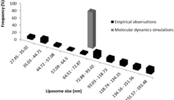
Fig 3. Histogram of liposome size based on molecular
dynamics simulations Fig 4.empirical observations Histogram of liposome size based on the
Fig 5. Comparisons between the liposome sizes resulted from the empirical observations (colored in black) and from
the molecular dynamics simulations (colored in grey)
Phospholipid bilayer is far more stable than the dissolved phospholipid, as can be seen from the very steep energy change in the first 10 ns, which also result in relatively fast bilayer patch formation (Fig. 1). The next process was the slow merging of the patches to form larger patches. Due to the random nature of the phospholipid placement, the bilayer patches were located and oriented in various ways, thus many of the edge of the bilayer faces and close to another bilayer patch. The edge of the bilayer patch is highly unstable because this area was filled with relatively exposed hydrophobic tail, which in turn tends to merge with the surrounding bilayer patch to reduce such exposure. As shown in Fig. 1, the process of curving and forming sphere took about 260 ns (60-320 ns). The first liposome structure was identified at 320 nm (Fig. 2).
The dynamic of liposome self-assembly can be seen in Fig. 2. Our benchmarking shows that the time required for 1 ns of this simulation was 348 min, which roughly translate to 2 days and 10 h per 10 ns. Thus, the
whole simulation process takes about 3 months to complete. Liposome size measurement shows that the mean (standard deviation) size of liposome based on computational is 71.22 (± 2.54) nm while the liposome size based on empirical observation is 95.99 (± 43.02) nm (Table 1).
Fig. 6. Liposomes morphology observed using TEM (A-D) and molecular dynamics simulations (E-H)
Fig. 5 indicates that the preparation of liposome formulations has many factors influencing either directly or indirectly to the quality of the resulting preparation. The influences of the chemical interactions that occur between the components of the liposomes and the influences of factors lipid and water composition to the results could be predicted by using molecular dynamics simulations approach. Based on the computational approach, the liposome size should be 72 nm and notably the liposome size resulted in empirical observations is 95.99 (± 43) nm. These results suggest that the formulation of liposomes with phospholipid and water ratio based on molecular dynamics simulations could produce liposomes with a size that is in the size range of liposomes empirical results (Fig. 5). However, the empirical observations resulted in significantly wider standard deviation of the liposome sizes compared to the results from molecular dynamics simulations (Fig. 5) owing to the simplicity of the molecular dynamics simulations compared to the system used in empirical observations.
The molecular dynamics simulations used DLPE as the phospholipids components, while the soy lecithin used in the empirical observations contains PE (including DLPE), PD, and PI. Different phospholipids components in the membrane bilayers structure of the liposomes could result in the different sizes and morphology of liposomes [10]. Interestingly, in this research the liposomes morphology observed using TEM showed similarity in term of morphology and size to the liposomes resulted in molecular dynamics simulations (Fig. 6). This indicates DLPE properties play an important role in liposomes formation employing soy lecithin as the source of phospholipids. Therefore,
further investigation on the correlation of liposomes physical properties between results from molecular dynamics simulations and empirical observations is of considerable interest.
CONCLUSION
The liposome size resulted from the molecular dynamic simulations (71.22 ± 2.54 nm) was located within the range of the liposome size (Fig. 5) resulted from the empirical observations (95.99 ± 43.02 nm) with similar morphology (Fig. 6). Molecular dynamics simulations using CGMD methods on DLPE could therefore predict in the atomic level of liposomes preparation using soy lecithin, especially the particle size and the morphology of liposomes.
ACKNOWLEDGEMENT
This research was financially supported by Doctoral Dissertation Research Grant funds in 2016 (Grant No.: 027a/Penel.LPPM USD/IV/2016); “Sanata Dharma” Foundation (Grant No.: K-1160/Yys/3-15/IX/2013 to Rini Dwiastuti) and Indonesian Directorate General of Higher Education (BPPDN Program No. 1251.15/E4.4/2012 to Rini Dwiastuti).
REFERENCES
1. Rahimpour, Y., and Hamishehkar, H., 2012, Expert Opin. Drug Delivery, 9 (4), 443–455.
2. Maghraby, G.M.M.E, Williams, A.C., and Barry, B.W., 1999, J. Pharm. Pharmacol., 51 (10), 1123–
1134.
Indones. J. Chem., 2016, 16 (2), 222 - 228 228
Rini Dwiastuti et al.
3. Maghraby, G.M.M.E., Williams, A.C., and Barry, B.W., 2001, J. Pharm. Pharmacol., 53 (6), 1069–
1077.
4. Wang, S., Zhang, J., Jiang, T., Zheng, L., Wang, Z., Zhang, J., and Yu, P., 2011, Int. J. Pharm., 403
(1-2), 219–229.
5. Zhao, L., Wei, Y.M., Zhong, X.D., Liang, Y., Zhang, X.M., Li, W., Li, B.B., Wang, Y., and Yu, Y., 2009, J. Pharm. Biomed. Anal., 49 (4), 989–996.
6. Liu, D., Hu, H., Lin, Z., Chen, D., Zhu, Y., Hou, S., and Shi, X., 2013, J. Photochem. Photobiol., B, 127,
8–17.
7. Laouini, A., Jaafar-Maalej, C., Limayem-Blouza, I., Sfar, S., Charcosset, C., and Fessi, H., 2012, J. Colloid Sci. Biotechnol., 1 (2), 147–168.
8. Eloy, J.O., de Souza, M.C., Petrilli, R., Barcellos, J.P.A., Lee, R.J., and Marchetti, J.M., 2014, Colloids Surf., B, 123, 345–363.
9. Yokomizo, Y., and Sagitani, H., 1996, J. Controlled Release, 38 (2-3), 267–274.
10. Risselada, H.J., and Marrink, S.J., 2009, Phys. Chem. Chem. Phys., 11 (2), 2056–2067.
11. Akbarzadeh, A., Rezaei-Sadabady, R., Davaran, S., Joo, S.W., Zarghami, N., Hanifehpour, Y., Samiei, M., Kouhi, M., and Nejati-Koshki, K., 2013,
Nanoscale Res. Lett., 8, 1–9.
12. Prates Ramalho, J.P., Gkeka, P., and Sarkisov, L., 2011, J. Surf. Colloids, 27 (7), 3723–3730.
13. Pickholz, M., and Giupponi, G., 2010, J. Phys. Chem. B, 114 (20), 7009–7015.
14. Marrink, S.J., Risselada, H.J., Yefimov, S., Tieleman, D.P., and de Vries, A.H., 2007, J. Phys. Chem. B, 111 (27), 7812–7824.
15. Humphrey, W., Dalke, A., and Schulten, K., 1996,
J. Mol. Graphics, 14 (1), 33–38.
16. Chng, C.P., and Yang, L.Y, 2008, Bioinf. Biol. Insights, 2, 171–185.
17. Martínez, L., Andrade, R., Birgin, E.G., and Martínez, J.M., 2009, J. Comput. Chem., 30 (13),
2157–2164.
18. Hudiyanti, D., Radifar, M., Raharjo, T.J., Narsito, N., and Noegrohati, S., 2014, J. Chem., 2014, 1–6.
19. Istyastono, E.P., and Setyaningsih, D., 2015,
Indones. J. Pharm., 26, 20–28.
20. Phillips, J.C., Braun, R., Wang, W., Gumbart, J., Tajkhorshid, E., Villa, E., Chipot, C., Skeel, R.D., Kalé, L., and Schulten, K., 2005, J. Comput. Chem., 26 (16), 1781–1802.
21. Hupfeld, S., Holsaeter, A.M., Skar, M., Frantzen, C.B., and Brandl, M., 2006, J. Nanosci. Nanotechnol., 6 (9-10), 3025–3031.
22. Jahadi, M., Khosravi-Darani, K., Ehsani, M.R., Mozafari, M.R., Saboury, A.A., Seydahmadian, F., and Vafabakhsh, Z., 2012, Asian J. Chem., 24 (9),
3891–3894.
23. Badran, M., Shalaby, K., and Al-Omrani, A., 2012,
![Fig 2. The molecular dynamics simulation snapshots every 40 ns, from 0 ns (randomly placing 8200 coarse-grained DLPE molecule in a 38 x 38 x 38 nm cubic box using Packmol 14.702 [19]) to 360 ns](https://thumb-ap.123doks.com/thumbv2/123dok/873659.821237/4.595.91.523.112.361/molecular-dynamics-simulation-snapshots-randomly-grained-molecule-packmol.webp)