Pulmonary Function of Tuberculosis Patients in Medication at Dr.
Hasan Sadikin General Hospital Bandung 2013–2014
Shalahuddin Galih Pradipta,1 Hendarsyah Suryadinata,2 Setiawan3
1Faculty of Medicine, Universitas Padjadjaran, 2Departement of Internal Medicine Faculty of Medicine Universitas Padjadjaran/Dr. Hasan Sadikin General Hospital, Bandung, 3Departement of
Anatomy, Cell Biology and Physiology Faculty of Medicine Universitas Padjadjaran
Abstract
Background: Schizophrenia is a severe and chronic mental disorder that needs a long term treatment and social support. This condition may results in burden and disturbance in the family and society A number of studies have investigated some environmental factors that may potentially lead to schizophrenia. One of many suspected environmental factors is place of born or grew up. This study was conducted to investigate association between place of born or grew up and age of onset of schizophrenia.
Methods: This study was conducted from August–October 2014 at DOTS Policlinic of Dr. Hasan Sadikin General Hospital Bandung. The study population was lung TB patients. The inclusion criteria were 1st category lung TB patients with anti-TB drug treatment on intensive phase. The exclusion criteria were extrapulmonary TB patients, patients with lung surgery history, and patients with asthma or chronic obstructive pulmonary disease (COPD). This study used purposive sampling. The subjects were given a spirometry test where the forced expiratory volume 1 second (FEV1), forced vital capacity (FVC), and their ratio were collected and then interpreted.
Results: Among the 60 subjects included in this study, the data showed that 83.4% of the subjects had a decreased pulmonary function consisting of obstructive (6.7%) and restrictive patterns (76.7%).
Conclusions: The majority of pulmonary TB patients treated with 1st category anti-TB drugs during intensive phase have a decrease in pulmonary function and most of them have restrictive pattern of pulmonary function.
Keywords: Lung, restrictive, spirometry, tuberculosis
Correspondence: Shalahuddin Galih Pradipta, Faculty of Medicine, Universitas Padjadjaran, Jalan Raya Bandung-Sumedang Km.21, Jatinangor, Bandung-Sumedang, Indonesia, Email: [email protected]
Introduction
Tuberculosis is a dangerous global disease that has infected millions of people. It is the second most frequent death cause by infectious diseases. The World Health Organization (WHO)1 2013 report stated that globally, there are 8.6 millions new cases of TB and 1.3 millions of death because of TB during 2012. Based on WHO1 data, Indonesia is at the 4th position among 5 countries with the highest TB incidence. The data stated that there are 0.4–0.5 millions TB cases in 2012 in Indonesia.1 Indonesia Basic Health Research in 2010 showed that Indonesia has the prevalence of TB as much as 0.725% and TB suspects as much as 2.728%. Java island is one of the regions with the most prevalent TB rate in Indonesia.2 This shows that TB is the main problem in Indonesia.
Tuberculosis attacks various human organs and tissues. An organ that is most frequently attacked by TB is lung. Pulmonary infection by
M. tuberculosis causes inflammation that will
eventually destroy lung tissues.3 This condition is shown by a decrease in Force Expiration Volume in 1 Second (FEV1), Forced Vital Capacity (FVC), and the ratio of FEV1/FVC. According to some studies regarding decrease in pulmonary function due to TB, it is found that the decrease of pulmonary function can be caused by obstructive, restrictive, or mixed lung disorders.4-6 Based on previous studies, there has been no study conducted regarding lung function of pulmonary TB patients in intensive phase of treatment.
Methods
October of 2014 at DOTS Policlinic of Dr. Hasan Sadikin General Hospital Bandung. The study population was new pulmonary TB patients in DOTS policlinic. The inclusion criterion for this study was TB patient treated with 1st category anti-TB drugs during intensive phase of treatment. The exclusion criteria for this study were extrapulmonary TB patients, patients with lung surgery history, patients who smoke cigarettes, and patients with asthma or chronic obstructive pulmonary disease (COPD). This study used purposive sampling and involved 62 subjects. Two subjects were excluded from this study for having asthma history since it could affect spirometry interpretation. The final number of subjects involved in this study were 60 subjects.
The data were collected using spirometry test. Then, the values of %FEV1, %FVC, and the ratio of FEV1/FVC were calculated and interpreted to determine subject’s pulmonary function. The tendency of the values of %FEV1, %FVC, and the ratio of FEV1/FVC towards age and sex characteristics were assessed. The data were collected by the permission of Health Research Ethics Committee of Dr. Hasan Sadikin General Hospital. The data were then processed using statistical software.
Results
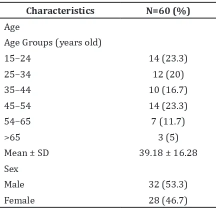
The subjects were classified based on the general characteristics consisting of age group and sex. Table 1 shows general characteristics of the subjects. The subjects were mostly in 15–24 year old and 45–54 year old age groups
Table 1 General Characteristics
Characteristics N=60 (%)
Age
Age Groups (years old)
15–24 14 (23.3)
25–34 12 (20)
35–44 10 (16.7)
45–54 14 (23.3)
54–65 7 (11.7)
>65 3 (5)
Mean ± SD 39.18 ± 16.28 Sex
Male 32 (53.3)
Female 28 (46.7)
Table 2 General Characteristics of FEV1, FVC, and the Ratio of FEV1/FVC
Variables n=60 (%)
%FEV1
0–39 9 (15)
40–49 5 (8.3)
50–59 12 (20)
60–69 15 (25)
70–79 8 (13.3)
>80 11 (18.3)
Mean ± SD 62.03 ± 19.5 %FVC
0–39 6 (10)
40–49 10 (16.7)
50–59 15 (25)
60–69 10 (16.7)
70–79 9 (15)
>80 10 (16.7)
Mean ± SD 61.7 ±17.95 FEV1/FVC
0–0.39 0 (0)
0.40–0.49 1 (1.7) 0.50–0.59 2 (3.3)
0.60–0.69 0 (0)
>0.70 57 (95)
Mean ± SD 0.88 ± 0.10
Note: *FEV1: Forced Expiratory Volume 1 second, **FVC: Forced Vital Capacity
with the mean age of 39.18 years old. Male subjects were more than female subjects.
The subjects were classified based on their FEV1, FVC, and FEV1/FVC value. Table 2 shows the overview of subjects’ %FEV1, %FVC, and the ratio of FEV1/FVC. Most of the subjects had decreased values of FEV1 and FVC with normal value of FEV1/FVC ratio. The subjects’ FEV1, FVC, and the ratio of FEV1/FVC were classified based on age and sex.
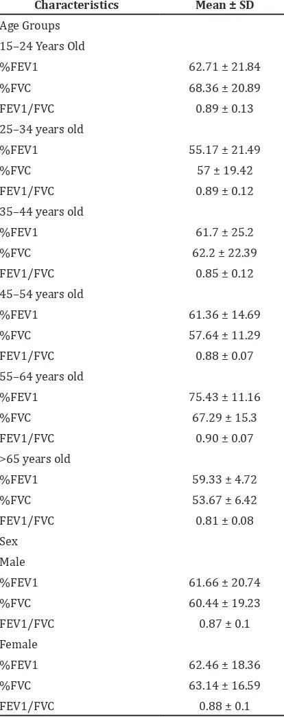
Table 3 shows the overview of %FEV1, %FVC and the ratio of FEV1/FVC based on subjects’ age and sex. All age groups underwent a decrease in the mean values of FEV1 and FVC. However, the ratio of FEV1/FVC remained normal. Male subjects’ %FEV1 and %FVC values were lower than female subjects’.
classified into normal, obstructive lung disorder, and restrictive lung disorder.
Table 4 shows the overview of subjects’ pulmonary function. Most subjects had
Table 3 Overview of Subjects’ %FEV1, %FVC and Ratio of FEV1/FVC based on Age and Sex
Characteristics Mean ± SD
Age Groups
15–24 Years Old
%FEV1 62.71 ± 21.84
%FVC 68.36 ± 20.89
FEV1/FVC 0.89 ± 0.13
25–34 years old
%FEV1 55.17 ± 21.49
%FVC 57 ± 19.42
FEV1/FVC 0.89 ± 0.12
35–44 years old
%FEV1 61.7 ± 25.2
%FVC 62.2 ± 22.39
FEV1/FVC 0.85 ± 0.12
45–54 years old
%FEV1 61.36 ± 14.69
%FVC 57.64 ± 11.29
FEV1/FVC 0.88 ± 0.07
55–64 years old
%FEV1 75.43 ± 11.16
%FVC 67.29 ± 15.3
FEV1/FVC 0.90 ± 0.07
>65 years old
%FEV1 59.33 ± 4.72
%FVC 53.67 ± 6.42
FEV1/FVC 0.81 ± 0.08
Sex
Male
%FEV1 61.66 ± 20.74
%FVC 60.44 ± 19.23
FEV1/FVC 0.87 ± 0.1
Female
%FEV1 62.46 ± 18.36
%FVC 63.14 ± 16.59
FEV1/FVC 0.88 ± 0.1
Note: *FEV1: Forced Expiratory Volume 1 second, **FVC: Forced Vital Capacity
restrictive lung disorder.
The subjects’ pulmonary function classification was classified based on the age group.
Table 5 shows the overview of pulmonary function based on subjects’ age. Obstructive lung disorder was mostly found in subjects aged 55−64 years old while restrictive lung disorder was most likely found in subjects aged 45–54 years old.
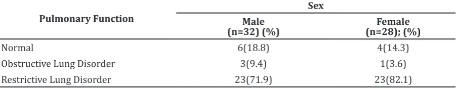
The subjects’ pulmonary function classification was classified based on sex.
Table 6 shows the overview of subjects’ pulmonary function based on sex. Based on subjects’ sex, the results were most likely the same.
Discussion
Subjects were mostly in 15–24 and 45–54 age groups with the mean age of 39.18 years old. This result was consistent with studies conducted by Wu et al.7 and Abioye et al.8 Wu’s7 study was a cohort analytical study regarding age period of tuberculosis notifications in Hongkong from 1961–2005. Wu et al.7 stated that people aged 20–24 years old have higher risk of suffering TB. The study conducted by Abioye et al.8 was a cross-sectional study involving TB patients in Nigeria. Abioye et al.8 stated that the mean age of TB patients is 32.9 years old and they are mostly 21–30 years old.
Most subjects in this study were male. This result was similar to the study conducted by Neyrolles et al.9 in France. Neyrolles’9 study compared TB incidence with previous studies. Neyrolles et al.9 stated that TB rate is higher in males than in females with various ratio in each region.
Most subjects had %FEV1 at the range of 60−90, %FVC at the range of 40–49 and 60–69, and the ratio of FEV1/FVC at approximately above 0.7. This result was consistent with a comparison study conducted by Paspinodya et al.5 Paspinodya et al.5 conducted a study regarding pulmonary function of pulmonary TB patients who have undergone treatment
Table 4 Overview of Subjects’ Pulmonary Function
Category Amount (%)
Normal Pulmonary Function 10 (16.7)
Obstructive Lung Disorder 4 (6.7)
Restrictive Lung Disorder 46 (76.7)
for 20 weeks compared to latent TB patients. Paspinodya’s5 study stated that most of the subjects have %FEV1 at the range of 60−90, %FVC at the range of 40–49 and 60–69, and the ratio of FEV1/FVC at approximately above 0.7.
All age groups in this study underwent a decrease of %FEV1and %FVC. This result was consistent with the study conducted by Paspinodya et al.5 Paspinodya et al.5 stated that pulmonary TB patients who have undergone treatment for 20 weeks have a decrease in pulmonary function in all age groups with mean %FEV1of 77.77 and %FVC of 76.07. This study did not show any decrease of pulmonary function in patients with latent TB.5
Among subjects who had decreased %FEV1 and %FVC values, male subjects had higher %FEV1and %FVC values. This finding was similar to a study done by Pereira et al.10 regarding reference value of pulmonary function between male and female in Brazil. Pereira et al.10 stated that %FEV1 and %FVC values and ratio of FEV1/FVC of male subjects are higher than of females.
Most subjects had restrictive lung disorder, similar to study conducted by Paspinodya et al.5 This study showed that restrictive lung disorder was the most common finding in pulmonary TB patients who have undergone treatment for 20 weeks or more.5 This finding was also similar to a study conducted by Dheda et al.11 Dheda et al.11 stated that lesion healing post-TB will cause restrictive lung disorder characteristic findings in spirometry test. Some differences were found by Menezes et al.4
in a study conducted in some big cities in Latin America. Menezes et al.4 stated that obstructive lung disorder is the most common finding in patients with pulmonary TB history. The result of Menezes’4 study was supported by a study conducted by Baig et al.12 in Pakistan. Baig et al.12 conducted a study regarding pulmonary function of people with cured TB and found out that most subjects have obstructive lung disorder. A study conducted by Ramos et al.13 regarding the sequela of pulmonary TB showed different results with Menezes et al.4 and Baig’s12 studies. Ramos’13 study found that most pulmonary TB patients have mixed lung disorder.
Obstructive lung disorder was mostly found in patients aged 55–64 years old. Meanwhile, restrictive lung disorder was mostly found in patients aged 45–54 years old. A study by Lamprecht et al.14 showed similar result. Lamprecht stated that the age group of 60–69 years old is one of the most prevalent age groups suffering from obstructive lung disorder. A study conducted by Paspinodya et al.5 showed that the mean age of subjects with obstructive lung disorder is 47 years old.
Most subjects had restrictive lung disorder. This result was similar yet different with a study conducted by Manino et al.15 Mannino et al.15 stated that obstructive lung disorder is mostly found in males while restrictive lung disorder is mostly found in females.
The high number of restrictive lung disorder in this study was most likely affected by some factors such as nutrition and treatment. A study conducted by Pakasi et al.16 in Nusa Table 5 Overview of Subjects’ Pulmonary Function based on Age
Pulmonary Function
Age Group (years old)
15–24
Normal 4(28.6) 2(16.7) 2(20) 1(7.1) 1(14.3) 0(0)
Obstructive Lung Disorder 1(7.1) 1(8.3) 1(10) 0(0) 1(14.3) 0(0)
Restrictive Lung Disorder 9(64.3) 9(75) 7(70) 13(92.9) 5(71.4) 3(100)
Table 6 Overview of Subjects’ Pulmonary Function based on Sex
Pulmonary Function
Tenggara found that nutrition is an important factor in TB natural history. This result stated that severe deficiency of vitamin A is associated with severe TB.16 A study conducted by Menzies et al.17 regarding the effectivity of TB treatment stated that treatment using rifampin in short duration or intermittently will produce worse outcome of TB treatment. Both studies showed that restrictive lung disorder is most likely caused by a longer duration of inflammation. Moreover, it is also affected by nutrition and TB treatment.
The conclusion of this study is the majority of pulmonary TB patients treated with 1st category anti-TB drugs during intensive phase undergo a decreased pulmonary function and restrictive lung disorder. This study has some limitations. Data homogenization cannot be done due to limited time and cost. It is suggested for further studies to provide larger sample size and comparison of pulmonary function of patients in treatment and cured pulmonary TB patients. It is also suggested to do a routine pulmonary function examination and specialized treatment to minimize the decrease of pulmonary function of pulmonary TB patients.
References
1. WHO. Global tuberculosis report 2013. WHO; 2013.
2. Trihono. Riset kesehatan dasar 2010. Jakarta: Departemen Kesehatan Republik Indonesia; 2010.
3. Fishman AP, Elias JA, Fishman JA, Grippi MA, Kaiser LR, Senior RM. Fishman’s manual of pulmonary diseases and disorders. 3th ed. Senior RM, Pack AI, editors. New York: McGraw-Hill; 2008. p. 2734.
4. Menezes AMB, Hallal PC, Perez-Padilla R, Jardim JRB, Muino A, Lopez MV, et al. Tuberculosis and airflow obstruction: evidence from the PLATINO study in Latin America. Eur Respir J. 2007;30(6):1180–5. 5. Paspinodya JG, Miller TL, Vecino M,
Munguia G, Garmon R, Bae S, et al. Pulmonary impairment after tuberculosis. Chest. 2007;131(6):1817–24.
6. Chung K, Chen J, Lee C, Wu H, Wang J, Lee L, et al. Trends and predictors of changes in pulmonary function after treatment for pulmonary tuberculosis. Clinics (Sao Paulo). 2011;66(4):549–56.
7. Wu P, Cowling BJ, Schooling CM, Wong IOL, Johnston JM, Leung CC, et al. Age-period-cohort analysis of tuberculosis notifications in Hong Kong from 1961 to 2005. Thorax. 2008;63(4):312–6.
8. Abioye LA, Omotayo MO, Alakija W. Socio-demographic determinants of stigma among patients with pulmonary tuberculosis in Lagos, Nigeria. Afr Health Sci. 2011;11(Suppl 1):S100–4.
9. Neyrolles O, Murci LQ. Sex inequality in tuberculosis. PLoS Medicine. 2009;6(12):1–5.
10. Pereira CAdC, Sato T, Rodrigues SC. New reference values for forced spirometry in white adults in Brazil. J Bras Pneumol. 2007;33(4):397–406.
11. Dheda K, Booth H, Hugget JF, Johnson MA, Zumla A, Rook GA. Lung remodeling in pulmonary tuberculosis. J Infect Dis. 2005;192(7):1201–9.
12. Baig IM, Saeed W, Khalil KF. Post-tuberculous chronic obstructive pulmonary disease. J Coll Physicians Surg Pak. 2010;20(8):542–4.
13. Ramos LMM, Sulmonett N, Ferreira CS, Henriques JF, Miranda SSD. Functional profile of patients with tuberculosis sequelae in a university hospital. J Bras Pneumol. 2006;32(1):43–7.
14. Lamprecht B, McBurnie MA, Vollmer WM, Gudmundsson G, Welte T, Nizankowska-Mogilnicka E, et al. COPD in never smokers: results from the population-based burden of obstructive lung disease study. Chest. 2011;139(4):752–63.
15. Mannino DM, McBurnie MA, Tan W, Kocabas A, Anto J, Vollmer WM, et al. Restricted spirometry in the burden of lung disease study. Int J Tuberc Lung Dis. 2012;16(10):1405–11.
16. Pakasi T, Karyadi E, Wibowo Y, Simanjuntak Y, Suratih N, Salean M, et al. Vitamin A deficiency and other factors associated with severe tuberculosis in Timor and Rote Islands, East Nusa Tenggara Province, Indonesia. Eur J Clin Nutr. 2009;63(9):1130–5.