MASSIVE GOITRE (STRUMA PARENCHYMATOSA) IN GEESE
Éva IVANICS1*, P. RUDAS2, G. SÁLYI1and R. GLÁVITS11Central Veterinary Institute, H-1149 Budapest, Tábornok u. 2, Hungary; 2Department of Physiology and Biochemistry, University of Veterinary Science,
Budapest, Hungary
(Received November 18, 1998; accepted January 12, 1999)
In a goose flock consisting of 2300 birds of 6 months of age severe goitre was diagnosed. To the best of our knowledge this is the first report of naturally occurring goitre in geese, which is not related to the feeding of rapeseed meal. The major pathological findings included retarded growth and plumage development, significantly (300%) increased relative thyroid weight, fat accumulation in the mesenteric and abdominal region, and lipid infiltration of liver and kidney cells. Subsequent hormone analysis showed undetectable thyroxine (T4) levels and a dramatic drop in triiodothyronine (T3) plasma levels of the diseased geese. Thy-roidal histology displayed the typical signs of struma parenchymatosa. In order to get more information about the possible causes of the goitre, 10 geese from the af-fected farm were transferred into the laboratories of the Central Veterinary Insti-tute. The geese were allotted into two groups. Group I received iodine supple-mentation for 55 days, while the other group served as sick control (Group S). Io-dine treatment caused a dramatic improvement in the birds clinical condition ex-cept in plumage growth in Group I, while the clinical and main pathological signs of goitre remained unchanged or worsened in the untreated Group S. Contrary to this, the serum levels of thyroid hormones and responsiveness to thyrotropin re-leasing hormone (TRH) improved not only in Group I but also in Group S. Almost euthyroid biochemical parameters were found after 55 days of iodine treatment in Group I and, surprisingly, a considerable improvement (especially in serum T3 levels) occurred also in Group S. These findings confirm the diagnosis of goitre but also call attention to the fact that iodine deficiency was not the only factor eliciting the disorder. The underlying possible goitrogenic substance could not be traced down.
Key words: Goitre, goose, pathological findings, hormone analysis
Goitre is a non-inflammatory, non-neoplastic enlargement of the thyroid gland that occurs in both humans and animals, in mammals and birds alike (Kapp, 1952; Bokori, 1956; Baker and Lindsey, 1968; Rac et al., 1968; Antal, 1972; Lissitzky et al., 1973; Kardeván, 1976; de Vijlder et al., 1978; Nesbitt et
al., 1980; Gosselin et al., 1980, 1981; Milne and Hayes, 1981; Robinson et al., 1988; Capen, 1993). Goitre may develop most often as a result of insufficient amounts of iodine in the diet or drinking water (Bokori, 1956; Kardeván, 1976; Antal, 1972; Capen, 1993). Other causative factors are the presence of goitro-genic substances in the feed (Kardeván, 1976; Capen, 1993), less frequently ex-cess iodine in the feed (Baker and Lindsey, 1968) or rarely the genetically de-termined lack of thyroid hormone production (Rac et al., 1968; Lissitzky, 1973; Kardeván, 1976; de Vijlder et al., 1978; Pammenter et al., 1978; Capen, 1993).
In all the above cases a major physiological factor leads to the enlarge-ment of the thyroid gland. The thyroid is unable to produce enough hormones and thus the level of circulating thyroxine (T4) and triiodothyronine (T3) drops in
the plasma (Belshaw and Rijnberk, 1979; Capen, 1993). This in turn elicits an excess production of the thyroid stimulating hormone (thyrotropin, TSH) from the pituitary. TSH then causes all thyroidal functions to increase: hypertrophy and hyperplasia of the follicular epithelial cells occurs but due to the underlying factors hormone production is not possible. This leads to a vicious circle where TSH effects on the thyroid may lead to the total disappearance of follicular col-loid, leaving behind hyperplastic follicles: struma parenchymatosa develops (Kardeván, 1976; Gosselin et al., 1980; Capen, 1993).
The major factor which causes the clinical symptoms in goitre to appear is the lack of thyroid hormones in the blood (hypothyroidism). In birds one of the major effects of hypothyroidism is retarded growth, feathering disorders and ac-cumulation of adipose tissue in the abdomen (for a summary see Janan and Ru-das, 1998). There are, however, several complications which may arise from the enlargement of the thyroid tissue itself: the enlarged mass compresses the sur-rounding tissues (trachea, oesophagus, vessels and nerves) in the cervical area (Kardeván, 1976; Capen, 1993).
Reports on endemic animal goitre are available from the most diverse geographical territories such as North America, Europe and Australia, mostly pinpointing well-defined districts of certain countries, e.g. Germany, the Neth-erlands, Norway, Russia (the Ural Mountains), Serbia and Switzerland (Karde-ván, 1976; Capen, 1993). In Hungary three mountainous areas (Bükk, Mátra, Mecsek) display regions where goitre occurs quite frequently (Kardeván, 1976).
Sporadic goitre was reported in many bird species including parrots and doves (Kardeván, 1976), as well as in birds kept in zoos (32 such species were described by Wisser and Ippen, 1991). To our knowledge, naturally occurring goitre has not yet been reported in geese.
The present paper describes one of the rare natural occurrences of goitre in geese kept on a large-scale farm. The nature of this goitre was further investigated in our laboratory under experimental conditions, which is also reported here.
Materials and methods Clinical signs and observation of the goose flock
The goitre occurred on a large-scale goose farm in Kecskemét, where 2600 geese aged 6.5 months were kept in an adobe roost. The birds were allowed to pas-ture regularly. As a supplement, the geese were regularly turned out to paspas-ture on the local rye field. They were kept on a starter and grower feed until 9 weeks of age and then on grain until 5 months of age. Subsequently fresh maize was offered. Water from a bored-type well served as drinking water throughout the raising period.
The birds were treated with Clamoxil at the age of 13 days. Within the plucking periods SCP 625 (containing sulfachlorpyridazine and trimethoprim) was applied for 3 days. After the third plucking the former treatment was sup-plemented by adding Trierra (containing furazolidone, zinc-bacitracin and oxy-tetracycline) for a 7-day period.
The geese were plucked first at 2 months of age and every 45 days there-after, altogether four times.
Clinical signs of the illness were investigated at the site of raising and, in the case of 10 geese, at the Central Veterinary Institute (CVI) where these ani-mals were transferred to for experimental purposes.
Pathological, histological and bacteriological findings
The eight 8 geese that died on the farm and the birds transferred to the CVI were subjected to postmortem examination. The thyroid gland, parathyroid gland, liver, kidney, testis, ovary and, in some instances, the ultimobranchial or-gan and adrenals of the birds were examined histologically. Tissue samples were fixed in 10% neutral (pH 7.2) formaldehyde and processed by freezing and em-bedding in paraffin. For general investigations the samples were stained with haematoxylin and eosin. Special staining involved the fat-red method to indicate the presence of lipids in the liver and kidney samples.
Bacteriology was done according to the standard protocols of the CVI from the heart blood and liver of the geese.
Animal experiments
Six geese (Group A) out of the 10 birds showing clinical signs of plumage problems and transported from the farm to the Institute were blood sampled on Day 1. On Day 2 the birds were randomly allotted into two groups of 5 individuals each. The geese in Group I (iodine-treated group) were treated for 4 weeks with Lugols solution via an oesophageal tube in a dose equivalent to 2 µg iodine/kg body weight/day. The daily iodine dose was increased to 50 µg between week 5 and week 8 of the experiment. The geese of Group S (sick control birds) were left
untreated throughout the period of experiment. In the first 6 weeks of the experi-ment these 10 geese were fed ad libitum with the original maize obtained from the farm. Later on maize was obtained from other sources. In the assessment of the biochemical parameters of thyroid activity five additional geese of the same age from an unaffected farm were used as normal controls (Group N).
Biochemical parameters of thyroid status
Plasma thyroxine (T4) and triiodothyronine (T3) concentrations were first
measured in 6 geese (Group A) randomly selected from the 10 animals one day after arrival at the Institute, before allotting them into Group I and Group S. Further hormone measurements were carried out on Day 25 and Day 55 from Group I and Group S. The measurements of hormone concentrations were car-ried out by radioimmunoassay (Pethes et al., 1978a, 1978b). In the animals transferred to the laboratory TRH (thyrotropin releasing hormone) provocation test was performed (in the case of Groups A and N on Day 0 and in the case of Groups I and S on Day 25) to check the responsiveness of the pituitarythyroid axis and the possible effectiveness of iodine supplementation. This entailed the following. After drawing 0 time blood sample, geese were given 10 mg TRH/ kg body weight (Sigma Chemical Co., St. Louis, MO, USA) intravenously. Forty minutes thereafter a blood sample was taken again. The changes in the concen-tration of T4 and T3 in the blood sample taken before and after TRH injection
were expressed in percentage of the original hormone concentrations. Results
Clinical symptoms and the course of the disease
Signs of a disease were first discovered in the stock at 6 months of age after the fourth plucking in mid-October. At that time the body weight dropped to an average of 4.7 kg as compared to the 5.0 kg measured at the previous plucking. The overall amount of down gained (130140 g per animal) was also way below the 170 g average obtained at the previous time. All birds displayed lack of appetite and retarded plumage growth even after 5 weeks in the post-plucking period. Deaths began to occur and at that time 68 animals died per day. Contrary to our advice, iodine supplementation was not given to the flock and the situation worsened day by day. Finally the whole flock had to be slaughtered and sold as table goose in December, since no hope for recovery from the plumage problems and weight loss could be foreseen.
Pathological, histological and bacteriological findings
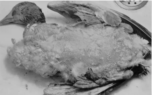
One of the main pathological findings was the uneven growth and devel-opment of geese of the same age. Body weight ranged between 35505800 g and considerable underdevelopment or lack of plumage was seen on the breast, ab-domen and back of the birds (Fig. 1). The integument remained normal. Within the subcutaneous connective tissue and also in the intraabdominal and mesen-teric region a large amount of fat had accumulated. Livers and kidneys had a yellowish colour and the surface of the sectioned organs displayed fatty shine. The texture of the tissues was easy to tear apart.
Fig. 1. Retarded plumage development was seen even 5 weeks after plucking
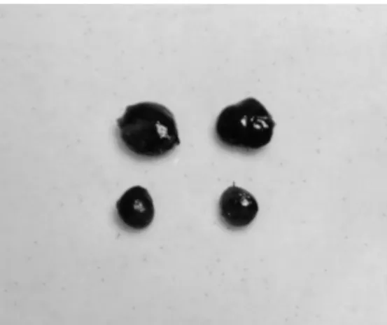
On the two sides of the trachea a proportional and symmetrical enlarge-ment of the thyroid glands was found. The brownish-red enlarged mass of the glands was dense to the touch (Figs 2 and 3). The total mass of the thyroids in these birds was twice larger than that measured in normal flocks of geese of the same age from other farms. When the weight of the thyroids was expressed in percentage of body weight, it was found to be three times higher than in normal geese (Table 1). No pathological signs were found in any other tissue.
Fig. 2. The thyroid glands of diseased geese are swollen, brownish-red in colour and protruding from their surrounding around the trachea
Fig. 3. Dramatically enlarged thyroid glands in sick birds as compared to thyroids of healthy geese of the same age (lower row)
Table 1
Panel A: The absolute and relative mass of the thyroid glands in 5 healthy geese from an unaf-fected farm and in geese displaying goitre at the time of discovering the abnormality in the afunaf-fected farm. Panel B: The same parameters of iodine-treated (Group I) and untreated sick (Group S) geese at the end of the laboratory experiment. Absolute mass in gram, relative mass in % of body weight,
average±SEM (N). Groups with same superscripts (athroughd) differ significantly, P < 0.05
Panel A Healthy geese Geese displaying goitre
Thyroid mass (g) 0.644±0.061(5)a 1.304±0.103(5)a
Thyroid mass/body weight % 0.0099±0.0007(5)b 0.0290±0.0016(5)b
Panel B Group I Group S
Thyroid mass (g) 0.323±0.054(5)c 0.830±0.162(5)c
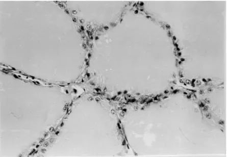
Thyroid mass/body weight % 0.0076±0.0006(5)d 0.0213±0.0039(5)d In the affected geese the histology of the thyroid gland as compared to the control (Figs 4 and 5) showed a considerably decreased follicular colloid content or sometimes a total lack of colloid in the follicles. The epithelial layer of the follicles grew to cubic or cylindrical in appearance and in many cases the cells occupied the follicular lumen in several layers (Figs 6 and 7). In some in-stances one could detect vacuoles in the cytoplasm of the follicular epithelial cells. In the connective tissue of the thyroid gland, among the follicles the num-ber and diameter of blood vessels were increased.
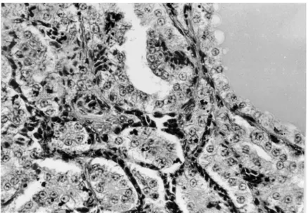
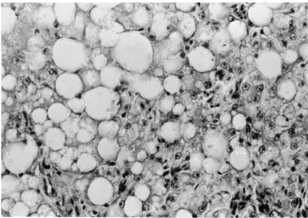
Vacuoles in the cytoplasm of liver cells occupied the whole intracyto-plasmic space, pushing the nucleus of cells towards the plasma membrane (Fig. 8). Within the vacuoles lipid droplets appeared as demonstrated by specific staining. Fat appeared either in stripes or in a diffuse manner within the liver tis-sue, exhibiting great individual variance. Lipid-containing vacuoles could also be detected in the tubular system of some of the kidneys examined.
The extent of liver and kidney damage was proportional to the severity of thyroidal findings.
By systematic investigations from heart blood and liver the presence of no pathogenic bacterium could be substantiated.
Mycological investigation of the maize originating from the farm showed an elevated mould concentration (16,000 CFU/g). All moulds belonged to the Acremonium species.
Fig. 4. Section of the thyroid gland from a healthy goose. The uniformly spacious follicles are filled with a vast amount of colloid. Haematoxylin and eosin (H.-E.), × 40
Fig. 5. Section of the thyroid gland from a healthy goose. Follicles are lined by flat or cuboidal epithelium. H.-E., × 200
Fig. 6. Parts from a section of the thyroid gland of a diseased goose. The shrunken follicles display no colloid or only traces of colloid. The epithelial layer is thickened. H.-E., × 40
Fig. 7. Parts from a section of the thyroid gland of a diseased goose. The shrunken follicular space does not contain colloid and has multilayered epithelium. The number and diameter of vessels are
Fig. 8. Parts from a section of the liver of a sick goose. Vacuoles of large diameter with giant lipid droplets are apparent in the hepatocytes. H.-E., × 200
Laboratory animal experiments
In Group I (birds receiving iodine supplementation) the most apparent clinical signs of the disease, namely the poor development of the plumage, did not disappear during the treatment period. The pathological and histological ex-aminations, however, showed a considerable improvement of the hypothyroid goitre as a response to iodine supplementation. In these birds (bled at the end of the experimental iodine supplementation period) no excess fat accumulation was seen either in the subcutaneous connective tissue or in the mesenteric and ab-dominal regions. Also the outer appearance and mass of the thyroid glands were very similar to those seen in normal individuals (Table 1). The histology of the thyroid gland also resembled the situation in normal animals. A decreased fol-licular and colloidal space with multilayered epithelium of cuboidal or cylindri-cal shape could be found only in about 1530% of the thyroidal sections. In this group (Group I) the livers were almost free of fat accumulation and only 25 30% of the hepatocytes showed signs of slight fat infiltration. No kidney dam-ages were seen.
Group S (geese that received no iodine supplementation in the laboratory) exhibited similar clinical and histological symptoms that were described earlier for the geese affected on the farm. Both the absolute and the relative weight of the thyroid glands decreased in this group (Table 1) but were still significantly
Thyroxine (T4) and triiodothyronine (T3) plasma levels
The comparison of plasma T4 and T3 levels in 5 normal control geese
(Group N) and 6 clinically affected birds (Group A) at the beginning of the labo-ratory experiments showed a dramatic difference in hormone levels (Panel A of Table 2). There was no detectable T4and only hypothyroid amounts of T3in the
plasma of sick animals (Group A), while the T4 and T3 values obtained in the
five healthy control geese were in the euthyroid physiological range.
The hormone levels changed during the course of the laboratory experi-ment (Panel B of Table 2) not only in the iodine-suppleexperi-mented Group I, but also in the non-supplemented Group S. On Day 25 of iodine supplementation T4and
T3levels were well detectable in Group I and their concentration was about
one-third of that in the euthyroid controls. In the non-supplemented group (Group S) T4and T3 levels were still very low but above the detection limit of the assay.
On Day 55 T4 levels in Group I increased to two-thirds of the euthyroid level,
while T3 values were very close to the euthyroid hormone concentrations. The
thyroid hormone levels in the non-supplemented (Group S) animals also in-creased. This increase was not very much pronounced for T4, but for T3the
con-centrations reached the lowest normal T3concentrations.
Table 2
Panel A: The concentration of thyroxine (T4) and triiodothyronine (T3) in the blood plasma of 5 healthy geese from a non-affected farm (Group N) and of 6 geese displaying goitre (Group A) at the time of discovering the abnormality in the affected farm. Panel B: The same parameters of
io-dine-treated (Group I) and non-treated sick (Group S) geese at Day 25 and Day 55 of the labora-tory experiment [nmol/l, average ± SEM (N)]. Groups with same superscripts (athroughd) differ
significantly, P < 0.05
Healthy geese Geese displaying goitre
Panel A Group N Group A
Thyroxine (T4) 17.9 ± 1.11(5)a non-detectable(6)a
Triiodothyronine (T3) 1.69 ± 0.09(5)b 0.26 ± 0.09(6)b
Panel B Group I Group S
Day 25 Thyroxine (T4) 5.98 ± 1.41(3)c 0.79 ± 0.27(3)c Triiodothyronine (T3) 2.02 ± 0.24(3) 1.15 ± 0.36(3) Day 55 Thyroxine (T4) 11.45 ± 1.32(3)d 1.32 ± 0.36(3)d Triiodothyronine (T3) 1.99 ± 0.05(3) 1.20 ± 0.21(3)
The TRH provocation test on Day 0 showed a total lack of thyroidal re-sponsiveness in the diseased animals (Group A) as compared to the five healthy geese (Group N) (Panel A of Table 3). Later on, on Day 25, the TRH provoca-tion test revealed a close to normal pattern in the iodine-supplemented birds (Group I), and also in the supplemented animals there was a small but non-negligible response to TRH provocation (Panel B of Table 3).
Table 3
Panel A: The results of the TRH provocation test (10 µg TRH / kg body weight, i.v.) in 5 healthy geese from a non-affected farm (Group N) and of 6 geese displaying goitre (Group A) at the time of transferring the goitrous geese from the affected farm to the laboratory. Panel B: The same pa-rameters of iodine-treated (Group I) and of untreated sick (Group S) geese at Day 25 of the labo-ratory experiment. [% increase of hormone concentrations 40 min after injecting TRH related to 0
time hormone concentrations, average ± SEM (N)]. Groups with same superscripts (athroughd) differ significantly, P < 0.05
Healthy geese Geese displaying goitre
Panel A Group N Group A
Thyroxine (T4) 201 ± 7.4 (5)a no detectable change (6)a
Triiodothyronine (T3) 108 ± 16 (5)b no detectable change (6)b
Panel B Group I Group S
Thyroxine (T4) 165 ± 45 (3)c 47 ± 10 (3)c
Triiodothyronine (T3) 17 ± 5 (3)d 1.9 ± 0.5 (3)d
Discussion
From the previous sections it is clear that the occurrence of goitre (struma parenchymatosa) under normal raising conditions in a geese farm was detected and diagnosed. The underlying clinical symptoms were retarded growth and poor plumage development in a very high percentage of the 2300 geese kept to-gether on the affected farm. Pathological and histological findings as well as the biochemical parameters of the blood plasma provided the final evidence for goi-tre. Significant enlargement of the thyroids, accumulation of fat in the subcuta-neous connective tissue and in the mesenteric region, lipid deposition in liver cells, together with the undetectable thyroxine concentrations, all support this diagnosis.
Even if the diagnosis is supported by the above sound evidence, the cause of goitre development remains unclear. The literature contains numerous reports
Baker and Lindsey, 1968; Rac et al., 1968; Antal, 1972; Lissitzky et al., 1973; Kardeván, 1976; de Vijlder et al., 1978; Nesbitt et al., 1980; Gosselin et al., 1980; Milne and Hayes, 1981; Robinson et al., 1988; Capen, 1993) where the major causative factor is the limited access to iodine (Bokori, 1956; Antal, 1972; Capen, 1993). The question arises whether or not the lack of iodine was a causative factor of the goitre in our case. At the first glance the laboratory ex-periments speak in favour of the supposition that the lack of iodine might have been the major eliciting factor of the goitre. Iodine treatment normalised almost all clinical, pathological and histological symptoms, except plumage growth. However, on closer scrutiny of the laboratory experiments and especially the biochemical parameters thereof (Table 2) it has to be realised that the inability of the thyroid to incorporate iodine into hormones might have been a more im-portant causative factor than the lack of iodine in the diet. This is so because thyroxine and triiodothyronine levels and TRH responses improved not only in the iodine-supplemented group (Group I) but also in the untreated sick geese (Group S) (see Tables 2 and 3). The tendency of biochemical thyroid parameters to normalise in Group S can be realised even better when we compare T4and T3
serum levels. T4levels were undetectable in diseased birds at the beginning, but
increased to detectable (although low) levels by Day 25 in Group S animals. At the same time, T3levels in these sick geese not receiving iodine supplementation
returned to almost normal levels. It is known that in birds the major hormone produced by the thyroid gland is T4 which is later transformed into its active
hormone form, triiodothyronine, in the peripheral tissues including the brain, liver, kidney, and other organs (Rudas, 1986). It was also shown that the rate at which the activation of thyroxine into triiodothyronine occurs is determined by the need of the body for T3(Rudas et al., 1993). In the present case an increased,
compensatory T4to T3conversion is able to produce close to normal levels of T3
in the blood. This can, however, only happen when a minimum amount of T4 is
produced by the thyroid. This is clearly the case in Group I and also in Group S. It was seen already at the end of the first 25 days of the laboratory trial, before changing the original diet from the affected farm to maize and before increasing the dose of iodine supplementation. Thus the improvement witnessed in Group S (sick geese not treated with iodine) after transferring them into the laboratory can only be attributed to the release of these animals from an unknown factor present on the affected farm that caused the thyroid to stop its hormone produc-tion. Endemic goitre (regional lack of iodine in the feed and in the drinking wa-ter) could easily be ruled out, since no goitre occurred in the region of the af-fected farm. If not the lack of iodine then other factors may have had a possible role in the development of the disease. What are those factors?
Goitre may be elicited by special goitrogens (Kardeván, 1976; Capen, 1993) and by excess iodine delivery (Baker and Lindsey, 1968; Antal, 1972) but
these factors may be excluded in our case, since the diet was the same for group I and Group S in the first 25 days of the laboratory experiments. Plus, if excess iodine had been the cause of goitre then supplementation with iodine should have worsened the condition of the animals, which was apparently not the case. It is also known that glucosinolates in mill cake extracts of rape species may also cause goitre in animals (Wight and Shannon, 1985) but in our case it was known that neither the starter nor the raising feed contained rape. We can rule out the possible goitrogenic effects of the Acremonium species detected in the feed in quite high concentrations. Firstly because these species (Acremonium coeno-phialum) produce ergotamic toxins not known as goitrogens, and secondly be-cause the same feed was available for both laboratory groups.
One should think rather of a factor that was present on the farm, but not in the laboratory. Among these factors, environmental lead pollution is the one that might cause goitre and decrease the level of thyroid hormones in the blood plasma as shown in ducks (Goldman and Dillon, 1982).
Another possible goitre-eliciting factor that was present on the farm but not in the laboratory might have been the sulphochlorpromazine applied on the farm every six weeks. It is known that this drug may provoke goitre in newborn pig (Blackwell et al., 1989). A proof of overdosing this substance is, however, lacking.
In conclusion: a rare disorder, goitre was diagnosed in a geese flock. The disease may have been elicited by two factors: a relative deficiency of iodine and an undetermined factor that depressed the hormone-producing ability of the geese kept at the farm. An important conclusion from this paper is that the bio-chemical parameters of thyroid activity, such as plasma levels of thyroxine and triiodothyronine and the results of the TRH provocation test, can signal changes in thyroid status much earlier than pathological and histological investigations.
Acknowledgements
The authors would like to thank Mrs Zs. Szikora for technical assistance. This work was partly made possible by grant no. 16545 of OTKA (Hungarian Scientific Re-search Fund) to P. R.
References
Antal, Á. (1972): Innate struma in lambs (in Hungarian, with English abstract). Magyar Állator-vosok Lapja 27, 544548.
Baker, H. J. and Lindsey, J. R. (1968): Equine goiter due to excess dietary iodide. J. Am. Vet. Med. Assoc. 153, 616630.
Belshaw, B. E. and Rijnberk, A. (1979): Radioimmunoassay of plasma T4and T3in the diagnosis of primary hypothyroidism in dogs. J. Am. Vet. Med. Assoc. 15, 1523.
Blackwell, T. E., Werdin, R. E., Eisenmenger, M. C. and FitzSimmons, M. A. (1989): Goitrogenic effects in offspring of swine fed sulfadimethoxine and ormetoprim in late gestation. J. Am. Vet. Med. Assoc. 194, 519523.
Bokori, J. (1956): Innate struma in piglets (in Hungarian, with English abstract). Magyar Állator-vosok Lapja 11, 364369.
Capen, C. C. (1993): The Endocrine Glands. In: Jubb, K. V. F., Kennedy, P. C. and Palmer, N. (eds) Pathology of Domestic Animals. Fourth edition. Volume 3, Chapter 3, pp. 267347. de Vijlder, J. J. M., van Voorthuizen, W. F., van Dijk, J. E., Rijnberk, A. and Tegelaers, W. H.
(1978): Hereditary congenital goiter with thyroglobulin deficiency in a breed of goats. En-docrinology 102, 12141222.
Janan, J. and Rudas, P. (1998): Interrelationship of thyroid activity with fat metabolism in poultry (in Hungarian, with English abstract). Magyar Állatorvosok Lapja 120, 339342.
Goldman, M. and Dillon, R. D. (1982): Interaction of selenium and lead on several aspect of thy-roid function in Peking ducklings. Res. Comm. Chem. Pathol. Pharmacol. 37, 487490. Gosselin, S. J., Capen, C. L., Martin, S. L. and Targowski, S. P. (1980): Biochemical and
immu-nological investigation of hypothyroidism in dogs. Can. J. Comp. Med. 44, 158168. Gosselin, S. J., Capen, C. C. and Martin, S. L. (1981): Histopathologic and ultrastructural
evalua-tion of thyroid lesions associated with hypothyroidism in dogs. Vet. Pathol. 18, 299309. Kapp, P. (1952): Innate struma in three kids (in Hungarian). Magyar Állatorvosok Lapja 7, 151153. Kardeván, A. (1976): Pathology of Domestic Animals (in Hungarian). Mezõgazdasági Kiadó,
Bu-dapest.
Lissitzky, S., Bismuth, J., Jaquet, P., Castay, M., Michel-Bechet, M., Koutras, D. A., Pharmakivtis, A. D., Moschas, A., Psarras, A. and Malamos, B. (1973): Congenital goiter with impaired thyroglobulin synthesis. J. Clin. Endocrinol. Metab. 36, 1729.
Milne, K. L. and Hayes, H. M. (1981): Epidemiologic features of canine hypothyroidism. Cornell. Vet. 71, 314.
Nesbitt, G. H., Izzo, J., Peterson, L. and Wilkins, R. J. (1980): Canine hypothyroidism: A retro-spective study of 108 cases. J. Am. Vet. Med. Assoc. 177, 11171122.
Pammenter, M., van der Walt, B. J. and van Jaarsveld, P. P. (1978): Afrikander cattle congenital goiter: Characteristics of its morphology and iodoprotein pattern. Endocrinology 102, 954965. Pethes, G., Losonczy, S. and Rudas, P. (1978a): Measurement of serum triiodothyronine by
ra-dioimmunoassay (in Hungarian, with English abstract). Magyar Állatorvosok Lapja 33, 172176.
Pethes, G., Losonczy, S. and Rudas, P. (1978b): Short periodical changes in the concentration of hormones of the thyroid gland in the serum of bulls and cows (in Hungarian, with English abstract). Magyar Állatorvosok Lapja 33, 237242.
Rac, R., Hill, G. N., Pain, R. W. and Wulhearn, C. J. (1968): Congenital goitre in Merino sheep due to an inherited defect in the biosynthesis of thyroid hormone. Res. Vet. Sci. 9, 209223. Robinson, W. F., Shaw, S. E., Stanley, B. and Wyburn, R. S. (1988): Congenital hypothyroidism
in Scottish deerhound puppies. Aust. Vet. J. 65, 368389.
Rudas, P. (1986): Comparison of type I 5-deiodination of thyroxine and of reverse triiodothy-ronine in rat and chicken liver homogenates. Gen. Comp. Endocrinol. 63, 657660. Rudas, P., Bartha, T. and Frenyó, V. L. (1993): Thyroid hormone deiodination in the brain of
young chickens acutely adapts to changes in thyroid status. Acta Vet. Hung. 41, 381393. Wight, P. A. L. and Shannon, D. W. F. (1985): The morphology of the thyroid glands of quails and
fowls maintained on diets containing rapeseed. Avian Pathol. 14, 383399.
Wisser, J. and Ippen, R. (1991): Zum Vorkommen von Strumen bei Zoovögeln. Wien. Tierärztl. Mschr. 78, 362365.