A STUDY ON
KALANJAGAPADAI
Dissertation Submitted To
THE TAMIL NADU Dr. M.G.R. Medical University
Chennai – 32
For the Partial fulfillment for the Award of Degree of
DOCTOR OF MEDICINE (SIDDHA)
(Branch – III, SIRAPPU MARUTHUVAM)
DEPARTMENT OF SIRAPPU MARUTHUVAM
ACKNOWLEDGEMENT
First, I am extremely grateful to the divine power enlightened my path with
blessings and strengthened me in all aspects towards the milestone successfully.
I
express
my
deep
sense
of
gratitude
to
our
Principal
Dr.N.Chandra Mohan Doss. M.D(S).,
and
Dr.S.Soundararajan. M.D(S).,
Vice
Principal, Government Siddha Medical College, Palayamkottai for granding
permission to execute this dissertation work in the college premises.
It is a honour for me to express my sincere thanks and gratitude to Asso.
Prof.
Dr.S.Kaniraja. M.D(s).,
Head of the department, Post Graduate Sirappu
Maruthuvam for his support and guidance to do this study.
It is my pleasure to express grateful thanks to
Dr.D.Rajasekar. M.D(S).,
lecturer and
Dr.A.S. Poongodi Kanthimathi. M.D(S).,
Lecturer Post Graduate
Sirappu Maruthuvam for rendering their valuable suggestion and guidance.
I convey my sincere thanks to
Dr.S.Ramaguru. B.Sc., M.B.B.S.,
M.S(ortho)., D.Ortho.,
Professor, Department of Orthopaedics, Department of
Sirappu Maruthuvam, Govt Siddha Medical College, Palayamkottai.
My sincere thanks to Prof.
N. Nagaprema. M.Sc.,
Head of the department,
Department of Bio-chemistry and
Mr. M. Kalaivanan. M.Sc.,
Lecturer,
Department
of
Pharmacology,
Government
Siddha
Medical
College,
Palayamkottai.
It is my duty to express my cheerful thanks to all those who encouraged and
supported during the whole study period.
CONTENTS
PG.NO
INTRODUCTION
1
AIM AND OBJECTIVES
7
REVIEW OF LITERATURES
I.
REVIEW OF SIDDHA LITERATURES
9
II.
REVIEW OF MODERN LITERATURES
41
MATERIALS AND METHODS
66
OBSERVATION AND RESULTS
71
DISCUSSION
99
SUMMARY
105
CONCLUSION
107
ANNEXURE
ANNEXURE I
108
ANNEXURE II
125
ANNEXURE III
128
ANNEXURE IV
138
BIBLIOGRAPHY
157
INTRODUCTION
Before entering the study let us know the basic concepts like origin of earth, origin of life in it, origin of medicine in mankind. Knowing these basics concepts reveals us some truths which glorify our great siddhars that they are the pioneers to all modern scientist invariable to the department they belong, and also invariable to the country they belong.For many scientific evolutions even for todays nano concepts our siddhar thoughts are the basics tools for the modern scientist which help them in their researches.
The age of earth still is a made of great debate. Some used Bible to calculate that the earth was created in 4004BC. Later on in the middle of the nineteenth century, the great scientist, Charles Darwin believed that the earth must be extremely old because he rcognised that natural, selection and evolution required vast amounts of time. it was well explained by our beloved siddhars which is quoted in our text THOTRA KIRAMA ARAICHI.
The history of earth describes the most important events and fundamental stages in the development of the planet. Nearly all the branches of Natural science contribute in understanding the Earth’s history. Biological and geological changes have been constantly occurring on our planet since the time of its formation. Organisms continuously evolve, taking on new forms or going extinct in response to an ever changing planet. This is quoted by our siddhars as “ khw;wj;jhy; MaJ cyfk;”. Life on earth starts as small and microscopic and then into complex multicellular life’s and experienced a rapid diversification into the most major phyla and then into chimpanzees to modern humans it was quoted as
Gy;yhfp g; G+lha;g; G*tha; kukhfpg; gy;tpUf khfpg; gwitaha;g; ghk;ghfpf; fy;yha; kdpjuha;g; Ngaha;f; fzq;fsha; ty;yRuuhfp Kdptuha;j; Njtuha;r; nry;th m epd;w ,j;jgtu rq;fkj;Js; vy;yug; gpwg;Gk; gpwe;jpisj;Njd;
(jpUthrfk;- rpt Guhzk;-26-31)
As soon as the humans originated in the earth many dramatic changes occurred in the aspects of knowledge of the living things. Sympathy, caring and love all these contribute more to show the difference between the mankind and their primitives. These feelings particularly sympathy on others pay the way for the origin of medicines. Thus sympathy is the basic step to treatment.
Crystallization of knowledge of early humans about plants and minerals helps to invent medicines. As soon as the man moves against the nature the word “disease” invades the mankind. According to who the definition of health is “health is a state of complete physical, mental and social well being” . So any system of medicine which fulfills this definition of health is said to be the best system of medicine. In this way our siddha systems is found to the best system of medicine as it heals the man in all aspects by its many wings like internal medicine, external medicine, varma, yoga. noi illa neri(social and preventive measures by holistic way).
rpj;j kUj;JtKk; kw;w kUj;Jtq;fSf;F Kjd;ik kUj;Jtkhf ,Uf;f Ntz;Lk; vd;gjw;F ,JNt jf;f rhd;W.
ekJ rpj;j kUj;Jtk; gQ;rG+jj;jpd; mbg;gilapNyNa mike;J cs;sJ. gQ;rG+jj;jpd; Nrh;f;ifahy; Ritfs; cz;lhfpwJ. (Ritfs; ghl;L)
kz;ZlNd Gdy;jPf;fhy;
Liwahfr; Nrh;e;jpl;lhy; tUNk ,dpg;G jpz;zkpyk; Jth;g;gpurk;
rjhfjpNah lhh;jPapd; jplkh Kiwg;Gk; vz;zhpa frg;GKz;lhe;
jz;zPhpy; fdypizg;gh nyOkh Kth;g;G cz;zhpa mWRitapd;
gpwg;gpnjDk; FUrpj;j Uiuj;j kiwNa
gQ;rG+jk; Ritfs; mtw;wpd; khWghL Fw;wq;fspd; khWghL Neha;fs; vf;Fw;wk; Nflile;jJ vd;W FwpFzk; ehbahy; mwpjy; rkdgLj;Jk; Rit cs;s kUe;Jfs; Neha; ePq;fy;
with the fungus infection. Researchers believe that inheritance, atmosphere, and the immune system may also play a primary role in psoriasis.
If you have psoriasis it will affects your immune system results an abnormally hasty skin cell cycle and usually itchy and feel sore. The process of having psoriasis begins at the bottom layer of the epidermis, where keratinocytes are completed. Keratinocytes are juvenile skin cells that fabricate keratin, a strong protein that helps the structure of hair, nails and skin.
Generally, skin cells that are produced in the deepest layers of our skin make their way to the outside in just a week or less. They are full-grown, that sloughed off the skin, and replaced with novel skin cells from underneath. Our skin cannot get rid of these cells as much as necessary speed, so they fabricate up and doing, leading to chunky, dry patches, or plaques, silvery, crumbling areas of dead skin.
They usually rise in your elbows, feet, palms, legs, face wrists, lower back and knees but can also affect any area such as our scalp and genitals but overall it can also affects our whole body. In relation with eczema, psoriasis is more prone to be found on the peripheral portion of the joint (psoriatic arthritis). Our fingernails and toenails can also affect with psoriasis (psoriatic nail dystrophy or nail lesions).
Psoriasis is not curable skin disease from time to time improving and worsening especially if you were triggered to scratch them. Some people with psoriasis can rise in the colder winter months while others in the warmer months in increased sunlight exposure. Patients with psoriasis
can explode by changes in climate, stress, infections, a drug-related rash and dry skin and excess in alcohol.
Psoriasis can infected worldwide, whatever your gender and race, either you are baby, teen or an adult but most of the patients can only diagnosed in their early adult years. People with rigorous psoriasis may have collective awkwardness, job strain, expressive anguish and other delicate issues for the reason that the outward show of their skin.
About 25-40% of patients with psoriasis can also develop psoriatic arthritis and still cannot be diagnosed especially if the symptoms are placid. They usually develops between the early 30’s to the late 40’s; on the other hand, as mentioned on the previous paragraph that it can affect of any age of any gender worldwide and 5.7 to 7.5 millions of people in the United States or 2 to 2.6 percent of the total population suffers psoriasis.
There are different types of psoriasis but regardless of what type you have it usually causes you a discomfort life. And because of psoriasis, most patients can be awake even at night because of the itchy feeling. The pain can be difficult to handle and some even think to finish their life because of discomfort.
Being a patient with psoriasis will be suffers a lifelong treatment and therapy and can losing all your financial investment just for the medications. so for this reason the author had selected a efficient
AIM AND OBJECTIVES Aim:
The aim of this dissertation is to bring out the effectiveness of a compound herbal drug against autoimmune diseases like kalanjaga padai.(Psoriasis) without the side effects from siddha system of medicine.
OBJECTIVES:
Primary objectives:
To evaluate the clinical efficacy of Neeradi muthu vallathy melugu as internal medicine and kalappai kizhangu velipoochu as external medicine For the diseases kalanjaga padai ( Psoriasis)
Secondary objectives:
- To study the efficacy of additional therapies like yoga and azanas
In heeling the disease along with the internal and external medicine.
- To discuss the various literary evidence in both siddha and modern text books for the disease kalanjaga padai.
- To confirm the diagnosis in siddha system with the help of modern parameters
- To know the extent of correlation of features given under kalanjaga padai with the features of psoriasis. And there by enlightening the highness of siddha system.
- To know the chemical and bio chemical analysis of the selected drug.
- To study the Pharmacological analysis of the selected drug. - To have an idea of an incidence of kalanjaga padai with
reference of age, sex, socio- economic status, and family history related to any psychosomatic problems, land where they (Nilam) live and (Paruva Kalam) climatic changes.
- To make an awareness among the patients in order to avoid further recurrence of the disease.
REVIEW OF LITERATURE A. SIDDHA ASPECTS
fhshQ;rfg; gil
fhshQ;rfg; gil
fhshQ;rfg; gil
fhshQ;rfg; gil
NtW ngaHfs;:
NtW ngaHfs;:
NtW ngaHfs;:
NtW ngaHfs;:
‘nrk;nghl;L Njhyow;rp> nrjpy; cjpHTg; gil> rhk;gy; gil Ez;zpa njhw;W nrjpy;gil ”
---- mUq;fiyr; nrhy; mfu Kjyp mUq;fiyr; nrhy; mfu Kjyp mUq;fiyr; nrhy; mfu Kjyp mUq;fiyr; nrhy; mfu Kjyp ‘ntz;gUr; nrjpy;> nrjpy; cjpH Neha;”
---- rpj;j kUj;Jtk; rpwg;G rpj;j kUj;Jtk; rpwg;G rpj;j kUj;Jtk; rpwg;G rpj;j kUj;Jtk; rpwg;G
Definition
According to the textbook “Siddha Maruthuvam Sirappu” Kalanjaga padai is a chronic non - infectious, recurrent, inflammatory disorder of the skin characterized by reddish, slightly elevated patches covered with silvery white scales
In Siddha system, skin disorders are brought under the clinical entity “Kuttam”.
In the text book “Aathma Ratchamirthamennum Vaiththiya Saarasangiragam” the characteristics of Kuttam are mentioned as follows: White scaly patches will appear in Foot, Hand, Lips, Vertex, Finger and Wrists.
T.V. Sambasivam Pillai has mentioned that in Tamil medicine ‘Kuttam’ means cutaneous affections and so it’s a comprehensive term used for various skin diseases.
In this book, ‘Sorikuttam’ has been compared to Psoriasis (Sorikuttam - A Kind of leprosy with diffuse papillary eruptions without ulceration on the entire surface of the body marked by intense itching and burning sensation followed by exfoliation of the epidermis or brawny scales – eczematous psoriasis, lepra icthyosis)
Classifications
In the Siddha literature “Yugi Muni Vaidhya Chinthamani” the kuttam has been classified into 18 types:
‘Kj;jhFk; Fl;le;jhNd gjpndl;Lf;Fk; Kdpahd A+fp ehd; nrhy;yf; Nfsha; gj;jhFk; Gz;lhpff; Fl;lj;NjhL nghUfpd;w tpw;Nghlf Fl;lkhFk; gj;jhFk; ghkf;Fl;lk;> frrh;kFl;lk; ghpthd fh;zFl;lk;> rpFuFl;lk; fpj;jhFk; fpUl;bzf;Fl;lk;> mTJk;guf;Fl;lk; nfbahd kz;lyFl;lK khnkd;Nd Fl;lkhk; ];ghpr Fl;lnkhL Fbykhk; tprh;r;rpff; Fl;lkhk; tl;lkhk; tpghjp Fl;lNkhL kUtyh fPBg Fl;l rh;kjy jpl;lkhe; Njj;jpU Fl;lNkhL rpj;Jkh Fl;lk; rjhU Fl;lk; Jl;lkhU Fl;le; jd;Ndhnlhf;f
3. ghkk; - Sirangu perunoi 4. f[rh;kk; - Yaanai thol perunoi 5. fuzk; - Kaadhu perunoi 6. rpFuk; - Tholperunoi 7. fpUl;bzk; - Karuperunoi 8. mTJk;guk; – Athikkai perunoi 9. kz;lyk; - Valaya perunoi 10. mghprk; - Vali perunoi 11. tprh;r;rpfk; - Sori perunoi 12. tpghjpfk; - Senkuttam
13. rh;kjyk; - Tholvedippu perunoi 14. fpBgk; - Pandrithol perunoi 15. jj;JU - Thadippu perunoi 16. rpj;Jkh - Naa perunoi 17. rjhU - Purai perunoi 18. RNtjk; - Venkuttam
Among the eighteen types of skin diseases (kuttam) described by Yugi, clinical features of Virpodaga kuttam, Sadharu kuttam and Thethru kuttam resemble those of Kalanjaga padai.
jd;te;jphp itj;jpaNuhfk; -18 tif
1. fghyk; 2. rhh;Nkfk; 3. cJk;guk; 4. fpBgk; 5. myrk; 6. rjhPfk; 7. tprh;r;rpfk; 8. kz;lyk; 9. mFNeha;
10.tpthjpfk; 11.Njj;JU 12.Gz;lhpfk; 13.tpw;Nghlfk; 14.rh;kjyk; 15.ghkk; 16.ffhee;jp 17.ntz; 18.rpj;Jkh
Classification by Anubava Vaithiya Deva Ragasiyam;
Based on Three thodams 18 types of kuttram has been classified as follows:
Vatham Kabala Kuttam
Pitham Avuthumbara Kuttam
Kabam Mandala Kuttam
Visharchiga Kuttam
VathaPitham Rusiya Jimmiga Kuttam
PithaKabam Saruma Kuttam Yega Kuttam Kidiba Kuttam Sithma Kuttam Alasa Kuttam
KabaVatham Vibathiga Kuttam
Thirithodam Thaththuru Kuttam Pundareega Kuttam
Kakasa Kuttam
In his dictionary T. V. Sambasivam Pillai has mentioned that, according to Tamil Medical Science kuttam is of 18 types;
1. ePh; - Leprosy with serous exudation 2. ntz; - White Leprosy
3. nrhwp - Psoriasis 4. fUq; - Black Leprosy 5. ngUk; - True Leprosy 6. nrq; - Macular Leprosy 7. nghwp - Leprosy with Granules
8. thp - Leprosy with Fissures
9. vhp - Leprosy with burning sensation 10.tpuy;Fiw - Lepra mutilans
11.ril - Leprosy with confluent ulcers 12.ahid - Thick skinned Leprosy
13.jpkph; - Anaesthetic Leprosy 14.tpuz - Ulcerated Leprosy 15.fha; - Nodular Leprosy
16.Rop - A form with sloughing ulcers 17.fpUkp - Leprosy with microbes 18.Mwh - Incurable Leprosy Mj;kul;rhkph;jk; itj;jpa rhurq;fhuk; -4 tif
1. ntz;Fl;lk; 2. nrq;Fl;lk; 3. fUq;Fl;lk; 4. ngUtpahjp jpU %yh; jpU %yh; jpU %yh;
jpU %yh; ---- 18 tif 18 tif 18 tif 18 tif
fpue;jp Nkfj;jhy; tUgit - 6
tz;bdhy; - 8
Aetiology
In “Yugi Muni Vaidhya Chinthamani” and “Thirumoolar Vaithiyam” we can find out the sources of information on aetiology and clinical features of the above disorders. In “Thirumoolar Vaithiyam”, the aetiology is given as follows;
“tpahjpAs; %thW tpsq;fpa Fl;lq;Nfs;
Rahjpf; fpue;jp Roy; Nkfj;jh yhWk;
gahjp kz;Zsg; gy tz;bdh nyl;Lk;
epahjp GOehyha; epd;wjpf; Fl;lNk”
---- jpU%yh; jpU%yh; jpU%yh; jpU%yh;
• Six types of skin diseases are caused by venereal disease
“fpue;jp Rod; Nkfj;jhyhWk;”
• Eight types of skin diseases are caused by insect bites
“gahjp kz;Zsg; gy tz;bdh nyl;Lk;”
• Four types are caused by worms infestation.
“epahjp GOehyha; epd;wjpf; Fl;lNk”
Guru Naadi Nool describes Aetiology as follows
„fpUkpahy; te;j Njhlk; ngUfTz;L Nfl;fp yjd; gphpTjid fpukkhfg; nghUkp tUk; thAnty;yhk; fpUkpahNy
#l;rKld; fphpirg;ghy; njhopy; nra;tPNu’ ---- FUehb FUehb FUehb FUehb The text book “Siddha Maruthuvam Sirappu” describes the aetiological factors of “Kalanjaga padai” as follows:
Unknown Aetiology Genetic Cause
“Agathiyar mentioned that Kanmam (genetic predisposition) is the main cause for kuttam in his Kanma Kandam as follows:
‘Nrh;e;j Fl;lnkhL FiwNehafs;; te;j NrjpNfs; kyuhj tUk;g nfha;jy; jhhpe;j rPh; nre;J tijfs; nra;jy;
jha; je;ij kdJ nehe;J Nuhfe;jhNd” ‘jhndd;w nja;tTUj; jidaopj;jy;
rhh;thd nghpNahh;fs; jikg; gopj;jy; fhndd;w ee;jtdk; G+Q;nrbfs; ntl;ly;
fUkklh rhPuj;jpw; fhR NghNy A+ndd;w Tlk;ngy;yhk; nkhl;L nkhl;lh
Ald; ntSj;J FiwNahAjpuQ; rpe;Jk; thndd;w fUkq;fs; jPh;g;gjw;F
tiunahd;W nrhy;Ntd; Nfs; ee;jtd;ikNa”
o Plucking the unflowered buds o Cruelty to animals
o Destroying statues of god o Teasing of elderly people o Destroying forests and gardens.
The text “Agathiyar paripoornam - 400” describes the Psycho-social causes (Kanma Varalaru)
“gotpidahy; tp\g;G+r;rp fbj;j Njh\k;
ghjfh;f;F xU ehSk; jPh;tjpy;iy cstpidah Yhlhgpf; nfhs;s te;j
cz;ikaJ mwpahky; %h;f;fQ; nra;thh; fstpidAe; jPh;tjpy;iy fbdnkj;j
fUizAs;s G+uzj;jpy; fz;fhl;rp mltpid eP fhZKd;Nd mfyr; nrhy;yp milahsk; tpuy; FWF kpd;dq;NfNs”
---- nra;As; 214 nra;As; 214 nra;As; 214 nra;As; 214
tpuy; FWFq;fhy jpkpUk; tp\k; NghNyWk; nka;aOe;Je; jiy RoYk; ntSf;F Nkdp gukhd Njfnky;yhe; jbj;J tPq;Fk;
ghjnky;yhk; ntbj;J kpf;F Gz;Z fhZk; rurKld; nrhhp fug;ghd; gzk; Nghy; NjhZk;
rhe;ijahNk tpe;ijnfLj; jbj;J tPq;Fk; ghUyfp ype;Neha;f;F kUjPahNj
In Agathiar Paripooranam 400 it has been mentioned that diseases which are caused due to sins committed in the previous birth will be cured only if kanmam is expiated.
‘Mr;nrd;w gjpndl;L Fl;le;jhDk;
mtuth;fs; nra;fpd;w mjh;kj;jhyhk; njr;nrd;w rpthyaj;jpYr; rpl;lq;fs;
nra;jth;fs; rpt epe;ij gz;zpdhh;fs; %r;nrd;w nghpNahiu Jbj;Njhh;fs;
%h;f;fkha; milf;fyj;ij vLf;fpd;whh;fs; Mr;nrd;w jpizastpd; Fiwe;j $yp
nfhLf;fpd;Nwhh; Fl;lj;jpw; $LthNdh”
---- A+fpKdp A+fpKdp A+fpKdp A+fpKdp----800800800 800
Excessive heat and cold, laziness, excessive sleep in day time, unbridled sexual indulgence, robbery etc..These habits are supposed to be the factors which lower the immune mechanism of the body (Iyarkkai vanmai) and makes the body liable to disease.
Excessive intake of food items which are hard to digest, imbalanced food, vomiting, frequent intake of food mixed with stone and hair.
Prolonged mental depression, intention to spoil others, greed, abusing God and noble people, neglecting orphans and beggars, cursing the elders and so on. These were the causes mentioned by Yugi.
In Agstiar Kanmakandam,
‘Nrh;e;j Fl;lnkhL FiwNeha;fs; te;j
jhhpj;j rpt nre;J tijfs; nra;jy;
jha; je;ij kdJ nehe;J Nuhfe;jhNd”
- mfj;jpah; fd;kfhz;lk;
• Plucking buds • Domestic violence • Hurting the parents
In Dhanvandhri Vaithiyarogam he Mentions
‘mwptpd;wp tghPj Nruhfhuk; Grpf;fyhYk;
Jiwapd;wp njhlhj njhd;iw njhl;litg; Grpf;fhYk; Fiwnfhz;l eprpj;jkhd Fykq;if aLr;rhYk;
epiwnfhz;l nghpNahh; jk;ik epe;jpj;Jg; NgryhYk;
epe;jpj;Jg; Gwj;jpahw; Nrhk epiy nflg; gphpf;fyhYk;
te;jpj;Jg; G+Uth nrd;khe;jpu ghtj;jhYQ;
re;jpf;ff;; fw;Gkhjh; jq;fisf; fUjyhYk;
njhe;jpj;j Fl;lNuhfe; njhLf;F nkd;Wiuj;jhh; Kd;Ndhh;”
- jd;te;jphp itj;jpaNuhfk;
• Intake of allergic foods • Intake of contaminated food • Thinking of females
In Manmurukiyam, he Mentions,
‘,lk; nghO JdT njhopNyhL gUtk;
vdkpit NtWgLj yhYk;
eQ;#wy; eQ;R $baJ nghWk;
gpzpAw yhWk; epwk; ngah;jLNk”
-khd; kWf;fpak;
• Change of place, food, work, climate and diurnal Toxins by animal bites
In Agasthiyar vaithyam he mentions,
‘Fay;tha; F\;lk; raq;Fd;kk; ePhpopT Ruf;fpuhzp ePuilg;G ghz;L %y tha;T
fay; thA tUq;fz;zpy; Fj;jha; fbe; jrthA
fzthf Kd; nra;j caph; tpidjhNd”
- mfj;jpah; itj;jpak; Skin diseases Tuberculosis Diabetes Fever Diarrhea
Triggering Factors of Kalanjaga Padai are:
a) Tonsilitis (Lasuna thabitham)
b) Respiratory disorders (Puppusa pinigal) c) Allergic disorders (Ovvamai)
d) Stress and strain e) Anxiety, Depression f) Seasonal variations.
g) Certain drugs (eg.) Thambira chendhooram, Chloroquin, Polio Vaccine
Clinical Features
In the text book “Siddha Maruthuvam Sirappu” written by Dr. R. Thiagarajan the clinical features of “Kalanjaga padai” have been given as follows:
Erythematous slightly elevated patches of different sizes and shapes (commonly coin shaped) covered with white silvery scales appear all over the body. It may also occur in scalp and face
Pinpoint bleeding appears on scratching the lesions In children the lesions may be like water drops In chronic cases;
Thickening of skin with fissures appears in both palms and soles
In one fourth of the patients, nails are affected (pitting, transverse ridges will be seen)
In 7% of the patients, joints are affected (Psoriatic arthropathy)
PSORIATIC ARTHROPATHY (Kalanjaga Vatham;):
Kalanjaga padai is often associated with painful joints known as
“Kalanjaga vatham”.It may affect any joint. The most often affected joints are inter-phalangeal joints. The terminal interphalangeal joints are usually involved as opposed to the proximal interphalangeal joints in
“Uthira vatha suronitham” which is identical with rheumatoid arthritis. In these cases the affected fingers show nail changes. This condition is termed as “Psoriatic Arthropathica”
The joints of fingers, ankles, knee and sacroiliac region are selectively affected. Those joints are swollen and painful with psoriatic lesions. Radiological changes are characteristic and consist of osteoporosis followed by increased density, diminished joint space, erosion of joint surfaces followed by eventual destruction of the ends of bones. Ultimately, the joints become deformed.
Yugi muni describes the clinical features of Kalanjaga vatham as follows;
“thjkhq; fhy; ifapy; Fuq;fpuz;Lk;
tUj;J re;J KWf;fpNa File;J nehe;J ehjkh eil jhDe;jhd; nfhlhky;
eype;JNk Klkhfpf; fuL fl;br; NrjkhQ; rle;jhD kpf ntSj;Jj;
jpdNthL rpuq;Fkha;r; Nrl;gkhfpf; fhjkh aUrpnahL kaf;fkhFk;
‘thjkhq; fhy; ifapy; Fuq;fpuz;Lk;
tUj;J re;J KWf;fpNa File;J nehe;J”
The joints of fingers, feet, ankles, knee and sacroiliac are selectively affected and these joints are painful.
‘ehjkh eil jhDe;jhd; nfhlhky; eype;JNk Klkhfpf; fuL fl;br;”
The deforming erosive arthritis targets fingers and toes. Marked cartilage destruction and bony articulation results in loss of joint space and marked instability.
“Nrjkhe; rle;jhD kpf ntSj;Jj; jpdNthL rpuq;Fkha;r; Nrl;gkhfpf; fhjkh aUrp nahL kaf;fkhFk;
fUjpNa fhshQ;rfkhk; thjkhNk”
The whole body becomes pale (anaemic). Well-defined erythematous papules which are sharply demarcated appear on the skin. There is also loss of taste and giddiness.
Neha; fzpg;G tpthjk;
Neha; fzpg;G tpthjk;
Neha; fzpg;G tpthjk;
Neha; fzpg;G tpthjk;
Among the eighteen types of skin diseases (kuttam) described by Yugi, clinical features of Virpodaga kuttam, Sadharu kuttam and Thethru kuttam resemble those of Kalanjaga padai.
tpw;Nghlff; tpw;Nghlff; tpw;Nghlff;
tpw;Nghlff; Fl;lk;:Fl;lk;:Fl;lk;:Fl;lk;:
“GJikaha;r; rhPunkq;Fe; jpdTz;lhFk;
nghUntbaha;j; jpf;nfdj;jPf; nfhOe;J Nghy nkJikaha; tpl;nlhpAk; ey;y ghk;gpd;
tp\g;glk; NghNy jbj;J ntSg;G khFk; RJikaha; kpfr; nrhhpAQ; rptg;GkhFk;
Jhf;fnkhL rQ;ryKk; kpfTz;lhFk; fJikaha; Njhnyy;yhe; jbg;Gz;lhFk;
fdj;jtpw; Nghlfkhd Fl;le;jhNd”
---- A+fp Kdp ngUEhy; 800 A+fp Kdp ngUEhy; 800 A+fp Kdp ngUEhy; 800 A+fp Kdp ngUEhy; 800
Characterized by elevated skin lesions with erythema and itching. Burning sensation will be present on and off. Usually these entities are associated with anxiety and despair.
rjhU rjhU rjhU rjhU Fl;lk; Fl;lk; Fl;lk; Fl;lk;: “vj;jhd nthpg;NghL jpdTkhFk; vspjhd Nrl;Lkthjj; Jw;gj;jp gj;jhd fl;bg; Gz;ZkhFk;
ghk;G Njhy; Nghw;wpiue;J gUj;Jf; fhZk;
tpj;jhd %f;NfhL fhJ fd;dk;
kpfj;Jbg;ghQ; rjhU F\;le;jhNd”
Characterized by skin lesions covered with silvery white scales, erythema, itching, burning sensation and thickening of ears, cheeks and nose Njj;JU Njj;JU Njj;JU Njj;JU Fl;lk; Fl;lk; Fl;lk; Fl;lk;: “rh;ke;jhd; rptg;ghf tl;lzpj;Jr; ryit Nghy; ntSf;FNk jpdTz;lhFk; th;ke;jhd; NuhfkJ kpfTz;lhFk;
kapnuy;yhQ; RUz;LNk cz;ilahFk;
fh;ke;jhd; gpj;j Nrl;Lk kpFf;Fk;
fhae;jhd; fjpj;JNk jpkpUz;lhFk;
jh;ke;jhd; rlnky;yh %jyhFk;
jhf;fhd Njj;jpUf; F\;le;jhNd”
---- A+fp Kdp itj;jpa rpe;jhkzp 800 A+fp Kdp itj;jpa rpe;jhkzp 800 A+fp Kdp itj;jpa rpe;jhkzp 800 A+fp Kdp itj;jpa rpe;jhkzp 800
Annular erythematous lesions with whitish appearance, itching, oedema of the body and rolling of hairs like balls are the characteristic clinical features in this entity.
Mukkuttram Verupadugal:
3. Samanan - due to other vayus, it is affected 4. kirukaran - loss of appetite
5. Devathathan - insomnia like condition are commonly affected
b. Pitham:
In case of kalanjaga padai
1. Anal pitham - indigestion of food
2. Ranjagam - paleness of the conjunctiva and tongue 3. Sathagam - difficulty to do the routine works
properly and sluggishness
4. Pirasagam - dryness and roughness of skin are commonly affected
c. Kabam:
In case of kalanjaga padai,
1. Kilethagam - loss of appetite
2. Tharpagam - burning sensation of eyes may be present
3. Santhigam - joint pain present in very few cases are commonly affected
Udal Kattugal
Our body consists of seven udal kattugal. It gives strength and structure to our body.In the case of Kalanjaga padai out of seven udal kattukkal saaram, senneer, oon and enbu are commonly affected.
Saaram : Dryness, roughness, tiredness Senneer : Erythematous patches present
Oon : Impairment of sense organ. Kozhupu : Synovial Fluid Secretion affected Enbu : joint pain present in few cases
UDAL VANMAI (Body Immunity)
The Udal Vanmai is classified into 3 types. They are, lyarkai Vanmai
Seyarkai Vanmai Kaala Vanmai
IYARKAI VAN MAI
Natural immunity of the body itself by birth.
SEYARKAI VANMAI
Improving the health by intake of nutritious food materials, activities and medicines.
KAALA VANMAI
Development of immunity according to age and the environment.
PINIYARI MURAIMAI
(Diagnostic methods adopted in Siddha system of Medicine)
Piniyari muraimai is a method of diagnosing a disease. It is based upon three main principles. They are,
Poriyalarithal (Inspection) Pulanalarithal (Palpation) Vinathal (Interrogation)
Physicians 'pori' and 'pulan' are used as tools for examine the 'pori pulan' of the patient. The above principles correspond to the methodology of
1. Inspection, 2. Interrogation
3. Palpation in modern medicine, in arriving a clinical diagnosis of the disease.
1. PORIYALARITHAL
Pori is considered as the five senses of perception namely, Nose
Tongue Eye Skin Ear
‘Poriyalarithal’ is examining the pori of the patient by the physician for diagnosing.
• Skin is commonly affected in kalanjagapadai.
• Ear lobes are rarely affected in kalanjagapadai.
• So skin and ear is used to diagnose the disease
2. PULANALARITHAL
'Pulan' is five object of senses. They are Smell
Taste Vision
Sensation to touch Hearing
Pulanalarithal' means examining the 'pulan' of the patient by the physician for diagnosing purpose.
♦ Dryness,roughness are elicited by touching the skin
3. VINATHAL
Vinathal is gathering the informations regarding the history of the disease, its clinical features etc., from the patient or his immediate relatives who are taking of him, when the patient is not in a position to speak or the patient is a child.
♦ Vinathal is use ful to know about the exacerbations based on seasonal variations
‘fhz;ly; fUjy; ciu mghtk; nghUs; xg;ghnwd;gh; msit NkYk; xopGz;ik ikjpfr; Njhbay; ngd ehd;
fsit fhz;gh; mitapw;wpd; NkYk; miwth; mitnay;yhk;
msit fhz;ly; fUjy; ciu vd;Dk; %d;wpylq;fpLNk”
rptrpj;jpahh; msit vz; rptrpj;jpahh; msit vz; rptrpj;jpahh; msit vz; rptrpj;jpahh; msit vz; 6666
Alavai divided in to ten types. They are,
• Observation - fhz;ly;
• Inference - fUjy;
• Authority, Literature - ciu
• Preception - mghtk; • Presumption - mUj;jhgj;jp • Comparison - cgkhdk; • Inference by elimination - ghhpNr\k; • Probability - rk;gtk; • Tradition - Ijpfk;
• Natural Inference - ,ay;G
The above mentioned "ten alavaigal" are included in three alavaigal.
They are,
• Kaandal (Inspection by Siddha method)]
Through 'kaandal' the physician can directly see the patient, hear all the complaints and at length concludes a diagnosis.
Karuthal (Through Siddha Investigation)
Through envagai thervu and neerkuri as well as neikuri, we can diagnose a disease by karuthal.
Urai (Text's evidence - Siddhar's)
Comparative study of the signs and symptoms of the patient with the reference of books and come to diagnosis.
Ubamanam
It is very use ful to differentiate kalanjapadai from other skin diseases
Iyalbu
It explains about the features of kalanjapadai
Ennvagai thervugal (eight diagnostic tools)
Siddhars have developed a unique method of diagnosing the disease by "Enn vagai thervugal'
“ehb ];ghprk; eh epwk; nkhop tpop
kyk; %j;jpuj;ij kUj;JtuhAjk;”
- Neha; ehly; Neha; Kjy; ehly; (Kjy; ghfk;)
1. Naadi (Pulse)
In kalanjaga padai the following types of naadi could be felt. They were,
a) Vathapitham b) Vathakabam c) Pithakabam
II. Sparism
In case of Kalanjaga padai slightly raised well defined dry erythematous macules or plaques, covered with white silvery scales can be noticed in affected areas.
III. Naa
In case of Kalanjaga padai no abnormality was seen in Naa.
IV. Niram
In case of Kalanjaga padai white patches with silvery scales could be noticed at affected areas.
V. Mozhi
In case of kalanjaga padai, no abnormalities were observed.
VI.Vizhi
In case of Kalanjaga padai, no abnormality was seen in vizhi.
VII.Malam
In case of Kalanjaga padai constipation was reported in some cases.
VIII.Moothiram
Collection of urine for the determination of Neerkkuri and Neikkuri, is an important dignostic method
NEERKKURI AND NEIKURI
“mUe;J khwpujKk; mtpNuhjkjha;
m////fy; myh;jy; mfhyT+d; jtph;e;jow;
Fw;ws tUe;jp cwq;fp itfiw
Mbf; fyrj; jhtpNa fhJnga;
NjhU K$h;j;jf; fiyFl; gLePhpd;
epwf;Fwp nea;f;Fwp epUkpj;jy; flNd”
---- rpj;j kUj;Jthq;f rpj;j kUj;Jthq;f rpj;j kUj;Jthq;f rpj;j kUj;Jthq;f RUf;fkRUf;fkRUf;fk; RUf;fk
Prior to the day of urine examination the patient is instructed to take a balanced diet. The patient should have good sleep. After waking up in the morning, the first urine voided is collected in a clear wide mouthed glass container and is subjected to analysis of “Neerkkuri and neikkuri” within one and a half hour
“te;j ePh;f;Fhp iail kzk; Eiu vQ;rnyd;
iwe;jpa Ysit aiwFJ KiwNa”
---- rpj;j kUj;Jthq;f rpj;j kUj;Jthq;f rpj;j kUj;Jthq;f rpj;j kUj;Jthq;f RUf;fk;RUf;fk;RUf;fk; RUf;fk;
1. Niram - Colour
2. Edai - Specific gravity 3. Manam - Smell
In Kalanjaga padai patients straw coloured urine was noticed.
NEIKKURI:
The speciality of neikuri is stated in in the text book ‘Siddha Maruthva Noi Naadal Noi Mudal Nadal Thirattu’ as follows:
“epwf;Fwpf; Fiuj;j epUkh zePhpw;
rpwf;f ntz;nza; Nahh; rpWJsp eLtpLj;
njd;Wwj; jpwe;njhyp Nafhjikj;jjp
dpd;wjptiy Nghk; newptpopawpaTk;
nrd;wJ GfYQ; nra;jpia AzNu”
The collected specimen as said above is to be analysed by following method. The specimen is kept open in a glass dish or china clay container.It is to be examined under direct sunlight, without any shaking of the vessel.
Then add one drop of gingelly oil through the side of vessel without disturbing the urinary specimen and the neikkuri was noted in direct sunlight, and conclude the diagnosis as follows:
““““muntd ePz;bd/Nj thjk;
MopNghw; gutpd; m/Nj gpj;jk;
Kj;njhj;J epw;fpd; nkhopt njd; fgNk
Mutpy; MopAk; muTk;
mutpy; Kj;Jk; Mopapy; Kj;Jk;”
-rpj;j kUj;Jt Neha; rpj;j kUj;Jt Neha; rpj;j kUj;Jt Neha; rpj;j kUj;Jt Neha; ehly; Neha; Kjdhly; ehly; Neha; Kjdhly; ehly; Neha; Kjdhly; ehly; Neha; Kjdhly; jpul;L jpul;L jpul;L jpul;L
In my Kalanjaha padai dissertation,
♦ 60 % cases have Vatha Neer ♦ 10% cases have pitha Neer ♦ 30 % cases have Kaba Neer.
Medicine like Pranayamam, Yoga.
LINE OF TREATMENT (gpzpePf;fk; (gpzpePf;fk; (gpzpePf;fk; (gpzpePf;fk; ---- ghpfhuk;) ghpfhuk;) ghpfhuk;) ghpfhuk;)
In accordance with the Siddha system of medicine, certain basic principles are developed before starting the specific drug therapy. These procedures are initially followed to balance, the deranged kutrams ie. Vatham, pitham and kabam.They are purgation, vomiting and application of drugs on the eyes to balance the deranged vatham, pitham and kabam respectively. This can be understood by the following verbs,
“tpNurdj;jhy; thjk; jhOk;
tkdj;jhy; gpj;jk; jhOk;
mQ;rdj;jhy; Iak; jhOk;”
In addition to this following medications are practiced in the Siddha system.
Aga marunthugal (Internal medicines) Pura marunthugal (External medicines)
Internal Medicine:
Neeradimuthu Vallathy Mezhugu 500 - 700mg twice daily
External Medicine:
Kalappaikizhangu Velipoochu
Paththiyam (Dietary Regimen):
SPECIAL NON - DRUG THERAPEUTICS
Several special medicaments of non-drug therapeutics like Yoga, Pranayamam, Asanas, Kalpa medicines are employed in Siddha system. These are employed during diseased state and also for the prevention of diseases during healthy days.
In Kalanjaha padai, patients are also advised to follow Pranayamam, Yoga and Asanas for the early cure, in order to avoid the remissions and exacerbation of this disease.
PRANAYAMAM
It is a form of Kayakalpa method. By practicing this one can prevent any disease. This is explained in the following verse,
nra;Kiw nra;Kiw nra;Kiw nra;Kiw:
Rfhrdj;jpypUe;jgb 2 cjLfSk; ntspNa nfhQ;rk; ehf;if kbj;Jf; Foy; Nghy; Mf;fpf; nfhz;L ‘,];” vd;w xspAld; thahy; fhw;iw ,Of;fNtz;Lk; Kbe;jtiu cs;Ns itj;jpUe;J %f;F topNa nky;y tplNtz;Lk;.
Ne Ne Ne
Neu msTu msTu msT u msT
10-30 epkplk; tiu gofyhk;
Vw;wfhyk; Vw;wfhyk; Vw;wfhyk; Vw;wfhyk; nta;apw;fhyj;jpy; gof Vw;wJ gad gad gad gad; : 1. clypd; ntg;gk; jzpAk; 2. ,uj;jk; Rj;jp ngWk; 3. grp ed;F vLf;Fk;
4. ehl;gl;l Njhy; tpahjp
5. mrPuzk;
6. kz;zPuy; Neha;
7. %yk; ,tw;iw Fzkhf;Fk;
8. eQ;Rjd;ikia nty;Yk; ty;yik ,jd; %yk; ngwg;gLtjhy; ,ijg; goFgth; ghk;G,Njs; fbahy; ghjpf;fgLtjpy;iy
YOGA:
Yoga is maintained by the body in a particular posture for a particular period of time. This is totally different from the ordinary
It maintains normal circulation to all the organs of the body. It is very safeguard for all the vital organs.
It avoids laziness, enhances pure mind and cleverness and memory power. There will be no problems like psychomotive disturbances if practised daily.
ASANAS
Regarding skin disease the following Asanas can be adviced 1. gj;khrdk; 2. rh;thq;fhrdk; 3. G+uz rtrhe;jp Mrdk; 1. 1. 1. 1. gj;khrgj;khrgj;khrgj;khrdk;dk;dk;dk;
rkjsj;jpy; rk;kzkpl;L cl;fhh;e;J tyg;ghjj;ij ,lj; njhilkPJk;> ,lg;ghjj;ij tyJ njhil kPJk; Vw;wp ,uz;L iffisAk; Kd; Gwk; xd;wpd; gpd; xd;whf Nfhh;j;J kyh;e;jpUf;FkhW ,Uj;jy;> ,jdhy; cly; eyKk; kd kfpo;r;rpAk; Vw;gLk;. gad;: gad;: gad;: gad;: nrhpkhd rf;jp cz;lhf;Fk;
cly; eyKk; kdkfpo;r;rpAk; Vw;gLj;Jk; Kf;Fw;wq;fSk; jd;dpiygLk;
euk;Gfs; tYtilAk; fPy;thA jPUk;
2. rh;thq;fhrdkrh;thq;fhrdkrh;thq;fhrdkrh;thq;fhrdk;
ky;yhe;J gLj;J fhy;fis nkJthf xl;bagbNa NkNy J}f;fpg; gpd; Gl;l ghfj;ijAk;> ,Lg;Gg; ghfj;ijAk; NkNy J}f;fpf; iffshy; KJFg; Gwj;jpy; jhq;fp epw;wy;.
gad;: gad;: gad;: gad;:
eiu> jpiu> %g;ig khw;wp ,sik cz;lhf;Fk; clypd; vy;yh cWg;GfSk; gyg;gLk;
tPjd Nfhsq;fs; J}z;lg;gLk; ngUk; Neha; jPUk;
fUg;ig NfhshW jPUk; kyr;rpf;fy; ehb gykpd;ik jiytyp mypj;jd;ik 3. 3. 3.
3. G+uz rtrhe;jp Mrdk;G+uz rtrhe;jp Mrdk;G+uz rtrhe;jp Mrdk;G+uz rtrhe;jp Mrdk;
ky;yhe;J gLj;Jf; fhy;fis NeuhfTk;> iffis clNyhL gf;fthl;bYk; itj;J Neuhfg; gLj;J ,Uj;jy;.
,J fisg;igf; Nghf;Fk; Gj;Jzh;r;rpia cz;lhf;Fk; ehb kz;lyq;fs; cWjp ngWk; kNdhrf;jp tsUk;
MODERN ASPECTS
ANATOMY OF THE SKIN
The skin is an ever-changing organ that contains many specialized cells and structures. The skin functions as a protective barrier that interfaces with a sometimes-hostile environment.
It is also very much involved in maintaining the proper temperature for the body to function well. It gathers sensory information from the environment, and plays an active role in the immune system protecting us from disease.
Understanding how the skin can function in these many ways starts with understanding the structure of the 3 layers of skin - the epidermis, dermis, and subcutaneous tissue
EPIDERMIS
The epidermis is the outer layer of skin. The thickness of the epidermis varies in different types of skin. It is the thinnest on the eyelids at 0.05 mm and the thickest on the palms and soles at 1.5 mm.
The epidermis contains 5 layers. From bottom to top the layers are named: o Stratum Basale o Stratum Spinosum o Stratum Granulosum o Stratum Lucidum o Stratum Corneum
Stratum Basale
The stratum basale is the bottom layer of keratinocytes in the epidermis and is responsible for constantly renewing epidermal cells. This layer contains just one row of undifferentiated columnar stem cells that divide very frequently. Half of the cells differentiate and move to the next layer to begin the maturation process. The other half stay in the basal layer and divide over and over again to replenish the basal layer.
Stratum Spinosum
Cells that move into the spinosum layer (also called prickle cell layer) change from being columnar to polygonal. In this layer the cells start to synthesize keratin.
Stratum Granulosum
The cells in the stratum granulosum, or granular layer, have lost their nuclei and are characterized by dark clumps of cytoplasmic material. There is a lot of activity in this layer as keratin proteins and water-proofing lipids are being produced and organized.
Stratum Lucidum
The stratum lucidum layer is only present in thick skin where it helps reduce friction and shear forces between the stratum corneum and stratum granulosum.
Stratum Corneum
Specialized Epidermal Cells
There are three types of specialized cells in the epidermis • The melanocyte produces pigment (melanin)
• The Langerhans' cell is the frontline defense of the immune system in the skin
• The Merkel's cell's function is not clearly known Epidermal Appendages
The following glands and structures are found in the epidermis:
Eccrine Glands
These are the sweat glands. They are found in abundance throughout the skin surface, except margins of the lip, labia minora , glans penis and inner aspect of the prepuce. These are also found in highest concentration in the palm, soles and the axillae. They secrete sweat that is a hypotonic solution, which permits evaporative cooling of the body.
Apocrine Glands
These are the scent glands .They are found primarily in the axillae and ano- genital region. Modified apocrine glands are found in the ear canal, eyelids and in the breasts.
Sebaceous Glands
These are found on all parts of the body except the palms and soles. They produce oil that lubricates and protects the skin and hair. This oil is called the sebum
Hair
Hair is found on almost every part of the body surface except on the palms, soles and the dorsal surface of the terminal phalanges. Hair differs in length, thickness and colour. It grows about 1-2 cm per month. The growth of Hair is controlled by endocrine.
Nails
These are semi transparent plate like horny structures, covering the dorsal surfaces of the distal phalanges of the fingers and toes. The proximal edge of the nail is known as the nail root; the visible portion of the nail is called the nail plate. It is semi transparent and looks red due to the abundant vascular supply in the nail bed. The more opaque and rather whitish semi lunar portion of the nail plate near its root is known as the lunula. The surface of the skin on which the nail rests is known as the nail bed.
DERMIS
The dermis varies in thickness depending on the location of the skin. It is 0.3 mm on the eyelid and 3.0 mm on the back. The dermis is composed of three types of tissues
o Collagen o Elastic tissue o Reticular fibers
o The lower reticular layer is thicker and made of thick collagen
fibers that are arranged parallel to the surface of the skin
Specialized Dermal Cells
The dermis contains many specialized cells and structures
• The hair follicles are situated here with the Arrector pili muscles that attaches to each follicle
• Sebaceous (oil) glands and apocrine (scent) glands are associated with the follicle.
• This layer also contains eccrine (sweat) glands, but they are not associated with hair follicles
• Blood vessels and nerves course through this layer. The nerves transmit sensations of pain, itch, and temperature
• There are also specialized nerve cells called Meissner's and Vater-Pacini corpuscles that transmit the sensations of touch and pressure
Subcutaneous Tissue
The subcutaneous tissue is a layer of fat and connective tissue that houses larger blood vessels and nerves. This layer is important in regulation of skin temperature and the body.
PHYSIOLOGY
1. Protective Function
It prevents the penetration of harmful substances and bacterial invasions and protects against sunlight by synthesis of melanin pigment.
2. Immunological Function
Langerhans cells play a crucial role in the contact sensitization, immuno surveillance against viral infections and neoplasms
3. Sensory Functions
The skin is richly supplied with nerves and various types of specialized sensory end-organs which provide information regarding environmental changes. By this, the body can adjust its activities accordingly.
4. Secretion and Excretion
The skin possesses various types of glands and the most important of them are Sweat and Sebaceous glands. These glands secrete Sweat and Sebum respectively. The substances excreted in Sweat are Sodium Chloride, Sodium Phosphate, Sodium Bicarbonate, Keratin and a small amount of Urea. The skin can also excrete certain drugs administered to the individual such as Mercury, Arsenic, Iodine etc.
Sebum is composed of Fatty acids, Cholesterol, Alcohol etc. The Sebum acts as a lubricant for the drying effects of atmosphere.
5. Synthesis of Vitamin D
Vitamin D is synthesized in the skin as a result of exposure to ultra violet ‘B’ (UVB) radiation.
6. Body heat regulation
7. Endocrine function
Hair follicles and Sebaceous glands are the targets for androgenic steroids secreted by the gonads
8. Storage function of skin
Blood is stored in the rich sub papillary plexus of the dermis (approximately 1 litre). The skin is also a good store house of ergosterol which is irradiated by the Ultra Violet light of the sun and converted into Vitamin D
9. Absorption
The skin can absorb substances dissolved in fatty solvents. This is the principle behind the local application and massaging of various ointments and creams dissolved in animal fats.
10. Gaseous exchange through skin
A small amount of gaseous exchange occurs through the skin. In man, the amount of CO2 exchanged through the skin is negligible when compared to the amount exhaled from the lungs
PSORIASIS
General Description:
Psoriasis is an immune mediated genetically determined common dermatological disorder which affects skin, nails, joints and has various systemic associations. There is evidence that the disease is associated with a high impact on the health-related quality of life and considerable cost. Large literature has been published focusing on its varied aspects. There is a no dearth of information available; however, many questions remain that are still unanswered.
Although the disease can develop at any time, 10-15% of all cases are diagnosed in children under 10, and the average age at the onset of symptoms is 28. Psoriasis is most common in fair-skinned people and extremely rare in dark-skinned individuals.
Normal skin cells mature and replace dead skin every 28-30 days. Psoriasis causes skin cells to mature in less than a week. Because the body can't shed old skin as rapidly as new cells are rising to the surface, raised patches of dead skin develop on the arms, back, chest, elbows, legs, nails, folds between the buttocks, and scalp.
Psoriasis is considered mild if it affects less than 5% of the surface of the body; moderate, if 5-30% of the skin is involved, and severe, if the disease affects more than 30% of the body surface.
Etymology:
The term psoro comes from the Greek word for itch; psoriasis corresponds to the term itchy. The term psoriasis was used as early as 1684 as the Latin term for Mange. [Mange is simply another word for scabies, a very itchy mite infestation]
Prevalence
There is a growing number of population-based studies providing worldwide prevalence estimates of psoriasis. Prevalence of psoriasis varies in different parts of the world. According to published reports, prevalence in different populations varies from 0% to 11.8%. For most of the data given, the range extends from around 0.5% to close to 2.5%. In the USA, the prevalence of psoriasis was estimated to be around 4.6% while in Canada it was 4.7%. Data from Europe show little variation in countries with a range from 1.4% (Norway), 1.55% (Croatia) and 1.6% (UK). In East Africa, the figure was 0.7% and in the Henan district of China only 0.7% were found affected.
Most of the data on prevalence has been derived from hospital-based studies while there are only few well-defined large population based studies done to find the exact prevalence of this dermatoses in the community.
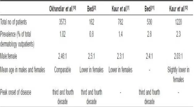
Prevalence studies from India are mostly hospital-based. presents the comparative data from various epidemiological studies on psoriasis from India. Okhandiar et al. collected a comprehensive data from various medical colleges located in Dibrugarh, Calcutta, Patna, Darbhanga, Lucknow, New Delhi and Amritsar. They found that the incidence of psoriasis among total skin patients ranged between 0.44 and 2.2%, with
overall incidence of 1.02%. They noted that the incidence in Amritsar (2.2%) was higher as compared to other centers in Eastern India and speculated that it may be related to different environmental conditions (extremes of temperature), dietary habits, and genetic differences. The ratio of male to female (2.46:1) was very high which could not be clearly accounted for. Highest incidence was noted in the age group of 20-39 years and the mean age of onset in males and females was comparable.
In another study from North India, Bedi reported the prevalence of psoriasis to be 0.8% among the skin patients but the sample size of the study was very small. Male to female sex ratio was 2.5:1. In this study, it was observed that females had lower mean age of onset compared to males. In a latter study by Bedi, which included larger number (530) of subjects, prevalence of psoriasis among dermatology outpatients was found to be 2.8% while male to female ratio continued to be the same.
In a study from tertiary health care center from North India, psoriasis patients accounted for 2.3% of the total dermatology outpatients. Of the total psoriasis patients, 67% were men and 33% were women, male to female ratio being 2.03:1. Ages of patients ranged from infancy to eighth decade, mean age being 33.6 years. Children accounted for 4.4% of total psoriasis patients. Women had slightly lower mean age of onset (27.6 years) compared to the men (30.9 years).
commonly accepted and validated diagnostic criteria. Furthermore, there is basically no reliable information on time trends of the disease.
Table 1 :Epidemiological studies on psoriasis from India
Genetic and familial incidence:
Psoriasis vulgaris is known to be associated with certain HLA antigens and complement factors but most of the studies published are in Western populations with only little information about Indians. Chablani
et al. in a study of 67 psoriasis subjects from Western India found association with the A1, B17 and Cw6, but not with B13 antigens. Pitchappan et al. reported association of HLA Bw57 and DR7 with psoriasis vulgaris in South India. Rani et al. showed that Cw *0602 was the main allele that had high frequency in psoriasis patients in North India.
Genetic predisposition has a significant role in the etiopathogenesis of psoriasis and familial clustering of the cases has been observed. Farber et al. reported familial occurrence in 36% of their patients. Familial incidence is greater in childhood psoriasis compared to adult onset psoriasis.
Indian studies report lower familial incidence of the disease. Bedi reported positive family history of psoriasis in 14% of their patients. While Kaur et al. reported family history in only 2% of their patients. First degree relatives were affected in 84% of the cases while second degree relatives in 12% cases. There are only few studies which have made record of family history of psoriasis in their patients, so definite statistical data on familial incidence is not available.
Quality of life
• 60 percent of people with psoriasis reported their disease to be a
large problem in their everyday life
• Nearly 40 percent with psoriatic arthritis reported their disease to
be a large problem in everyday life
• Patients with moderate to severe psoriasis experienced a greater
negative impact on their quality of life
• Psoriasis has a greater impact on quality of life in women and
younger patients
simply seen as a fault of the epidermis and its keratinocytes. The second hypothesis sees the disease as being an immune-mediated disorder in which the excessive reproduction of skin cells is secondary to factors produced by the immune system
Psoriasis - an Autoimmune Disorder
• As part of its defense against foreign invaders, our body's bone
marrow and thymus gland collaborate to pump out specialized white blood cell warriors called "T cells." Under normal circumstances, T cells are programmed to identify and coordinate an attack on enemy combatants.
• In psoriasis, T cells mistakenly identify skin cells as "other" and
attack them. This attack injures the skin cells, setting off a cascade of responses in immune system and in skin, resulting in skin damage (that is, swelling, reddening and scaling).
• In an effort to heal, skin cells begin reproducing rapidly. Activities
that should take a month take place in only days, and abnormally large numbers of new skin cells push their way to the surface of skin. This occurs so quickly that older skin cells and white blood cells aren't shed quickly enough. These discarded cells pile up on the surface of the skin, creating thick, red plaques with silvery scales on their surface: the hallmark of the classic form of plaque psoriasis
Factors that increase the risk of developing psoriasis include:
• family history • stress
• injury, illness, or infection • steroids and other medications • race
Trauma and certain bacteria may trigger psoriatic arthritis in patients with psoriasis.
Clinical Symptoms:
o A sharply delimited, hyperemic patch, covered with a silvery-white
layer of scales of variable thickness, is found as typical primary eruption
o With careful scratching, small silvery-white lamellar scales come
off, more or less as from a solidified candle strip (candle grease sign)
o Phenomenon of the last skin layer: if scratching continued, a thin,
glistening epithelial layer is revealed
o Auspitz sign: removal of this epithelial layer reveals punctuate
bleeding because of a lesion of the capillaries running out into the tips of the papillae.
o Koebner’s phenomenon: if the skin of a psoriatic patient is
irritated (e.g. scratching) in the phase of an acute episode of eruption, a new psoriatic focus is formed on the floor of this epithelial lesion
Types of psoriasis:
PLAQUE PSORIASIS.
Plaque psoriasis (psoriasis vulgaris), the most common form of the disease, is characterized by small, red bumps that enlarge, become inflamed, and form scales. The top scales flake off easily and often, but those beneath the surface of the skin clump together. Removing these scales exposes tender skin, which bleeds and causes the plaques (inflamed patches) to grow.
Plaque psoriasis can develop on any part of the body, but most often occurs on the elbows, knees, scalp, and trunk.
SCALP PSORIASIS.:
At least 50 of every 100 people who have any form of psoriasis have scalp psoriasis. This form of the disease is characterized by scale-capped plaques on the surface of the skull.
NAIL PSORIASIS:
The first sign of nail psoriasis is usually pitting of the fingernails or toenails. Size, shape, and depth of the marks vary, and affected nails may thicken, yellow, or crumble. The skin around an affected nail is sometimes inflamed, and the nail may peel away from the nail bed.
GUTTATE PSORIASIS.:
Named for the Latin word gutta, which means "a drop," guttate psoriasis is characterized by small, red, drop-like dots that enlarge rapidly and may be somewhat scaly. Often found on the arms, legs, and trunk and sometimes in the scalp, guttate psoriasis can clear up without treatment or disappear and resurface in the form of plaque psoriasis.
PUSTULAR PSORIASIS.
Pustular psoriasis usually occurs in adults. It is characterized by blister-like lesions filled with non-infectious pus and surrounded by reddened skin. Pustular psoriasis, which can be limited to one part of the body (localized) or can be widespread, may be the first symptom of psoriasis or develop in a patient with chronic plaque psoriasis.
Generalized pustular psoriasis is also known as Von Zumbusch pustular psoriasis. Widespread, acutely painful patches of inflamed skin develop suddenly. Pustules appear within a few hours, then dry and peel within two days.
Generalized pustular psoriasis can make life-threatening demands on the heart and kidneys.
Palomar-plantar pustulosis (PPP) generally appears between the ages of 20 and 60. PPP causes large pustules to form at the base of the thumb or on the sides of the heel. In time, the pustules turn brown and peel. The disease usually becomes much less active for a while after peeling.
Acrodermatitis continua of Hallopeau is a form of PPP characterized by painful, often disabling, lesions on the fingertips or the tips of the toes. The nails may become deformed, and the disease can damage bone in the affected area.
ERYTHRODERMIC PSORIASIS.
Characterized by severe scaling, itching, and pain that affects most of the body, erythrodermic psoriasis disrupts the body's chemical balance and can cause severe illness. This particularly inflammatory form of psoriasis can be the first sign of the disease, but often develops in patients with a history of plaque psoriasis.
PSORIATIC ARTHRITIS.
About 10% of partients with psoriasis develop a complication called psoriatic arthritis. This type of arthritis can be slow to develop and mild, or it can develop rapidly. Symptoms of psoriatic arthritis include:
• Joint discomfort, swelling, stiffness, or throbbing • Swelling in the toes and ankles
• Pain in the digits, lower back, wrists, knees, and ankles • Eye inflammation or pink eye (conjunctivitis).
PATHOGENESIS
The biochemical basis for the pathogenesis of psoriasis, which is as equally varied as the genetic basis, can be attributed to both overexpression and underexpression of certain proteins in psoriatic lesions. The anomalies in protein expression can be divided into three areas:
A. Abnormal keratinocyte differentiation B. Hyperproliferation of the keratinocyte
C. Infiltration of inflammatory elements
A. Keratinocyte Differentiation
At least six markers of abnormal keratinocyte differentiation have been found, and all have implications in the pathogenesis of the disease. These include aberrations of
• Keratinocyte Transglutaminase type I (TGase K) • Skin-derived Antileukoproteinase (SKALP)
• Migration inhibitory factor-related protein-8 (MRP-8) • Involucrin
• Filaggrin
• Keratin expression
TGase K - The overexpression of this enzyme causes the excessive cornification seen in psoriatic lesions.
MRP-8 - Its biochemical function is not completely understood. It has been found in psoriasis and other inflammatory diseases but not in normal skin.
Involucrin - A precursor protein that helps to stabilize the CE, is also upregulated in psoriasis vulgaris.
Keratin - Its expression is also disrupted in psoriasis. K6 and K16, markers of abnormal hyperproliferative conditions, are up-regulated in psoriatic epidermis, whereas K1 and K10, markers of terminal differentiation, are down-regulated.
Filaggrin -It is normally found in the granular layer of the skin and is absent in psoriaticlesions.
B. Hyperproliferation
It is the second category of anomalies that contributes to the symptoms of psoriasis vulgaris. Several possible biochemical causes for the overproduction of the keratinocytes have been found in psoriatic skin:
• Epidermal Growth Factor (EGF)
• Bone Morphogenetic Protein-6 (BMP-6) • Transforming Growth Factor-alpha (TGF-α) • Ornithine Decarboxylase
• Activating Protein (AP1)
• Mitogen-Activated Protein Kinase (MAPK)
EGF binding capacity is doubled in the upper layers of the epidermis of psoriatic skin. This increase in binding over-stimulates the growth of the keratinoctyes, causing hyperproliferation.
BMP-6, another growth factor, is present in newborns, but normally disappears by adulthood, except in psoriatic patients, which
makes it a prime candidate for a growth factor sponsoring the formation of psoriatic lesions in humans.
TGF-α expression is up-regulated in psoriatic skin.
Ornithine Decarboxylase, an essential enzyme in polyamine biosynthesis, is higher in lesional and nonlesional psoriatic skin.
AP1, a complex of the oncoproteins, stimulates the expression of many genes that are important in cell proliferation and inflammation. The last indicator, MAPK, helps to regulate cellular proliferation. Numerous growth factors and cytokines modulate MAPK's activity, which is higher in psoriatic fibroblasts.
C. Inflammatory Elements
The inflammatory aspect of psoriasis is physically evident by the redness of psoriatic plaques. The biochemical basis for this inflammation stems from several immune modulators including various cytokines released from keratinocytes and other proteins involved in the inflammatory response.
Among the interleukins (ILs), IL-1, IL-6, IL-7 and IL-8 are up regulated in psoriatic skin. In addition, IL-15, which reduces keratinocyte cell apoptosis, is elevated in psoriatic lesions. Pituitary Adenylate Cyclase Activating Polypeptide (PACAP) is an inflammatory mediator that is up regulated in psoriatic lesions.
All of these inflammatory factors exert specific effects on T cells, endothelial cells, macrophages and neutrophils, which in turn spawn the
disease where activation of T lymphocytes is central to the inflammation in the dermal microenvironment and the epidermal hyper proliferation is secondary to the inflammatory events that follow a Th1 type of immune response.
T Cells
In psoriasis, the activity of T cells is the driving force for induction and maintenance of the skin lesions.
T Cell Activation
Activation of T cells requires three steps • Binding
• Antigen specific activation, known as signal 1
• Non-antigen specific cell-cell interaction, known as signal 2 Binding
The T cell attaches to the Antigen Presenting Cell [APC] through surface adhesion molecules. In skin the Langerhans cell is the most efficient Antigen Presenting Cell.
Antigen specific activation
Once the T cell-APC binding has occurred through their respective surface adhesion molecules, the antigen is presented to the T cell by the APC. This antigen stimulated activation leads to conversion of the naïve T cell into an antigen specific cell that may further develop into a long lived memory cell that circulates in the body and can recognize the same antigen at a later date, even after several years.
Non antigen specific cell-cell interaction
This is also known as co-stimulation. If co-stimulation by other cell surface molecules does not occur following antigen presentation, the T
cell will not respond to the antigen and will undergo apoptosis or be rendered unresponsive to that antigen in the future (anergy).
Effector functions of activated T cells
Once the T cell is activated the next step is induction of inflammatory responses and tissue changes leading to the clinical picture of psoriasis.
TNF-a is strongly implicated in the pathogenesis of psoriasis and psoriatic arthritis. TNF-a functions in a positive feedback loop by recruiting more inflammatory cells and upgrading receptors on those cells. TNF-a levels are markedly increased in skin lesions, synovium and serum of patients with psoriasis and these correlate with the severity of the disease. Decreased levels are associated with clinical resolution.
DIAGNOSIS OF PSORIASIS
There are no laboratory tests which will positively identify psoriasis. The blood count, Urine analysis, ESR and other hematologic chemical and serologic studies are within normal limits in most cases of psoriasis.
The diagnosis of psoriasis is based upon:
1. The family history of psoriasis