www.elsevier.com / locate / bres
Research report
Convulsive seizures induced by N-methyl-
D
-aspartate microinjection
into the mesencephalic reticular formation in rats
*
Takahiro Ishimoto , Nobuyuki Omori, Fukuyasu Mutoh, Shigeru Chiba
Department of Psychiatry and Neurology, Asahikawa Medical College, Midorigaoka higashi 2-1-1-1, Asahikawa 078-8510, Japan Accepted 8 August 2000
Abstract
Effects of microinjections of a single 2 or 10 nmol dose of N-methyl-D-aspartate (NMDA) into the unilateral mesencephalic reticular
formation (MRF) on behavior and electroencephalogram were examined in rats (n518) during a 15 min period (Exp. 1), and subsequent effects of sound stimulation with key jingling applied at 15, 30, and 45 min after the injections were observed (Exp. 2). The microinjections of 2 nmol dose of NMDA (n510) induced hyperactivity (9 of 10 rats) and running / circling (8 of 10 rats) in Exp. 1, and hyperactivity (3 of 10 rats) in Exp. 2. Moreover, the microinjections of 10 nmol dose of NMDA (n58) induced not only hyperactivity (8 of 8 rats) and running / circling (7 of 8 rats) but also generalized tonic–clonic seizures (GTCS) (5 of 8 rats) in Exp. 1; these seizure patterns were also elicited by sound stimulation in Exp. 2. The seizure patterns were accompanied by electroencephalographic seizure discharges in the MRF and the motor cortex. In contrast, the control group rats (n510) which received a single dose of saline microinjection into the unilateral MRF showed no behavioral or electroencephalographic changes in both Exp. 1 and 2. These findings suggest that the MRF has an important role in the development of GTCS, which follows hyperactivity and running / circling, and that potentiation of excitatory neurotransmission in the MRF participates in the development of audiogenic seizures as well as GTCS. 2000 Elsevier Science B.V. All rights reserved.
Theme: Disorders of the nervous systems
Topic: Epilepsy: basic mechanisms
Keywords: Brainstem; N-methyl-D-aspartate; Convulsion; Epilepsy; Audiogenic seizures; Rat
1. Introduction brainstem reticular formation (RF) is considered to be
important for the development of audiogenic seizures in Previous experimental studies suggest that the brainstem genetically epilepsy-prone rats (GEPR) [5,21].
reticular formation, particularly the MRF, is involved in It has been suggested that an imbalance between inhib-the generation of primary generalized seizures [26,40] or in itory and excitatory neurotransmission participate in the the secondary generalization of partial seizures originating generation and expression of human [16,17] and animal from the forebrain [9–13]. Electrical stimulation of the epileptic seizures [2,30,34,36]. With respect to inhibitory unilateral MRF induces generalized convulsions in rats [6], neurotransmission, repeated administration of a GABA rabbits [4], and cats [27]. The MRF can be kindled with receptor antagonist, picrotoxin [7] or bicuculline [45], to the development of generalized convulsions in rats [10]. the unilateral amygdala induces the development of Local administration of bicuculline, ag-aminobutyric acid amygdala seizure in rats. Systemic administration of a (GABA) antagonist, to the unilateral MRF [9] induced GABA receptor agonist, muscimol, strongly suppresses generalized tonic seizures in rats. Moreover, propagation amygdaloid kindled seizuresin rats [35,36]. In contrast, in of seizure discharges from the inferior colliculus to the excitatory neurotransmission, systemic administration of excitatory amino acids such as NMDA [33]or kainic acid [43], or focal microinjection of NMDA into the unilateral *Corresponding author. Tel.: 181-166-68-2473; fax: 1
81-166-68-amygdala [14,15]or the massa intermedia [24,25], 2479.
E-mail address: [email protected] (T. Ishimoto). produces generalized convulsive seizures. In the hip-0006-8993 / 00 / $ – see front matter 2000 Elsevier Science B.V. All rights reserved.
pocampus and amygdala of the hippocampal kindled brain, saline. The sound stimulation was provided by a short increased release of glutamic acid during both ictal and manual shake of a bunch of keys (6 metal door keys on a interictal periods has been demonstrated in rats [44]. metal key-ring) held at 50 cm above the floor of an Furthermore, dizocilpine (MK-801) [42]and 3-(2-carbox- open-topped observation box (35 cm335 cm335 cm); the ypiperazine-4-yl)propyl-1-phosphonic acid (CPP) [38], frequency and intensity of the sound was measured by which is an NMDA receptor antagonist, have potent sound level meters (Leader Electronics Corp., Yokohama, inhibitory effects on the development of amygdala kindling Japan, NL-05A).
or kindled amygdala seizures in rats. Therefore, potentia- On completion of Exp. 2, the animals were deeply tion of excitatory neurotransmission by excitatory amino anesthetized and their brains were subjected to perfusion acids seems to play a crucial role in the development of fixation with 10% formalin, and then cut into frozen several experimental models of epilepsy. sections 10mm thick to histologically confirm the position To further clarify the role of MRF in the expression of of the depth electrode. Statistical comparisons were made epileptic seizures, we microinjected NMDA into the unila- using Fisher’s exact test.
teral MRF in rats and observed the behavioral and EEG changes for 15 min (Exp. 1). Subsequently, we examined
the effects of sound stimulation applied at 15, 30 and 45 3. Results
min after NMDA microinjections on the rat behaviors and
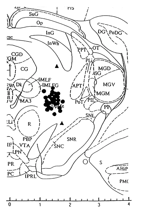
EEGs (Exp. 2). Histological examination revealed that the MRF elec-trodes were located in the intended area (within 0.5 mm of the target site) in all the rats except for those in 2 of the
2. Materials and methods Group A rats (Fig. 1); results from these two rats were
2.1. Experiment 1: NMDA injection
Thirty male adult Sprague–Dawley rats (2–3 months of age, weighing 250–400 g) were used The rats were randomly allocated to Group A (n510), B (n510), or C (n510). Under pentobarbital anesthesia, chemitrodes, i.e., 24G guide cannulas with bipolar electrodes made of twisted stainless steel wire (200 mm in diameter), were implanted stereotaxically [39] into the left MRF (the deep mesencephalic nucleus; 5.8 mm posterior, 1.7 mm lateral from bregma, and 6.6 mm ventral from the skull). The tip of bipolar electrodes extended 1.0 mm beyond the ends of the guide cannulas. Two surface electrodes (stainless steel screws) were driven into the skull: one for recording from the unilateral sensorimotor cortex, and the other, over the unilateral olfactory bulb, as the reference electrode
Seven days after operation a single 10 and 2 nmol dose of NMDA (Sigma, St Louis, MO, USA) was administered into the MRF in Groups A and B, respectively The dose of NMDA was dissolved in saline and delivered in a volume of 1.0ml at a rate of 1.0ml / min by a microsyringe pump (Eicom corp., Kyoto, Japan, EP-60) The microinjections were performed with 30G needles extending 1.0 mm beyond the ends of the guide cannulas The Group C rats received a saline injection (1.0 ml) into the MRF in a manner identical to that carried out in Groups A and B Behavioral and EEG changes were recorded for 15 min after the end of the NMDA microinjections in Exp. 1.
2.2. Experiment 2: sound stimulation
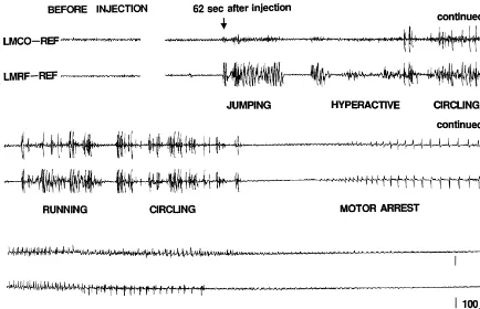
Fig. 2. EEGs of hyperactivity and running / circling patterns in a Group A rat. From 62 s after the NMDA injection, the rat displayed jumping, hyperactivity, and running / circling. NMDA, N-methyl-D-aspartate; LMCO, left motor cortex; LMRF, left mesencephalic reticular formation; REF, reference electrode.
therefore excluded from data analysis. One of them, with convulsive patterns in the following order: (1) hyperactivi-MRF electrode located far dorsally, showed only mild ty (a state in which a rat is frequently moving or restlessly hyperactivity and running / circling behavior while the walking around in the observation box), (2) running / other, with MRF electrode located ventrolaterally, showed circling, and (3) GTCS. The microinjections of 2 nmol mild hyperactivity without seizure discharges. dose of NMDA induced only hyperactivity, and running / The peak frequency of sound stimulation was around circling. As shown in Figs. 2 and 3, these seizure patterns 480 Hz, with a wide range of 300–2000 Hz. The intensity were accompanied by electroencephalographic seizure of the sound source ranged from 80 to 90 dB. discharges in the MRF and the motor cortex. The Group C rats did not show any behavioral or electroencephalog-3.1. Experiment 1 raphic changes during the 15 min after the end of the saline injection. The incidence of the seizure patterns The microinjections of 10 nmol dose of NMDA induced observed in each group is shown in Table 1. The
Table 1 Table 2
The incidence of the seizure patterns induced by NMDA injections in The incidence of the seizure patterns induced by sound stimulation in
a a
each Group (Exp. 1) each Group (Exp.2)
a a
Seizures were classified into 3 patterns: hyperactivity, running / circling Sound stimulation induced seizures were classified into 3 patterns: and GTCS (generalized tonic–clonic seizures), and the incidence was hyperactivity, running / circling, and GTCS. For each figure, the analyzed using Fisher’s exact probability test among Groups A, B and C. numerator indicates the total number of rats which showed the seizure NMDA, N-methyl-D-aspartate; GTCS, generalized tonic–clonic seizures. pattern induced by sound stimulation applied at either 15, 30, or 45 min *P,0.05, **P,0.01 by Fisher’s exact test. after the end of NMDA injections. The incidence was analyzed using Fisher’s exact probability test among the three groups. GTCS, generalized tonic–clonic seizures; NMDA, N-methyl-D-aspartate.*P,0.05, **P,0.01 by Fisher’s exact test.
dences of hyperactivity and running / circling were sig-nificantly higher in Groups A and B than in Group C. The incidence of GTCS was significantly higher in Group A
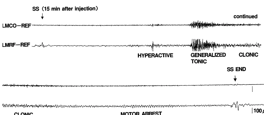
than in Groups B and C. and their EEG findings observed in Exp. 2 were almost identical to those observed in Exp. 1 (Fig. 4). In contrast, 3.2. Experiment 2 no behavioral or EEG changes were elicited by sound stimulation in Group C in either Exp. 1 or 2. The Although NMDA-induced seizure patterns were not incidences of the seizure patterns in Groups A, B, and C observed during the period from 15 to 45 min after the end are summarized in Table 2. The incidence of each seizure of NMDA injections, sound stimulation applied at 15, 30 pattern was significantly higher in Group A than in Groups and 45 min after NMDA injections could induce hyperac- B and C. However, there was no significant difference in tivity, running / circling, and GTCS. These seizure patterns the incidence of each seizure between Groups B and C.
4. Discussion continuous with the intermediate zone of the spinal cord, and rostrally continuous with the intralaminar nuclei of the Previous experimental studies have suggested that thalamus and zona incerta of the ventral thalamus, occupy-generalized convulsions may have a MRF origin ing the central portion of the brainstem. In addition, [4,6,9,10,27]. Electrical stimulation applied to the MRF complicated fiber connections are ventrically and horizon-triggers self-sustaining generalized tonic seizures in rats tally present among neurons in the brainstem RF [37]. [6], rabbits [4], and cats [27]. Injection of bicuculline, a Therefore, it is speculated that within the brainstem RF selective GABAA antagonist, into the unilateral MRF of epileptic discharges can easily propagate from one struc-rats results in fatal prolonged generalized tonic seizures, ture to the others. The fact that a GTCS induced by with the electroencephalographic seizure discharges pre- bucuculline microinjections into the MRF is almost identi-dominant in the subcortical MRF or amygdala rather than cal to one induced by the bicuculline microinjections into in the motor cortex [9]. In addition, repeated electrical the pontine RF [9]is not inconsistent with this possibility. MRF stimulation can produce ultimately prolonged GTCS In order to induce audiogenic seizures, an electric bell with afterdischarges in the MRF and the motor cortex [10]. [5,18–22,31] or key jingling [1,23,29,32] have commonly In Exp. 1, we observed that GTCS was induced by NMDA been used as sound sources. Interestingly, in our prelimin-injection into the unilateral MRF. The results further ary study, key jingling induced the seizures more effective-support the view that generalized seizures can originate ly than the electric bell. The reason for this remains to be from the MRF. clarified, but it is probably due to the difference in the peak In Exp. 1, the microinjection of 10 nmol dose of NMDA of frequency between the electric bell and key jingling; into the unilateral MRF induced varied seizure patterns, sound stimulation by the former bell has peaks at 1000 and i.e., hyperactivity, running / circling, and GTCS, in that 1600 Hz, while that by key jingling has a peak at around order. Injection of 2 nmol dose of NMDA also induced 480 Hz, with a range from 300 to 2500 Hz.
hyperactivity and running / circling, but did not produce A recent experimental study using brain chimera tech-GTCS. These findings suggest that potentiation of excitat- nology on 12-somite stage chick embryos with hereditary ory neurotransmission in the MRF participates in the reflex epilepsy (in response to either light or sound generation of varied convulsive seizures including GTCS, stimulations), demonstrated that the mesencephalon con-and that the severity of the seizure symptoms depends on tains the generator of the epileptic manifestations of the level of potentiation of excitatory neurotransmission. running and generalized convulsions [3]; the study sug-In Exp. 2 the results imply that the rats that received the gested that the brainstem is important as a possible focus injections of NMDA are susceptible to audiogenic seizures, of epileptic seizures. Our present study revealed that suggesting that potentiation of excitatory neurotrans- NMDA microinjection into the rat MRF induces varied mission in the MRF has a facilitory effect on the develop- seizure patterns, including GTCS, and also results in ment of audiogenic seizures. increased seizure susceptibility to sound stimulation. Our It is assumed that the brainstem RF participates in the findings provide an insight into the mechanism of epileptic development of audiogenic seizures. In GEPR, which seizures arising from the brainstem, suggesting that the shows running or generalized tonic seizures with sound potentiation of excitatory neurotransmission in the MRF stimulations, elevated levels of glutamate and aspartate are may play a facilitatory effect on the generation of epileptic observed in the inferior colliculus (IC) and the brainstem seizures.
RF [8,28,41]. Bilateral microinjection into the IC or the pontine RF of excitatory amino acid receptor antagonists
blocks audiogenic seizures in GEPR [22]. In addition, Acknowledgements neuronal recordings using microwire electrodes in the IC
and the pontine RF suggest that the IC serves as the The authors wish to thank Dr. Akihiko Nunomura for initiation site of the audiogenic seizures, and that the his advice in histological investigation and Prof. influence of the IC on the pontine RF neurons is magnified Yoshikatsu Mochizuki for statistical analyses. Part of the in association with the susceptibility to audiogenic sei- material in the manuscript was presented at the 33rd zures, with the neuronal firing rate of the pontine RF Congress of the Japan Epilepsy Society in Sendai, Japan increased markedly at the onset of audiogenic tonic seizure on October 22 1999.
[21]. Therefore, it is assumed that the propagation of seizure discharges from the IC to the brainstem RF, particularly to the pontine RF, is crucial for the
develop-References
ment of audiogenic seizures, and that the brainstem RF neurons play a major role in generation of audiogenic
[1] T. Asakura, J.A. Wada, Neurobiology of audiogenic seizure, Brain seizures [21]. and Nerve (Tokyo) 24 (1972) 513–534, (in Japanese).
feedback systems suggests a neurophysiological explanation of brief [22] C.L. Faingold, M.E. Randall, D.K. Naritoku, C.A. Boersma Ander-paroxysms, Brain Res. 52 (1973) 1–17. son, Noncompetitive and competitive NMDA antagonists exert anticonvulsant effects by actions on different sites within the [3] C. Batini, M.A. Teillet, R. Naquet, N.M. Le Douarin, Brain
neuronal network for audiogenic seizures, Exp. Neurol. 119 (1993) chimeras in birds application to the study of a genetic form of reflex
198–204. epilepsy, Trends Neurosci. 19 (1996) 246–252.
[23] W.J. Griffiths, Absence of audigenic seizures in wild Norway and [4] F. Bergmann, A. Costin, J. Gutman, A low threshold convulsive
Alexandrine rats, Science 99 (1944) 62–63. area in the rabbit’s mesencephalon, Electroencephalogr. Clin.
Neurophysiol. 15 (1963) 683–690. [24] Y. Hirayasu, J.A. Wada, Convulsive seizures in rats induced by
N-methyl-D-aspartate injection into the massa intermedia, Brain Res. [5] R.A. Browning, C. Wang, C.L. Faingold, Effect of norepinephrine
57 (1992) 36–40. depletion on audiogenic-like seizures elicited by microinfusion of an
excitant amino acid into the inferior colliculus of normal rats, Exp. [25] Y. Hirayasu, J.A. Wada, N-methyl-D-aspartate injection into the Neurol. 112 (1991) 200–205. massa intermedia facilitates development of limbic kindling in rats,
Epilepsia 33 (1992) 965–970. [6] W.M. Burnham, P. Albright, J. Schneiderman, P. Chiu, T. Ninchoji,
‘Centrencephalic’ Mechanisms in the Kindling Model, in: J.A. Wada [26] H.H. Jasper, Current evaluation of the concepts of centrencephalic (Ed.), Kindling 2, Raven Press, New York, 1981, pp. 161–178. and cortico-reticular seizures, Electroencephalogr. Clin.
Neuro-physiol. 78 (1991) 2–11. [7] D.P. Cain, Kindling by repeated intraperitoneal or intracerebral
injection of picrotoxin transfers to electrical kindling, Exp. Neurol. [27] A. Kreindler, E. Zuckermann, M. Steriade, D. Chimion, Electro-97 (1987) 243–254. clinical features of convulsions induced by stimulation of brain
stem, J. Neurophysiol. 21 (1958) 430–436. [8] A.G. Chapman, C.L. Faingold, G.P. Hart, H.M. Bowker, B.S.
Meldrum, Brain regional amino acid levels in seizure susceptible [28] S.M. Lasley, Roles of neurotransmitter amino acids in seizure rats: changes related to sound-induced seizures, Neurochem. Int. 8 severity and experience in the genetically epilepsy-prone rat, Brain
(1986) 273–279. Res. 560 (1991) 63–70.
[9] S. Chiba, S. Kamata, A. Nunomura, F. Mutoh, M. Matsumoto, T. [29] N.R.F. Maier, N.M. Glaser, Studies of abnormal behavior in the rat Miyagishi, Convulsive response induced by microinjections of II. A comparison of some convulsion-producing situations, Comp. bicuculline methiodide into the brainstem reticular formation in rats, Psychol. Monographs 16 (1940) 1–30.
Hokkaido Epilepsy Res. 15 (1994) 39–44. [30] H. Matsumoto, C. Ajmone-Marsan, Cortical cellular phenomena in [10] S. Chiba, N. Omori, S. Kamata, A. Nunomura, F. Mutoh, Kindling experimental epilepsy ictal manifestations, Exp. Neurol. 9 (1964)
of the mesencephalic reticular formation and its influence on 305–326.
subsequent amygdala kindling in rats, Epilepsia 37 (Suppl.3) (1996) [31] M.H. Millan, B.S. Meldrum, C.A. Boersma Anderson, C.L. Faing-116–117. old, Excitant amino acids and audgenic seizures in the genetically [11] S. Chiba, J.A. Wada, Amygdala kindling in rats with brainstem epilepsy-prone rat II Efferent seizure propagating pathway, Exp.
bisection, Brain Res. 682 (1995) 50–54. Neurol. 99 (1988) 687–698.
[12] S. Chiba, J.A. Wada, Kindling of the interpeduncular nucleus and its [32] Y. Mirovsky, G.C. Wagner, A. Sekowski, M. Goldberg, H. Fisher, influence on subsequent amygdala kindling in rats, Epilepsia 36 Stimultaneous changes in striatal dopamine serotonin and metabo-(1995) 410–415. lites after withdrawal seizures in rats from dependence on alcohol, [13] S. Chiba, J.A. Wada, The effect of electolytic lesioning of the Alcohol 12 (1995) 251–256.
midbrain prior to amygdala kindling in rats, Neurosci. Lett. 227 [33] N. Mori, J.A. Wada, Bidirectional transfer between kindling induced (1997) 83–86. by excitatory amino acids and electrical stimulation, Brain Res. 425 [14] M.J. Croucher, K.L. Cotterell, H. F Bradford, Amygdaloid kindling (1987) 45–48.
by repeated focal N-methyl-D-aspartate administration: comparison [34] K. Morimoto, S.E. Mason, G.V. Goddard, Kindling-induced changes with electrical kindling, Eur. J. Pharmacol. 286 (1995) 265–271. in the EEG recorded during stimulation from the site of stimulation. [15] M.J. Croucher, K.L. Ruffle, H.F. Bradford, The effects of focal II comparison between spontaneous and evoked potentials, Exp.
N-methyl-D-aspartate pretreatment on the parameters of amygdaloid Neurol. 97 (1987) 1–16.
electrical kindling, Eur. J. Pharmacol. 319 (1997) 207–213. [35] K. Morimoto, S.E. Mason, G.V. Goddard, Kindling-induced changes [16] M.J. During, K.M. Ryder, D.D. Spencer, Hippocampal GABA in the EEG recorded during stimulation from the site of stimulation. transporter function in temporal-lobe epilepsy, Nature 376 (1995) III direct pharmacological manipulations of the kindled amygdala,
174–177. Exp. Neurol. 97 (1987) 17–34.
[17] M.J. During, D.D. Spencer, Extracellular hippocampal glutamate [36] K. Morimoto, N. Yamada, Abnormal neurotransmission in ex-and spontaneous seizure in the conscious human brain, Lancet 341 perimental models of epilepsy, Shinkei Kenkyu no Shinpo, 38
(1993) 1607–1611. (1994) 823–837 (in Japanese).
[18] C.L. Faingold, M.J. Marcinczyk, D.J. Casebeer, M.E. Randall, S.P. [37] R. Nieuwenhuys, J. Voogt, C. van Huijzen (Eds.), The Human Arneric, R.A. Browning, GABA in the inferior colliculus plays a Nervous System A Synopsis and Atlas, 3rd Edition, Springer-Verlag, critical role in control of audiogenic seizures, Brain Res. 640 (1994) Berlin, 1988.
40–47. [38] M. Osonoe, N. Mori, S. Hoshino, Y. Yamada, K. Osonoe, H. Kittaka, [19] C.L. Faingold, M.H. Millan, C.A. Boersma, B.S. Anderson, Mel- Y. Iwata, The effects of N-methyl-D-aspartate (NMDA) and its drum, Induction of audiogenic seizures in normal and genetically competitive antagonist 3-(2-carboxypiperazine-4-yl)-propyl-1-phos-epilepsy-prone rats following focal microinjection of an excitant phonic acid (CPP) injected into caudate-putamen on kindled amino acid into reticular formation and auditory nuclei, Epilepsy amygdaloid seizures in rats, Brain Res. 728 (1996) 242–246. Res. 3 (1989) 199–205. [39] G. Paxinos, C. Watson (Eds.), The Rat Brain in Stereotaxic [20] C.L. Faingold, D.K. Naritoku, C.A. Copley, M.E. Randall, A. Riaz, Coordinates, 2nd Edition, Academic Press, Sydney, 1986.
C.A. Boersma Anderson, S.P. Arneric, Glutamate in the inferior [40] W. Penfield, H.H. Jasper (Eds.), Epilepsy and the Functional colliculus plays a critical role in audiogenic seizure initiation, Anatomy of the Human Brain Little, Brown and Company, Boston,
Epilepsy Res. 13 (1992) 95–105. 1954.
non-competitive antagonist of NMDA receptors MK-801 in the [44] Y. Ueda, N. Tsuru, Sequential change of glutamate release in kindling model of epilepsy, Brain Res. 463 (1988) 12–20. bilateral hippocampi in amygdaloid kindling rat, Jpn. J. Psychiatry [43] T. Tanaka, S. Tanaka, T. Fujita, K. Takano, H. Fukuda, K. Sako, Y. Neurol. 47 (1993) 225–228.
Yonemasu, Experimental complex partial seizures induced by a [45] S. Uemura, H. Kimura, Amygdaloid kindling with bicuculline microinjection of kainic acid into limbic structures, Prog. Neurobiol. methiodide in rats, Exp. Neurol. 102 (1988) 346–353.