Journal of Biology, Agriculture and Healthcare ISSN 2224-3208 (Paper) ISSN 2225-093X (Online) Vol.3, No.17, 2013
www.iiste.org
Vol.3, No.17, 2013
Dept. of Biotechnology, Ministry of Science and Technology, India Dr. Chiung Ting Chang
Maastricht University, Netherlands Prof. Dr. Venus S. Solar
Manila Central University, Philippines Dr. Eng. Rares Halbac-Cotoara-Zamfir
"Politehnica" University of Timisoara Romania Prof. Dr. P. Satheeshkumar
Central Marine Fisheries Research Institute, India Prof. Dr.Ibrahim Hassan
Alexabdria University, Egypt Prof. Dr.Jagruthi Joshi Novartis Healthcare, India Dr. Nabil Miled
Sfax University, Tunisia Prof. Dr. Carlos K B Ferrari
Federal University of Mato Grosso (UFMT), Brazil Prof. Dr. H. A. Ibrahim,
Suez Canal university, Egypt Dr. Arda YILDIRIM
Gaziosmanpasa University, Turkey Dr.Nexhbedin Beadini
Faculty of Medical Sciences, State University of Tetova, Macedonia Dr.Sheqibe Beadini
Faculty of Medical Sciences, State University of Tetova, Macedonia
Paper submission email: [email protected]
ISSN (Paper)2224-3208 ISSN (Online)2225-093X
Please add our address "[email protected]" into your email contact list.
This journal follows ISO 9001 management standard and licensed under a Creative Commons
Attribution 3.0 License.
Copyright © www.iiste.org
Vol 3, No 17 (2013)
Table of Contents
Articles
Effect of Regulated Deficit Irrigation on Growth and Yield of Sorghum
Adzemi Mat Arshad, Ibrahim W, Wan Zaliha W S
1-8
Journal of Biology, Agriculture and Healthcare ISSN 2224-3208 (Paper) ISSN 2225-093X (Online) Vol.3, No.17, 2013
www.iiste.org
Hospital of Osun State, Nigeria.
B. L. Ajibade, Oladeji M. O, E.A. Oyedele, Amoo P.O, Makinde O.Y
9-14
Feed Resources and Seasonal Nutrient Composition of Predominant Forages for Small
Ruminant Production in Iwo Local Government Area of Osun state, Nigeria
Funmilayo BAMIGBOYE, Olaniyi Babayemi, Adegboyega Adekoya
15-24
Impact of a Designed Nursing Intervention protocol on Myocardial Infarction Patient's
Outcome at a selected University Hospital in Egypt
Abdelhameed Mahros Abdelhameed, Warda Youssef Mohamed, Yousria Abd El-salam
Seloma, Hanan El-sayed Zaghla
25-35
Epidemiologic Attributes and Virulence Profile of Salmonella Tennessee isolates from
Infections associated with Peanut Butter National Outbreak
Chau H. Nguyen, Seongbeom Cho, Mahdi A. Saeed
36-42
Adaptive Mechanisms of Rural Fishermen Towards Climate Change On Quantity of Fish
Caught in Asari-toru Local Government Area of Rivers State Nigeria.
Henry Unaeze, Adaba Ibim
43-48
Heritabiliy Studies In Some Sweet Sorghum (Sorghum Bicolor. L. Moench) Genotypes
R. A. Sami, M. Y. Yeye, M. F. Ishiyaku, I. S. Usman
49-51
The Effect of Household Characteristics on Child Mortality in Ghana
Michael Ofori Fosu, Ir. Peter Romeo Nyarko
52-58
Identification of Badh2 Mutation Type among Indonesian Fragrant Rice Varieties
Djarot Sasongko Hami Seno, I Made Artika, Waras Nurcholis, Tri Joko Santoso,
Kurniawan R Trijatmiko, Bambang Padmadi, Dewi Praptiwi, Jap Mai Ching, Zainal Alim
Mas’ud
59-65
Glucosinolates, Glycosidically Bound Volatiles and Antimicrobial Activity of Brassica
oleraceae Var. Botrytis, (Soultany Cultivar)
Mohamed S. Hifnawy, Rabab M. Abdel Salam, Mohamed A. Rabeh, Mahmoud A.
Aboseada
66-81
Management of Root Knot Nematode Affecting Banana Crop by Using Organic
Amendment And Biological Products
Zahra Ferji, El Hassan Mayad, Mohamed Alfalah
82-85
New Agricultural Land Measurement Adapted in Langowan, Minahasa, North Sulawesi,
Indonesia
Loing, Jeane Catty
86-92
First Report on Fusarium solani, a Pathogenic Fungus Causing Stem Rot Disease on
Dragon Fruits (Hylocereus sp.) in Bali
Wiwik Susanah Rita, Dewa Ngurah Suprapta, I Made Sudana, I Made Dira Swantara
Vol.3, No.17, 2013
Mamman, T., Ipinjolu, J.K, I, Magawata
100-104
Effect of Some Thermal Processing Techniques on the Proximate, Mineral and
Anti-Nutrient Compositions of Bambara Groundnut (Voandzeia subterranea, (L) Thour) Meal
Isikwenu J. O, Nwanbe R . N, Elechi F. N
105-109
Adaptation, Biomass and Ethanol Yields of Sweet Sorghum (Sorghum bicolor (L.)
Moench) Varieties at Dryland Farming Areas of Jimbaran Bali, Indonesia
I Gusti Ayu Mas Sri Agung, I Ketut Sardiana, I Wayan Diara, I Gusti Made Oka Nurjaya
110-115
Assistive Technology For Hearing and Speech Disorders
Adedayo O. Olaosun, Olawale Ogundiran
116-120
Determinants of Output among Pig Farmers in Abia State, Nigeria
Kelechi Igwe, Amarachi Ifekaonwu, Samson Amao, Clara Igwe
121-126
Donkey-Cart Transport, a Source of Livelihood for Farmers in the Kassena Nankana
Municipality in Upper East Region of Ghana
Maurice M. Braimah, Issahaku Abdul-Rahaman, Daniel Oppong-Sekyere
127-136
Effect of Partial Rootzone Drying Technique on Growth Performance of Sorghum
Adzemi Mat Arshad, Ibrahim W, Wan Zaliha W S
137-142
Variations in Phosphatase Activity of Crude Oil and Used Crankase Oil Polluted
Agricultural Soil.
Reginald C. Ohiri, Agha, N. C., Nwachukwu, N.
143-149
Immune Response to Hepatitis B Virus Vaccine
AYAM MOHAMMED SALIH ALI
150-154
First Report on Fusarium solani, a Pathogenic Fungus Causing
Stem Rot Disease on Dragon Fruits (Hylocereus sp.) in Bali
Wiwik Susanah Rita1, Dewa Ngurah Suprapta2*, I Made Sudana2, I Made Dira Swantara3 1.Doctorate Program in Agricultural Science, School for Postgraduate Udayana University. 2.Laboratory of Biopesticide Faculty of Agriculture, Udayana University, Jl. PB. Sudirman Denpasar Bali
Indonesia.
3.Laboratory of Chemistry, Faculty of Natural Science and Mathematic, Udayana University *Email of corresponding author : [email protected]
Abstract
In recent years, dragon fruit crop (Hylocereus spp.) has become increasingly important in Bali Indonesia due to its high nutrient content and healing properties. However, the dragon fruit was reported to be seriously infected with several complex diseases caused by fungi and causing serious losses to the farmers. The study on morphological and molecular characterization the fungal pathogen was conducted to confirm the species of the fungi. Koch Postulate was applied to confirm the causal agent of the disease. There were two isolates of fungi isolated from the stems of diseased plants, namely isolate w1 (from stem of H. undatus) and isolate w2 (from stem of H. polyrhizus). Based on macroscopic and microscopic characteristics, and analysis of 18S rDNA, both of them were identified as Fusarium solani. This is the first report on the F. solani the cause of stem rot disease on dragon fruits in Bali.
Journal of Biology, Agriculture and Healthcare ISSN 2224-3208 (Paper) ISSN 2225-093X (Online) Vol.3, No.17, 2013
www.iiste.org
1. Introduction
Dragon Fruits (Hylocereus spp.), which are also known as pitaya, are the fruits of cactus species, especially of the genus Hylocereus. There are three species which have high commercial valuable fruits are the species of Hylocereus undatus, (red rind, white flesh), Hylocereus polyrhizus (red rind, red flesh), and Hylocereus costaricensis (red rind, super red flesh). Hylocereus spp. is grown commercially in Vietnam, Spain, Malaysia, Japan, Mexico and other tropical and subtropical areas because of its high nutrient content and healing properties. In recent years, this fruit has become increasingly important in Bali Indonesia. The dragon fruit is rich in vitamin, it helps the digestive process due to its fiber, prevent colon cancer and diabetes, neutralize toxic substances such as heavy metals, and helps to reduce cholesterol levels and high blood pressure (He et al., 2012; Zainoldin and Baba, 2009). Recently, dragon fruit has been reported to be seriously infected with several complex diseases caused by fungi and causing serious losses to farmers. Several dragon fruit plants grown in Sobangan Village, Bali Indonesia showed severe symptom of stem rot disease. The disease caused significant yield losses. Isolation of the fungi associated with the diseased-plants showed that Fusarium sp. was the most frequent found on the stems showing brown rot symptom.
Various diseases caused by fungi have been reported on dragon fruit in tropical and subtropical countries, such as fruit rot (Bipolaris cactivora) (Tarnowski et al., 2010; He et al. 2012), stem rot (Fusarium semitectum, Fusarium oxysporium, Fusarium moniliforme) (Hawa et al., 2010), anthracnose (Colletotrichum gloeosporioides) (Masyahit et al., 2009), brown spot (Botryodiplodia sp.), basal rot (Pythium sp.) (Lin et al., 2006), wilt (Fusarium oxysporium), stem blight (Diplodia sp., Ascochyta sp., and Phoma sp.), black spot (Alternaria sp.), speck blight (Nectriella sp.), (Wang et al.,2007), and stem lesion (Septogloeum sp.) (Zheng et al., 2009).
In order to control the disease, it is necessary to identify the causal agent of the disease. The fungal pathogen can be identified based on cultural and morphological characters. However it could be highly variable depending on the media and cultural conditions that could be the problems in fungal identification. In recent years, the increasing use of molecular methods in fungal identification has emerged as a possible answer to the problems associated with the existing phenotypic identification systems (Mishra et al., 2003). One of the molecular approaches to fungal identification is based on Polymerase Chain Reaction (PCR).
Vol.3, No.17, 2013
applications, most researchers use sequence alignments that are based on nucleotide similarity (Tippery and Les, 2008).
Suga et al. (2000) has investigated phylogenetic relationships of Fusarium solani using sequences from the rDNA-ITS region. Mishra et al. (2003) has developed a fluorescent-based polymerase chain reaction in ITS region to identify five toxigenic and pathogenic Fusarium species. Abd-Elsalam et al. (2003) have developed two taxon-selective primers for quick identification of the Fusarium genus. These primers, f and ITS-Fu-r weITS-Fu-re designed by compaITS-Fu-ring the aligned sequences of inteITS-Fu-rnal tITS-Fu-ranscITS-Fu-ribed spaceITS-Fu-r ITS-Fu-regions (ITS) of a ITS-Fu-range of Fusarium species. Zhang et al. (2006) and O'Donnell et al. (2008) have studied phylogeni of Fusarium solani Species Complex (FSSC) that cause infection in both humans and plants based on three genes of the ribosomal DNA. The ITS region including 5.8S rDNA sequence of 58 isolates Candida parapsilosis in Brazil and Japan was analyzed by Iida et al. (2005).
This paper reports on identification of fungal pathogen causing stem rot disease from dragon fruits planted in Bali based on morphological and molecular methods using sequences from rDNA-ITS region.
2. Materials and Methods
2.1. Fungal Pathogen Isolation and Virulence Test
Fungi were isolated from the diseased stem of H. undatus and H. polyrhizus from dragon fruit’s orchard in Sobangan village, Mengwi Bali. Surface sterilization was carried out by cleaning the symptom margins with 70% ethanol and cut into small blocks (ca 1.5 x 1.5 x 1.5 cm), soaked in 1% sodium hypochlorite (NaOCI) for 3 min, and rinsed in several changes of sterile distilled water (each 1 min). All sterilized samples were placed onto Potato Dextrose Agar (PDA) and incubated at 25 ± 2°C for 7 days. Mycelium growing after 3 days of incubation was transferred to a new PDA to obtain pure fungal cultures. The pure culture was inoculated on healthy dragon fruit stems to confirm the similarity of symptoms in the field. For this purpose the Koch's postulates test was carried out. Dragon fruit stems (30 days in age) were injured with a sterile needle and inoculated by spraying with a suspension of spores of 1.5x10⁶ spores/mL. Three plants were inoculated with a fungal isolate. For control, dragon fruit stems were pierced with a sterile needle and sprayed with sterile water. Symptoms were observed for a week after inoculation. The symptoms were compared with the symptoms of the disease in the field. After that, the isolation of pathogenic fungi on infected stem was performed again. Pure cultures of pathogenic fungi were inoculated again on dragon fruit plants and the same inoculation procedure was done to obtain the similarity of symptoms. Fungal isolates obtained can be regarded as the cause of the disease and were used for further identification.
2.2. Morphological Characterization
Characterization of the main fungal pathogens was carried out macroscopically, by observing the fungal colony color, colony reverse color, no lines or concentric radier, issued exudates or not, media pigmentation, colony surface, and how the growth of fungi (fast or slow). Microscopic identification was also carried out by observing the shape of hyphae or spores under a microscope, and then the results were confirmed using fungal identification book (Pit and Hocking, 1997).
2.3. DNA Extraction
Fusarium sp. isolates w1 and w2 were grown on potato dextrose agar (PDA) medium for 3 days at room temperature. The mycelium grown was harvested and grown to a fine powder in a sterile mortar with liquid nitrogen. DNA was extracted by using PhythopureTM DNA Extraction Kit (GE Healthcare, UK) according to manufacturer’s instructions.
2.4. Molecular identification and phylogenetic relationships of fungal pathogen
Identification of fungal isolates was performed based on molecular genetic analysis using the internal transcribed spacer region (ITS). PCR amplification used ITS 5 F: 5`- GGAAGTAAAAGTCGTAACAAGG - 3` and ITS 4 R: 5 `- TCCTCCGCTTATTGATATGC - 3 '(White et al. 1990). Amplification was performed on a volume of 25 L with the reaction mixture: 10 µ L nuclease free water, 12.5 µ L Go taq green master mixTM, ITS5 and ITS4 each 0.5 µ L, 0.5 µ L DMSO, and 1 µ L of DNA template. PCR amplification for regional ITS consists of: pre denaturation 95 ºC for 90 seconds, followed by 95 ºC for 30 seconds with 35 cycles, annealing 55 ºC in 30 seconds, extension 72 ºC in 90 seconds, and final extension 72 ºC for 5 minutes. The product was purified and then sequenced. The nitrogen base sequence was analyzed using automated DNA sequencer (ABI PRISM 3130 Genetic Analyzer) (Applied Biosystems).
Journal of Biology, Agriculture and Healthcare ISSN 2224-3208 (Paper) ISSN 2225-093X (Onlin Vol.3, No.17, 2013
3. Results and Discussion 3.1. Fungal Isolates
There are 10 fungal isolates were obtai fruits grown in Sobangan Village, Bali of Fusarium sp. namely isolate w1 (fro disease with the symptom similar to the brown stem rot as shown in Figure 1.
Figure 1. Symptoms of the
3.2. Macroscopic Characteristic Fusarium sp. isolate w1 grown on PDA condition), white colony appearance, c (3.75-4.45 cm in 3 days) whereas Fusar aerial), white colony appearance, yellow in 3 days).
In a similar study, Chandran and K of Fusarium solani (Mart.) Sacc., incita characterized as fast growing, moderate dusky red color pigmentation in PDA produced pale yellow to dark yellow pi others produced moderate sporulation. M of F. udum causing wilt of pigeon pea. pigmentation, and sporulation. Most o dusky red color pigmentation and moder
3.3. Microscopic Characteristics
Microscopic characters such as size, sh Fusarium sp. isolate w1 had longer mac hyaline in color, whereas Fusarium sp length), curved in shaped with hyaline Fusarium sp. isolate w2 produces abun shaped. Furthermore both the fungi had located in the middle of hyphae as prese Pitt and Hocking (1997) reported t size and shape of the macroconidia chlamydospora. Kawuri et al. (2012) rep four septates and foot cell, 31μ m length length. Chandran and Kumar (2012) re Fusarium solani (Mart.) Sacc. are 3-5 a is sickle shaped with blunt ends and m middle of hyphae (intercalary), on tip of macro conidia.
line)
tained from isolation of fungi associated with the stem dis ali Indonesia. Based on the Koch's postulates test, there w (from H. undatus) and isolate w2 (from H. polyrhizus) cau the symptom occurred in the field. The symptom appeared o
the stem rot diseases on Hylocereus sp. under field condition
DA produced abundant mycelium (sometimes in aerial, dep , cream colony reverse, yellow to brown pigmentations, a sarium sp. isolate w2 produced abundant-powdery mycelium low colony reverse, yellow pigmentations, and fast growin
d Kumar (2012) studied for cultural, morphological variabil citant of dry root-rot of citrus, they reported that F. sola rately growing, and slow growing. The five isolates produ
A medium and potato dextrose broth culture. The remai pigmentation. Most of the isolates produced profuse spor . Madhukeshwara (2000) has studied cultural variability am ea. All the isolates varied with each other in terms of grow t of the isolates produced cottony white raised mycelium, derate to profuse sporulation on PDA medium.
shape, septation, and color of conidia were studied using acroconidia (1-4 septates; 18.4-31.7 µ m in length), curved sp. isolate w2 had shorter macroconidia (1-3 septates; 1 line in color. Fusarium sp. isolate w1 produced less mic bundant microconidia (1 septate; 5.7- 8.5 µ m in length), r ad the septate hyphae and clamydospores in pairs with hya sented in Figure 2.
d that the main characters used to distinguish species of Fu idia; the presence or absence of microconidia; and t
reported that Fusarium oxysporum had macroconidia curve gth and smooth surface, whereas microconidia has rough su
reported that the number of septa in macro conidia and m 5 and 0-1 respectively and the color is hyaline. The shape o d micro conidia is round to oval shaped. The chlamydos
of the hyphae (terminal) and some chlamydospores were s
www.iiste.org
disease of dragon e were two isolates caused the stem rot d on stem as dark
tions.
epends on cultural , and fast growing lium (sometimes in ing (4.35-5.25 cm
bility in 13 isolates olani isolates were duced pale pink to aining all isolates orulation, whereas among six isolates
rowth, mycelium, m, pale yellow to
ng PDA medium. ved in shaped with ; 15.3-24.8 µ m in icroconidia while ), round to oval in yaline in color and
Vol.3, No.17, 2013
Figure 2. Microscopic characteristics of Fusarium sp. isolate w1 (A) macro conidia (B) septated hypae (C)
Chlamydospore. Bars = 10 m.
3.4. Molecular Characteristics of the Pathogenic Fungus
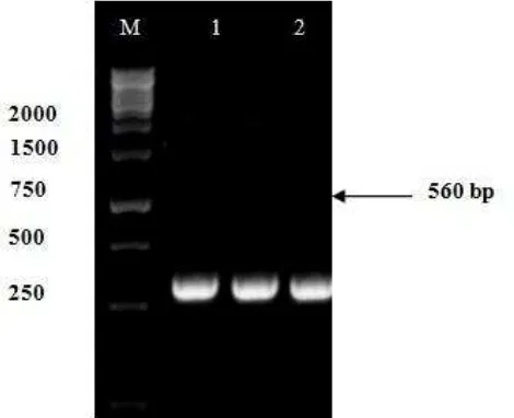
PCR amplification of 18S rDNA of Fusarium sp. isolates w1 and w2 with primers ITS5 (F: 5`-GGAAGTAAAAGTCGTAACAAGG -3`) and ITS4 (R: 5`-TCCTCCGCTTATTGATATGC- 3') produced about 560 bp of DNA fragments, it is corresponding to 18S rDNA (Figure 3). The fragments then sequenced to determine the species of fungus based on the similarity with other references of identified species.
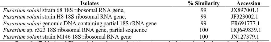
Based on the 18S rDNA analysis showed that Fusarium sp. isolates w1 and w2 have a close relationship with Fusarium solani. This can be seen in the phylogenetic tree shown in Figure 5. Fusarium sp. isolates w1 and w2 have 99% similarity with Fusarium solani strain 68 18S ribosomal RNA gene (Accession Number Gen Bank: JX897001.1), Fusarium solani strain H8 18S ribosomal RNA gene (Accesion Number Gen Bank: JF323002.1), and Fusarium solani genomic DNA containing partial 18S rRNA gene (Accession Number Gen Bank: FR691777.1). They have a larger similarity (100%) with Fusarium sp. r323 18S ribosomal RNA gene, partial sequence (Accession Number Gen Bank: HQ649839.1), and Fusarium solani strain M146 18S ribosomal RNA gene(Accession Number Gen Bank: JN127379.1) (Table 1).
Figure 3. PCR amplification of the ITS gene with primer ITS_5F and primer ITS_4R. M = marker 1 Kb
Journal of Biology, Agriculture and Healthcare ISSN 2224-3208 (Paper) ISSN 2225-093X (Online) Vol.3, No.17, 2013
www.iiste.org
100
Fus arium sp. isolat w1
Fus arium sp. isolat w2
Fus arium sp. r323
Fus arium solani strain 68
Fus arium solani strain M 146
Fus arium solani strain H8
Fus arium solani DNA containing
Fus arium sp. r104
Mel anelixia aff. Fuliginosa C respo 6005o
100 Mel anelixia Fuliginosa voucher Robertson 7140
100
Leptodiscella sp. FMR 10885
Lept odisc ella chlamydospora
0.05
Figure 4. Phylogenetic relationship constructed from ITS sequences of the characterized clone library of
Fusarium. Bootstrap value greater than 50% are shown at each node.
Table 1.comparisons of 18S rDNA gene similarity levels of Fusarium sp. isolates w1 and w2 with multiple sequences in GenBank using BLAST program
Isolates % Similarity Accession
Fusarium solani strain 68 18S ribosomal RNA gene, 99 JX897001.1
Fusarium solani strain H8 18S ribosomal RNA gene, 99 JF323002.1
Fusarium solani genomic DNA containing partial 18S rRNA gene 99 FR691777.1
Fusarium sp. r323 18S ribosomal RNA gene, partial sequence 100 HQ649839.1
Fusarium solani strain M146 18S ribosomal RNA gene 100 JN127379.1
From the Table 1 and Figure 4, it can be seen that Fusarium sp. isolates w1 and w2 are closely related to the Fusarium solani strain 68 18S ribosomal RNA gene, Fusarium solani strain H8 18S, Fusarium solani genomic DNA containing partial 18S rRNA gene, Fusarium sp. r323 18S ribosomal RNA gene, and Fusarium solani strain M146 18S ribosomal RNA gene. Ronquillo (2012) reported that the Fusarium solani strain H8 18S caused bud rot in the oil palm in Ecuador. Shahnazi et al. (2012) investigated that Fusarium solani strain H8 18S caused yellowing disease of black pepper (Piper nigrum L.) in Malaysia. Sarmiento-Ramirez et al. (2010) reported that Fusarium solani genomic DNA containing partial 18S rRNA gene was responsible for mass mortalities in nests of logger head sea turtle. Fusarium sp. r323 18S ribosomal RNA gene was associated with Roots of Halophytic and Non-halophytic Plant Species was reported by Macia-Vicente et al. (2012). In addition, Rosado-Rodriguez et al. (2011) reported that Fusarium solani strain M146 18S ribosomal RNA gene was associated with Leatherback Sea Turtle (Dermochelys coriacea) Nests in the Mayaguez-Anasco Bay Coast, Western Puerto Rico.
Fusarium solani is one of the most frequently isolated fungi from soil and plant material, where they act as decomposers, but they are also host-specific pathogens of a number of agriculturally important plants, including sweet potato, cucurbits, and pea. Moreover, they are increasingly associated with opportunistic infections of humans and other animals, causing systemic infections with a high mortality rate, as well as localized infections in the skin and other body parts (Zhanget al., 2006). Mycotoxin trichothecenes produced by Fusarium is very toxic for human (Miller and Trenholm, 1994). This toxin can cause cancer, hemorrhage, edema and immune deficiency (Alexoupolos et al., 1996). WHO (1979) reported that mycotoxins are hazardous to human and animal health.
4. Conclusion
Vol.3, No.17, 2013
University, Bali for financial support under research grant No. 05/biop-IV/2012.
References
Alexoupolos, C.J., C.W. Mims and M. Blackwell, 1996. Introductory Mycology.John Wiley and Sons.Inc. Singapore.
Atkins, S.D. and I.M. Clark, 2004. Fungal molecular diagnostics: a mini review, J. Appl. Genet., 45: 3-15. Chandran, M.R. and M.R. Kumar, 2012.Studies on cultural, morphological variability in isolates of Fusarium
solani(Mart.) Sacc., incitant of dry root-rot of Citrus. Current Biotica, 6: 152-162.
Felsenstein. J., 1985. Confidence limits on phylogenies: an approach using the bootstrap. Evolution, 39: 783–
791.
He, P.F., H. Ho, X. Wu, M.S. Hou, and Y.Q. He, 2012. Bipolaris cactivora causing fruit rot of dragon fruit imported from Vietnam. Plant Pathology & Quarantine, 2: 31-35.
Hawa, M.M, B. Salleh and Z. Latiffah, 2010. Characterization and intraspecific variation of Fusarium semitectum (Berkeley and Ravenel) associated with red-fleshed dragon fruit (Hylocereus polyrhizus [Weber] Britton and Rose) in Malaysia. African Journal of Biotechnology, 9: 273–284.
Iida, S., T. Imai, T. Oguri, K. Okuzumi, A. Yamanaka, M.L. Moretti-Branchini, K. Nishimura, and Y. Mikami, 2005.Genetic Diversity of the Internal Transcribed Spacer (ITS) and 5.8S sRNA Genes among The Clinical Isolates of Candida parapsilosis in Brazil and Japan.Jpn. J. Med., 46: 1333-137.
Kawuri, R., D.N. Suprapta, Y. Nitta, and T. Homma, 2012. Destructive Leaf Rot Disease Caused by Fusarium oxysporum on Aloe barbadensis Miller in Bali. Agricultural Science Research Journal, 2: 295–301. [cited: 2013 July 15]. Available online from http://www.resjournals.com/ARJ
Lin, C.C., W.B. Guo and S.F. Cai, 2006. Diseases of red dragon fruit in Taiwan. Good Year (Chinese), 56: 38–
42.
Macia-Vicente, J.G., V. Ferraro, S. Burruano and L.V. Lopez-Llorca, 2012. Fungal Assemblages Associated with Roots of Halophytic and Non-halophytic Plant Species Vary Differentially Along a Salinity Gradient. Microb. Ecol., 64: 668-679.
Madhukeshwara, S. S., 2000. Studies on variation and management of Fusarium wilt of igeonpea (Cajanus cajan). M.Sc., Thesis, UAS, GKVK, Bangalore pp: 85-94.
Masyahit, M., K. Sijam, Y. Awang, M. Ghazali and M. Satar, 2009. The First Report of the Occurrence of Anthracnose Disease Caused by Colletotrichum gloeosporioides (Penz.) Penz. & Sacc.on Dragon Fruit (Hylocereus spp.) in Peninsular Malaysia. American Journal of Applied Sciences, 6 : 902-912.
Mishra, P.K., R.T.V. Fox and A. Culham, 2003. Development of a PCR-based assay for rapid and reliable identification of pathogenic Fusaria. FEMS Microbiology Letters, 218: 329-332.
O’Donnell, K., D.A. Sutton, A. Fothergill, D. McCarthy, M.G. Rinaldi, M.E. Brandt, N. Zhang and D.M. Geiser, 2008. Molecular Phylogenetic Diversity, Multilocus Haplotype Nomenclature, and In Vitro Antifungal Resistance within the Fusarium solani Species Complex,J. Clin. Microbiol., 46 : 2477-2490.
Pit, J.I. and A.D. Hocking, 1997. Fungi and Food Spoiladge. 2nd Edition. Blackie Academic and Professional Press. Pp. 137-139.
Ronquillo, M.P., 2012. Etiology of Bud Rot in the Oil Palm in Ecuador. Crop Protection, 2.http://getentry.ddbj.nig.ac.jp/getentry/na/JX897001/?filetype =html.
Rosado-Rodriguez,G., 2011. Mycelial Fungal Diversity Associated with Leatherback Sea Turtle (Dermochelys coriacea) Nests in the Mayaguez-Anasco Bay Coast, Western Puerto Rico. Biology, http://getentry.ddbj.nig.ac.jp/getentry/na/JN127379/?filetype=html
Sarmiento-Ramirez, J.M., E. Abella, M.P. Martin, M.T. Telleria, L.F. Lopez-Jurado, A. Marco, and J. Dieguez-Uribeondo, 2010. Fusarium solani is responsible for mass mortalities in nests of loggerhead sea turtle, Caretta caretta, in Boavista, Cape Verde. FEMS Microbiol Lett. 312 : 192-200.
Shahnazi, S., S. Meon, G. Vadamalai, K. Ahmad and N. Nejat, 2012. Morphological and molecular characterization of Fusarium spp. associated with yellowing disease of black pepper (Piper nigrum L.) in Malaysia. J. Gen Plant Pathol.,78: 160-169.
Suga, H., T. Hasegawa, H. Mitsui, K. Kageyama and M. Hyakumachi, 2000. Phylogeneticanalysis of the phytopathogenic fungus Fusarium solani based on the rDNA-ITS region. Mycol. Res., 104: 1175-1183. Tamura, K., D. Peterson, N. Peterson, N. Stecher, M. Nei and S. Kumar, 2011. MEGA5: Molecular
Evolutionary Genetics Analysis Using Maximum Likelihood, Evolutionary Distance, and Maximum Parsimony Methods. Mol Biol Evo. [cited 2013 July 15]. Available online at: http://mbe.oxfordjournals.org/content/early/2011/08/17/.
Tarnowski, T. L. B., A.J. Palmateer and J.H. Crane, 2010. First Report of Fruit Rot on Hylocereusundatus Caused by in South Florida. The American Phytopathological Society, 94: 1605.2.
Journal of Biology, Agriculture and Healthcare ISSN 2224-3208 (Paper) ISSN 2225-093X (Online) Vol.3, No.17, 2013
www.iiste.org
Menyanthaceae using predicted secondary structure. Molecular Phylogenetics and Evolution, 49: 526–
537.
Wang, D.F., Q. Wei, R. Yang, W.J. Sang, G.Q. Fan and Y.L. Jin, 2007–Preliminary identification of disease of pitaya in Luodian Country. Journal of Mountain Agriculture and Biology (Chinese), 26: 267–270.
White, T.J., T. Bruns, Lee and S.J. Taylor, 1990. Amplification and direct sequencing of fungal ribosomal RNA genes for phylogenetics. In MA Innis, DH Gelfand, JJ Sninsky; T. J. White, (Eds). PCR Protocols: a Guide to Methods and Applications. Pp. 315-322. Academic Press, San Diego, CA.
World Health Organization (WHO). 1979. Mycotoxins, Environment, Health Criteria No. 11, Geneva.
Zainoldin, K.H. and A.S. Baba, 2009. The Effect of Hylocereus polyrhizus and Hylocereus undatuson Physicochemical, Proteolysis, and Antioxidant Activity in Yogurt. World Academy of Science, Engineering and Technology, 60: 361-366.
Zhang, N., K. O’Donnell, D.A. Sutton, F.A. Nalim, R.C. Summerbell, A.A. Padhye and D.M. Geiser, 2006. Members of the Fusarium solani Species Complex That Cause Infections in Both Humans and Plants Are Common in the Environment. J. Clin. Microbiol., 44: 2186-2190.
Vol.3, No.17, 2013
This academic article was published by The International Institute for Science, Technology
and Education (IISTE). The IISTE is a pioneer in the Open Access Publishing service based
in the U.S. and Europe. The aim of the institute is Accelerating Global Knowledge Sharing.
More information about the publisher can be found in the IISTE’s homepage:
http://www.iiste.org
CALL FOR JOURNAL PAPERS
The IISTE is currently hosting more than 30 peer-reviewed academic journals and
collaborating with academic institutions around the world. There’s
no deadline for
submission. Prospective authors of IISTE journals can find the submission instruction
on
the following
page:
http://www.iiste.org/journals/
The IISTE editorial team
promises to the review and publish all the qualified submissions in a fast manner. All the
journals articles are available online to the readers all over the world without financial, legal,
or technical barriers other than those inseparable from gaining access to the internet itself.
Printed version of the journals is also available upon request of readers and authors.
MORE RESOURCES
Book publication information:
http://www.iiste.org/book/
Recent conferences:
http://www.iiste.org/conference/
IISTE Knowledge Sharing Partners
Journal of Biology, Agriculture and Healthcare ISSN 2224-3208 (Paper) ISSN 2225-093X (Online) Vol.3, No.17, 2013
Journal of Biology, Agriculture and Healthcare ISSN 2224-3208 (Paper) ISSN 2225-093X (Online) Vol.3, No.17, 2013
Journal of Biology, Agriculture and Healthcare ISSN 2224-3208 (Paper) ISSN 2225-093X (Online) Vol.3, No.17, 2013