Journal
of
BiologV
Agriculture
and
Healthcare
l;-i.l :'
"*
"l-1,:l ,111r."r la-''""n' :^ -'.rrtrrt{ a ".i..; r;ri^. .
!. /+: 'l iri-- : l rrt; ); .;!r :
-About .Iournal of Biology, Agriculture and Healthcare
Journal of Biology, Agriculture and Healthcare is a peer reviewed joumal published by IISTE. The joumal publishes original papers at the forefront of Agriculture, Biology and Healthcare research. The journal is published in both printed and online versions.
Joumal of Biology, Agriculture and Healthcare is published by IISTE and follows a quarterly publication schedule. General inquiries and Paper zubmission: [email protected] or [email protected]
Index of this jorirnal
INDEX
COPER.NICUS iNrEf,LAftoi.at,ULRIcHSWEB
JournalTOCslr,.r@ ZeitschriFtenbi bliothek
#.s
i'=r'ced! wDf{dtsttknowledgements to the supports liom eo-hosdng uuiversities worldwide
Ilniversity ofNorth Carolina at Charlqtte, United States Califonia State University, United States
Tb
City University of New Yorlg United States Aristotle Universrty of Thessaloniki, Greece Ihiversiteit Leiden, Netherl andsVol 3, No
17
(2013)
Table
of
Contents
Artlcles
Effect of Regulated Deficit Irrigation on Growth and Yield of Sorghum
Adzemi Mat Arshad, Ibrqhim W, Wan Zaliha W S
Factors Influencing Initiation of Breast Feeding among Post-Partum Mothers
in a Teaching Hospital of Osun State, Nigeria.
B. L. Ajibade, Oladeji M. O, E.A. Oyedele, Amoo P.0, Makinde O.Y
Feed Resources and Seasonal Nutrient Corrposition of Predominant Forages
6r
Small Ruminant Production in Iwo Local Government Area of Osun state,Nigeria
Funmilayo BAMIGBOYE, Olantyi Babryemi, Adegboyega Adekoya
of a Designed Nursing Intewention protocol on Myocardial Infarction
Outcome at a selected University Hospital in Egypt
Abdelhameed Mahros Abdelhameed, Warda Youssef Mohamed, Yousia
Abd El-salam Seloma, Hanan El-sayed Zaghla
isloeic Attributes and Virulence Profile of Salmonella Tennessee from Infections associated with Peanut Butter National Outbreak
Chou H- Nguyen, Seongbeom Cho, Mahdi A. Saeed
Mechanisms of Rural Fishermen Towards Climate Change On
ofFish Caught in Asari-toru Local Govemment Area of Rivers State
futry
Unaeze, Adaba IbimStudies In Some Sweet Sorghum (Sorghum Bicolor. L- Moench)
L
fumi, M. Y. Yeye, M. F. Ishiyaku, I. S- Usmanof Household Characteristics on Child Mortality in Ghana Ofori Fosu, Ir. Peter Romeo Nyarko
of Badh2 Mutation Tlpe among Indonesian Fragrant Rice
Sasongko Hami Seno, I Made Artika, Waras Nurcholis, Tri Joko
Kurniawqn RTrijatmiko, Bambang Padmadi, Dewi Praptiwi,
ki
Ching, Zainal Alim Mas'udGlycosidically Bound Volatiles and Antimicrobial Activity
of
Var. Botqrtis, (Soultany Cultivar)
S. Hifuawy, Rabab M. Abdel Salam, MohamedA. Rabeh, A. Aboseada
of Root Knot Nematode Affecting Banana Crop by Using And Biological Products
Faji,
EI Hassan Mayad, Mohamed AlfulahI-and Measurement Adapted in Langowan, Minahasa, North
l-8
9-14
l5-24
25-35
3642
4348
49-5t
52-58
66-81
82-85 59-65
First Report on Fusarium solani, a Pathogenic Fungus Causing Stem Rot
Disease on Dragon Fruits (Hylocereus sp.) in Bali
Wiwik Susanah Rila, Daua Ngurah Suprapta, I Made Sudana, I Made
Dira Swantara
Hematological Indices of Clarias griepinus (Burchell, 1882) Fingerlings Fed
Diet Containing Graded Levels of Calabash (Lagenaria vulgaries) Seed Meal
Mamman,7., Ipinjolu, J.K, I, Magawata
Effect of Some Thermal Processing Techniques on the Proximate, Mineral and
Anti-Nutrient Compositions of Bambara Groundnut (Voandzeia subterranea, (L) Thour) Meal
Isila'venu J. O, Nwanbe R. N, Elechi F. N
Adaptation, Biomass and Ethanol Yields of Sweet Sorghum (Sorghum bicolor (L.) Moench) Varieties at Dryland Farming Areas of Jimbaran Bali, Indonesia
I
Gusti Ayu Mas Sri Agung, I Ketut Sardiana,I
l[/ayan Diara,I
Gusti Made Oka NurjayaAssistive Technology For Hearing and Speech Disorders Adedayo O. Olaosun, Olawale Ogundiran
Determinants of Output among Pig Farmers in Abia State, Nigeria
Kelechi lgwe, Amorachi lfekaonwu, Samson Amao, Clara lgwe
Donkey-Cart Transport, a Source of Livelihood for Farmers in the Kassena
Nankana Municipality in Upper East Region of Ghana
Maurice M. Braimah, Issahaht Abdul-Rohaman, Daniel
Opporg-Sel<yere
Effect of Partial Rootzone Drying Technique on Growth Performance
of
Sorghum
A&emi Mat Arshad, IbrahimW, Wan Zaliha W S
Variations in Phosphatase Activity of Crude Oil and Used Crankase Oil
Polluted Agricultural Soil.
Reginald C. Ohin, Agha, N. C., Nwachuh,w, N.
Immune Response to Hepatitis B Virus Vaccine AYAM MOHAMMED SALIH ALI
93-99
100-104
r05-109
I
l0-l
15tr6-t20
t2t-126
127 -136
r37-t42
t43-r49
Journal of Biology, Agriculture and Healthcare www.iiste.org
ISSN 2224-3208 (Paper) ISSN 2225-093X (Online) Vol.3, No.17, 2013
93
First Report on Fusarium solani, a Pathogenic Fungus Causing
Stem Rot Disease on Dragon Fruits (Hylocereus sp.) in Bali
Wiwik Susanah Rita1, Dewa Ngurah Suprapta2*, I Made Sudana2, I Made Dira Swantara3
1.Doctorate Program in Agricultural Science, School for Postgraduate Udayana University. 2.Laboratory of Biopesticide Faculty of Agriculture, Udayana University, Jl. PB. Sudirman Denpasar Bali
Indonesia.
3.Laboratory of Chemistry, Faculty of Natural Science and Mathematic, Udayana University *Email of corresponding author : [email protected]
Abstract
In recent years, dragon fruit crop (Hylocereus spp.) has become increasingly important in Bali Indonesia due to
its high nutrient content and healing properties. However, the dragon fruit was reported to be seriously infected with several complex diseases caused by fungi and causing serious losses to the farmers. The study on morphological and molecular characterization the fungal pathogen was conducted to confirm the species of the fungi. Koch Postulate was applied to confirm the causal agent of the disease. There were two isolates of fungi
isolated from the stems of diseased plants, namely isolate w1 (from stem of H. undatus) and isolate w2 (from
stem of H. polyrhizus). Based on macroscopic and microscopic characteristics, and analysis of 18S rDNA, both
of them were identified as Fusarium solani. This is the first report on the F. solani the cause of stem rot disease
on dragon fruits in Bali.
Keywords : stem rot disease, dragon fruit, Fusarium solani
1. Introduction
Dragon Fruits (Hylocereus spp.), which are also known as pitaya, are the fruits of cactus species, especially
of the genus Hylocereus. There are three species which have high commercial valuable fruits are the species of
Hylocereus undatus, (red rind, white flesh), Hylocereus polyrhizus (red rind, red flesh), and Hylocereus costaricensis (red rind, super red flesh). Hylocereus spp. is grown commercially in Vietnam, Spain, Malaysia, Japan, Mexico and other tropical and subtropical areas because of its high nutrient content and healing properties. In recent years, this fruit has become increasingly important in Bali Indonesia. The dragon fruit is rich in vitamin, it helps the digestive process due to its fiber, prevent colon cancer and diabetes, neutralize toxic
substances such as heavy metals, and helps to reduce cholesterol levels and high blood pressure (He et al., 2012;
Zainoldin and Baba, 2009). Recently, dragon fruit has been reported to be seriously infected with several complex diseases caused by fungi and causing serious losses to farmers. Several dragon fruit plants grown in Sobangan Village, Bali Indonesia showed severe symptom of stem rot disease. The disease caused significant
yield losses. Isolation of the fungi associated with the diseased-plants showed that Fusarium sp. was the most
frequent found on the stems showing brown rot symptom.
Various diseases caused by fungi have been reported on dragon fruit in tropical and subtropical countries,
such as fruit rot (Bipolaris cactivora) (Tarnowski et al., 2010; He et al. 2012), stem rot (Fusarium semitectum,
Fusarium oxysporium, Fusarium moniliforme) (Hawa et al., 2010), anthracnose (Colletotrichum gloeosporioides) (Masyahit et al., 2009), brown spot (Botryodiplodia sp.), basal rot (Pythium sp.) (Lin et al.,
2006), wilt (Fusarium oxysporium), stem blight (Diplodia sp., Ascochyta sp., and Phoma sp.), black spot
(Alternaria sp.), speck blight (Nectriella sp.), (Wang et al.,2007), and stem lesion (Septogloeum sp.) (Zheng et
al., 2009).
In order to control the disease, it is necessary to identify the causal agent of the disease. The fungal pathogen can be identified based on cultural and morphological characters. However it could be highly variable depending on the media and cultural conditions that could be the problems in fungal identification. In recent years, the increasing use of molecular methods in fungal identification has emerged as a possible answer to the
problems associated with the existing phenotypic identification systems (Mishra et al., 2003). One of the
molecular approaches to fungal identification is based on Polymerase Chain Reaction (PCR).
Journal of Biology, Agriculture and Healthcare www.iiste.org
ISSN 2224-3208 (Paper) ISSN 2225-093X (Online) Vol.3, No.17, 2013
94
applications, most researchers use sequence alignments that are based on nucleotide similarity (Tippery and Les, 2008).
Suga et al. (2000) has investigated phylogenetic relationships of Fusarium solani using sequences from the
rDNA-ITS region. Mishra et al. (2003) has developed a fluorescent-based polymerase chain reaction in ITS
region to identify five toxigenic and pathogenic Fusarium species. Abd-Elsalam et al. (2003) have developed
two taxon-selective primers for quick identification of the Fusarium genus. These primers, f and
ITS-Fu-r weITS-Fu-re designed by compaITS-Fu-ring the aligned sequences of inteITS-Fu-rnal tITS-Fu-ranscITS-Fu-ribed spaceITS-Fu-r ITS-Fu-regions (ITS) of a ITS-Fu-range of
Fusarium species. Zhang et al. (2006) and O'Donnell et al. (2008) have studied phylogeni of Fusarium solani
Species Complex (FSSC) that cause infection in both humans and plants based on three genes of the ribosomal
DNA. The ITS region including 5.8S rDNA sequence of 58 isolates Candida parapsilosis in Brazil and Japan
was analyzed by Iida et al. (2005).
This paper reports on identification of fungal pathogen causing stem rot disease from dragon fruits planted in Bali based on morphological and molecular methods using sequences from rDNA-ITS region.
2. Materials and Methods
2.1. Fungal Pathogen Isolation and Virulence Test
Fungi were isolated from the diseased stem of H. undatus and H. polyrhizus from dragon fruit’s orchard in
Sobangan village, Mengwi Bali. Surface sterilization was carried out by cleaning the symptom margins with 70% ethanol and cut into small blocks (ca 1.5 x 1.5 x 1.5 cm), soaked in 1% sodium hypochlorite (NaOCI) for 3 min, and rinsed in several changes of sterile distilled water (each 1 min). All sterilized samples were placed onto Potato Dextrose Agar (PDA) and incubated at 25 ± 2°C for 7 days. Mycelium growing after 3 days of incubation was transferred to a new PDA to obtain pure fungal cultures. The pure culture was inoculated on healthy dragon fruit stems to confirm the similarity of symptoms in the field. For this purpose the Koch's postulates test was carried out. Dragon fruit stems (30 days in age) were injured with a sterile needle and inoculated by spraying
with a suspension of spores of 1.5x10⁶ spores/mL. Three plants were inoculated with a fungal isolate. For
control, dragon fruit stems were pierced with a sterile needle and sprayed with sterile water. Symptoms were observed for a week after inoculation. The symptoms were compared with the symptoms of the disease in the field. After that, the isolation of pathogenic fungi on infected stem was performed again. Pure cultures of pathogenic fungi were inoculated again on dragon fruit plants and the same inoculation procedure was done to obtain the similarity of symptoms. Fungal isolates obtained can be regarded as the cause of the disease and were used for further identification.
2.2. Morphological Characterization
Characterization of the main fungal pathogens was carried out macroscopically, by observing the fungal colony color, colony reverse color, no lines or concentric radier, issued exudates or not, media pigmentation, colony surface, and how the growth of fungi (fast or slow). Microscopic identification was also carried out by observing the shape of hyphae or spores under a microscope, and then the results were confirmed using fungal identification book (Pit and Hocking, 1997).
2.3. DNA Extraction
Fusarium sp. isolates w1 and w2 were grown on potato dextrose agar (PDA) medium for 3 days at room temperature. The mycelium grown was harvested and grown to a fine powder in a sterile mortar with liquid nitrogen. DNA was extracted by using PhythopureTM DNA Extraction Kit (GE Healthcare, UK) according to manufacturer’s instructions.
2.4. Molecular identification and phylogenetic relationships of fungal pathogen
Identification of fungal isolates was performed based on molecular genetic analysis using the internal transcribed spacer region (ITS). PCR amplification used ITS 5 F: 5`- GGAAGTAAAAGTCGTAACAAGG - 3` and ITS 4
R: 5 `- TCCTCCGCTTATTGATATGC - 3 '(White et al. 1990). Amplification was performed on a volume of 25
L with the reaction mixture: 10 µL nuclease free water, 12.5 µL Go taq green master mix
TM
, ITS5 and ITS4 each 0.5 µL, 0.5 µL DMSO, and 1 µL of DNA template. PCR amplification for regional ITS consists of: pre denaturation 95 ºC for 90 seconds, followed by 95 ºC for 30 seconds with 35 cycles, annealing 55 ºC in 30 seconds, extension 72 ºC in 90 seconds, and final extension 72 ºC for 5 minutes. The product was purified and then sequenced. The nitrogen base sequence was analyzed using automated DNA sequencer (ABI PRISM 3130 Genetic Analyzer) (Applied Biosystems).
Sequencing raw data were trimmed and assembled using ChromasPro program version 1.5. The assembled data were BLASTED with genomic data that has been registered in NCBI / National Center for Biotechnology Information (http://www.ncbi.nlm.nih.gov/BLAST/). Some data sequence which is a result of blast nearest species and is a type strains of each species were taken from the data in the NCBI gene bank. Then the data were
analyzed again with the sequence aligment using MEGA version5.0 program (Tamura et al. 2011) and boostrap
Journal of Biology, Agriculture and Healthcare www.iiste.org
ISSN 2224-3208 (Paper) ISSN 2225-093X (Online) Vol.3, No.17, 2013
95
3. Results and Discussion 3.1. Fungal Isolates
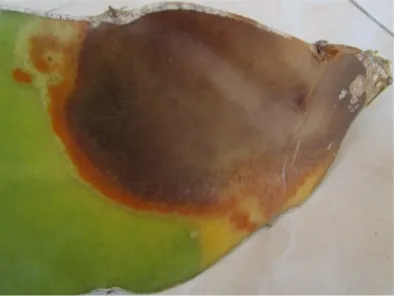
[image:7.595.202.403.192.340.2]There are 10 fungal isolates were obtained from isolation of fungi associated with the stem disease of dragon fruits grown in Sobangan Village, Bali Indonesia. Based on the Koch's postulates test, there were two isolates of Fusarium sp. namely isolate w1 (from H. undatus) and isolate w2 (from H. polyrhizus) caused the stem rot disease with the symptom similar to the symptom occurred in the field. The symptom appeared on stem as dark brown stem rot as shown in Figure 1.
Figure 1. Symptoms of the stem rot diseases on Hylocereus sp. under field conditions.
3.2. Macroscopic Characteristic
Fusarium sp. isolate w1 grown on PDA produced abundant mycelium (sometimes in aerial, depends on cultural condition), white colony appearance, cream colony reverse, yellow to brown pigmentations, and fast growing
(3.75-4.45 cm in 3 days) whereas Fusarium sp. isolate w2 produced abundant-powdery mycelium (sometimes in
aerial), white colony appearance, yellow colony reverse, yellow pigmentations, and fast growing (4.35-5.25 cm in 3 days).
In a similar study, Chandran and Kumar (2012) studied for cultural, morphological variability in 13 isolates of Fusarium solani (Mart.) Sacc., incitant of dry root-rot of citrus, they reported that F. solani isolates were characterized as fast growing, moderately growing, and slow growing. The five isolates produced pale pink to dusky red color pigmentation in PDA medium and potato dextrose broth culture. The remaining all isolates produced pale yellow to dark yellow pigmentation. Most of the isolates produced profuse sporulation, whereas others produced moderate sporulation. Madhukeshwara (2000) has studied cultural variability among six isolates of F. udum causing wilt of pigeon pea. All the isolates varied with each other in terms of growth, mycelium, pigmentation, and sporulation. Most of the isolates produced cottony white raised mycelium, pale yellow to dusky red color pigmentation and moderate to profuse sporulation on PDA medium.
3.3. Microscopic Characteristics
Microscopic characters such as size, shape, septation, and color of conidia were studied using PDA medium.
Fusarium sp. isolate w1 had longer macroconidia (1-4 septates; 18.4-31.7 µm in length), curved in shaped with
hyaline in color, whereas Fusarium sp. isolate w2 had shorter macroconidia (1-3 septates; 15.3-24.8 µm in
length), curved in shaped with hyaline in color. Fusarium sp. isolate w1 produced less microconidia while
Fusarium sp. isolate w2 produces abundant microconidia (1 septate; 5.7- 8.5 µm in length), round to oval in shaped. Furthermore both the fungi had the septate hyphae and clamydospores in pairs with hyaline in color and located in the middle of hyphae as presented in Figure 2.
Pitt and Hocking (1997) reported that the main characters used to distinguish species of Fusarium are the
size and shape of the macroconidia; the presence or absence of microconidia; and the presence of
chlamydospora. Kawuri et al. (2012) reported that Fusarium oxysporum had macroconidia curved in shaped with
four septates and foot cell, 31μm length and smooth surface, whereas microconidia has rough surface at 4.6 μm
length. Chandran and Kumar (2012) reported that the number of septa in macro conidia and micro conidia of
Fusarium solani (Mart.) Sacc. are 3-5 and 0-1 respectively and the color is hyaline. The shape of macro conidia is sickle shaped with blunt ends and micro conidia is round to oval shaped. The chlamydospores located in middle of hyphae (intercalary), on tip of the hyphae (terminal) and some chlamydospores were seen in middle of macro conidia.
Journal of Biology, Agriculture and Healthcare www.iiste.org
ISSN 2224-3208 (Paper) ISSN 2225-093X (Online) Vol.3, No.17, 2013
96
Figure 2. Microscopic characteristics of Fusarium sp. isolate w1 (A) macro conidia (B) septated hypae (C)
Chlamydospore. Bars = 10 m.
3.4. Molecular Characteristics of the Pathogenic Fungus
PCR amplification of 18S rDNA of Fusarium sp. isolates w1 and w2 with primers ITS5 (F:
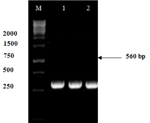
5`-GGAAGTAAAAGTCGTAACAAGG -3`) and ITS4 (R: 5`-TCCTCCGCTTATTGATATGC- 3') produced about 560 bp of DNA fragments, it is corresponding to 18S rDNA (Figure 3). The fragments then sequenced to determine the species of fungus based on the similarity with other references of identified species.
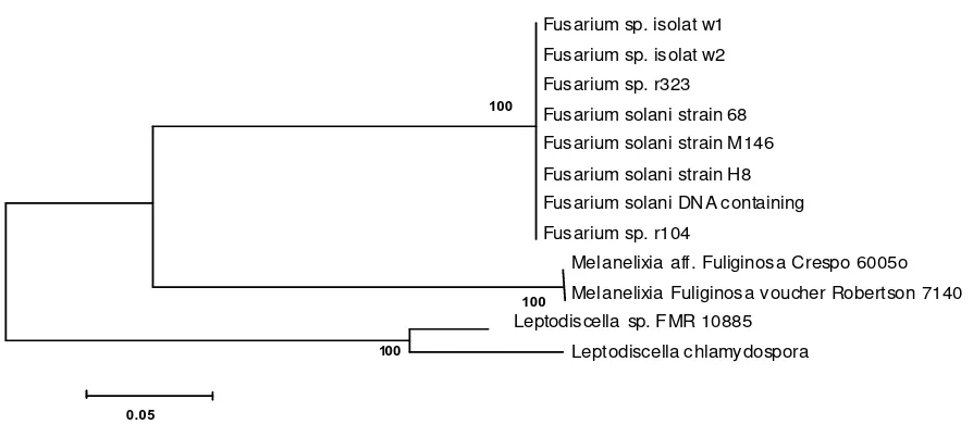
Based on the 18S rDNA analysis showed that Fusarium sp. isolates w1 and w2 have a close relationship
with Fusarium solani. This can be seen in the phylogenetic tree shown in Figure 5. Fusarium sp. isolates w1 and
w2 have 99% similarity with Fusarium solani strain 68 18S ribosomal RNA gene (Accession Number Gen
Bank: JX897001.1), Fusarium solani strain H8 18S ribosomal RNA gene (Accesion Number Gen Bank:
JF323002.1), and Fusarium solani genomic DNA containing partial 18S rRNA gene (Accession Number Gen
Bank: FR691777.1). They have a larger similarity (100%) with Fusarium sp. r323 18S ribosomal RNA gene,
partial sequence (Accession Number Gen Bank: HQ649839.1), and Fusarium solani strain M146 18S ribosomal
[image:8.595.84.521.87.255.2]RNA gene(Accession Number Gen Bank: JN127379.1) (Table 1).
Figure 3. PCR amplification of the ITS gene with primer ITS_5F and primer ITS_4R. M = marker 1 Kb
ladder (Fermentas), 1 = PCR product of Fusarium sp. isolate w2, and 2 = PCR product of Fusarium sp.
[image:8.595.177.426.461.664.2]Journal of Biology, Agriculture and Healthcare www.iiste.org
ISSN 2224-3208 (Paper) ISSN 2225-093X (Online) Vol.3, No.17, 2013
97
Figure 4. Phylogenetic relationship constructed from ITS sequences of the characterized clone library of
Fusarium. Bootstrap value greater than 50% are shown at each node.
Table 1.comparisons of 18S rDNA gene similarity levels of Fusarium sp. isolates w1 and w2 with multiple
sequences in GenBank using BLAST program
Isolates % Similarity Accession
Fusarium solani strain 68 18S ribosomal RNA gene, 99 JX897001.1
Fusarium solani strain H8 18S ribosomal RNA gene, 99 JF323002.1
Fusarium solani genomic DNA containing partial 18S rRNA gene 99 FR691777.1
Fusarium sp. r323 18S ribosomal RNA gene, partial sequence 100 HQ649839.1
Fusarium solani strain M146 18S ribosomal RNA gene 100 JN127379.1
From the Table 1 and Figure 4, it can be seen that Fusarium sp. isolates w1 and w2 are closely related to
the Fusarium solani strain 68 18S ribosomal RNA gene, Fusarium solani strain H8 18S, Fusarium solani
genomic DNA containing partial 18S rRNA gene, Fusarium sp. r323 18S ribosomal RNA gene, and Fusarium
solani strain M146 18S ribosomal RNA gene. Ronquillo (2012) reported that the Fusarium solani strain H8 18S
caused bud rot in the oil palm in Ecuador. Shahnazi et al. (2012) investigated that Fusarium solani strain H8 18S
caused yellowing disease of black pepper (Piper nigrum L.) in Malaysia. Sarmiento-Ramirez et al. (2010)
reported that Fusarium solani genomic DNA containing partial 18S rRNA gene was responsible for mass
mortalities in nests of logger head sea turtle. Fusarium sp. r323 18S ribosomal RNA gene was associated with
Roots of Halophytic and Non-halophytic Plant Species was reported by Macia-Vicente et al. (2012). In addition,
Rosado-Rodriguez et al. (2011) reported that Fusarium solani strain M146 18S ribosomal RNA gene was
associated with Leatherback Sea Turtle (Dermochelys coriacea) Nests in the Mayaguez-Anasco Bay Coast,
Western Puerto Rico.
Fusarium solani is one of the most frequently isolated fungi from soil and plant material, where they act as decomposers, but they are also host-specific pathogens of a number of agriculturally important plants, including sweet potato, cucurbits, and pea. Moreover, they are increasingly associated with opportunistic infections of humans and other animals, causing systemic infections with a high mortality rate, as well as localized infections
in the skin and other body parts (Zhanget al., 2006). Mycotoxin trichothecenes produced by Fusarium is very
toxic for human (Miller and Trenholm, 1994). This toxin can cause cancer, hemorrhage, edema and immune
deficiency (Alexoupolos et al., 1996). WHO (1979) reported that mycotoxins are hazardous to human and
animal health.
4. Conclusion
Based on the results of present study, it can be concluded that the causal agent of the stem rot disease on dragon
fruits (Hylocereus sp.) in Bali is identified as Fusarium solani. Hard efforts must be done to control the disease
in order to reduce the losses of dragon fruit production and the risk of mycotoxins contamination which are
probably produced by Fusarium solani.
Acknowledgement
Authors wish to express their appreciation to the Laboratory of Biopesticide, Faculty of Agriculture Udayana
100
100
100
Fusarium sp. isolat w1
Fusarium sp. isolat w2
Fusarium sp. r323
Fusarium solani strain 68
Fusarium solani strain M146
Fusarium solani strain H8
Fusarium solani DNA containing
Fusarium sp. r104
Mel anelixia aff. Fuliginosa Crespo 6005o
Mel anelixia Fuliginosa voucher Robertson 7140
Leptodiscella sp. FMR 10885
Leptodiscella chlamydospora
[image:9.595.71.518.89.284.2]Journal of Biology, Agriculture and Healthcare www.iiste.org
ISSN 2224-3208 (Paper) ISSN 2225-093X (Online) Vol.3, No.17, 2013
98
University, Bali for financial support under research grant No. 05/biop-IV/2012.
References
Alexoupolos, C.J., C.W. Mims and M. Blackwell, 1996. Introductory Mycology.John Wiley and Sons.Inc. Singapore.
Atkins, S.D. and I.M. Clark, 2004. Fungal molecular diagnostics: a mini review, J. Appl. Genet., 45: 3-15.
Chandran, M.R. and M.R. Kumar, 2012.Studies on cultural, morphological variability in isolates of Fusarium
solani (Mart.) Sacc., incitant of dry root-rot of Citrus. Current Biotica, 6: 152-162.
Felsenstein. J., 1985. Confidence limits on phylogenies: an approach using the bootstrap. Evolution, 39: 783–
791.
He, P.F., H. Ho, X. Wu, M.S. Hou, and Y.Q. He, 2012. Bipolaris cactivora causing fruit rot of dragon fruit
imported from Vietnam. Plant Pathology & Quarantine, 2: 31-35.
Hawa, M.M, B. Salleh and Z. Latiffah, 2010. Characterization and intraspecific variation of Fusarium
semitectum (Berkeley and Ravenel) associated with red-fleshed dragon fruit (Hylocereus polyrhizus
[Weber] Britton and Rose) in Malaysia. African Journal of Biotechnology,9: 273–284.
Iida, S., T. Imai, T. Oguri, K. Okuzumi, A. Yamanaka, M.L. Moretti-Branchini, K. Nishimura, and Y. Mikami, 2005.Genetic Diversity of the Internal Transcribed Spacer (ITS) and 5.8S sRNA Genes among The
Clinical Isolates of Candida parapsilosis in Brazil and Japan.Jpn. J. Med., 46: 1333-137.
Kawuri, R., D.N. Suprapta, Y. Nitta, and T. Homma, 2012. Destructive Leaf Rot Disease Caused by Fusarium
oxysporum on Aloe barbadensis Miller in Bali. Agricultural Science Research Journal, 2: 295–301. [cited: 2013 July 15]. Available online from http://www.resjournals.com/ARJ
Lin, C.C., W.B. Guo and S.F. Cai, 2006. Diseases of red dragon fruit in Taiwan. Good Year(Chinese), 56: 38–
42.
Macia-Vicente, J.G., V. Ferraro, S. Burruano and L.V. Lopez-Llorca, 2012. Fungal Assemblages Associated with Roots of Halophytic and Non-halophytic Plant Species Vary Differentially Along a Salinity Gradient. Microb. Ecol., 64: 668-679.
Madhukeshwara, S. S., 2000. Studies on variation and management of Fusarium wilt of igeonpea (Cajanus
cajan). M.Sc., Thesis, UAS, GKVK, Bangalore pp: 85-94.
Masyahit, M., K. Sijam, Y. Awang, M. Ghazali and M. Satar, 2009. The First Report of the Occurrence of
Anthracnose Disease Caused by Colletotrichum gloeosporioides (Penz.) Penz. & Sacc.on Dragon Fruit
(Hylocereus spp.) in Peninsular Malaysia. American Journal of Applied Sciences, 6 : 902-912.
Mishra, P.K., R.T.V. Fox and A. Culham, 2003. Development of a PCR-based assay for rapid and reliable identification of pathogenic Fusaria. FEMS Microbiology Letters, 218: 329-332.
O’Donnell, K., D.A. Sutton, A. Fothergill, D. McCarthy, M.G. Rinaldi, M.E. Brandt, N. Zhang and D.M. Geiser,
2008. Molecular Phylogenetic Diversity, Multilocus Haplotype Nomenclature, and In Vitro Antifungal
Resistance within the Fusarium solani Species Complex, J. Clin. Microbiol., 46 : 2477-2490.
Pit, J.I. and A.D. Hocking, 1997. Fungi and Food Spoiladge. 2nd Edition. Blackie Academic and Professional
Press. Pp. 137-139.
Ronquillo, M.P., 2012. Etiology of Bud Rot in the Oil Palm in Ecuador. Crop Protection,
2.http://getentry.ddbj.nig.ac.jp/getentry/na/JX897001/?filetype =html.
Rosado-Rodriguez,G., 2011. Mycelial Fungal Diversity Associated with Leatherback Sea Turtle (Dermochelys
coriacea) Nests in the Mayaguez-Anasco Bay Coast, Western Puerto Rico. Biology, http://getentry.ddbj.nig.ac.jp/getentry/na/JN127379/?filetype=html
Sarmiento-Ramirez, J.M., E. Abella, M.P. Martin, M.T. Telleria, L.F. Lopez-Jurado, A. Marco, and J.
Dieguez-Uribeondo, 2010. Fusarium solani is responsible for mass mortalities in nests of loggerhead sea turtle,
Caretta caretta, in Boavista, Cape Verde. FEMS Microbiol Lett. 312 : 192-200.
Shahnazi, S., S. Meon, G. Vadamalai, K. Ahmad and N. Nejat, 2012. Morphological and molecular
characterization of Fusarium spp. associated with yellowing disease of black pepper (Piper nigrum L.) in
Malaysia. J. Gen Plant Pathol.,78: 160-169.
Suga, H., T. Hasegawa, H. Mitsui, K. Kageyama and M. Hyakumachi, 2000. Phylogeneticanalysis of the
phytopathogenic fungus Fusarium solani based on the rDNA-ITS region.Mycol. Res., 104: 1175-1183.
Tamura, K., D. Peterson, N. Peterson, N. Stecher, M. Nei and S. Kumar, 2011. MEGA5: Molecular Evolutionary Genetics Analysis Using Maximum Likelihood, Evolutionary Distance, and Maximum
Parsimony Methods. Mol Biol Evo. [cited 2013 July 15]. Available online at:
http://mbe.oxfordjournals.org/content/early/2011/08/17/.
Tarnowski, T. L. B., A.J. Palmateer and J.H. Crane, 2010. First Report of Fruit Rot on Hylocereusundatus
Caused by in South Florida. The American Phytopathological Society, 94: 1605.2.
Journal of Biology, Agriculture and Healthcare www.iiste.org
ISSN 2224-3208 (Paper) ISSN 2225-093X (Online) Vol.3, No.17, 2013
99
Menyanthaceae using predicted secondary structure. Molecular Phylogenetics and Evolution, 49: 526– 537.
Wang, D.F., Q. Wei, R. Yang, W.J. Sang, G.Q. Fan and Y.L. Jin, 2007 – Preliminary identification of disease of pitaya in Luodian Country. Journal of Mountain Agriculture and Biology (Chinese), 26: 267–270. White, T.J., T. Bruns, Lee and S.J. Taylor, 1990. Amplification and direct sequencing of fungal ribosomal RNA
genes for phylogenetics. In MA Innis, DH Gelfand, JJ Sninsky; T. J. White, (Eds). PCR Protocols: a
Guide to Methods and Applications. Pp. 315-322. Academic Press, San Diego, CA.
World Health Organization (WHO). 1979. Mycotoxins, Environment, Health Criteria No. 11, Geneva.
Zainoldin, K.H. and A.S. Baba, 2009. The Effect of Hylocereus polyrhizus and Hylocereus undatuson
Physicochemical, Proteolysis, and Antioxidant Activity in Yogurt. World Academy of Science, Engineering and Technology, 60: 361-366.
Zhang, N., K. O’Donnell, D.A. Sutton, F.A. Nalim, R.C. Summerbell, A.A. Padhye and D.M. Geiser, 2006.
Members of the Fusarium solani Species Complex That Cause Infections in Both Humans and Plants Are
Common in the Environment. J. Clin. Microbiol., 44: 2186-2190.
Zheng, W., B. Wang and Y.Q. Cai, 2009. Inhibitory test for Septogloeum sp. causing stem lesion of pitaya with
This academic article was published by The International Institute for Science,
Technology and Education (IISTE). The IISTE is a pioneer in the Open Access
Publishing service based in the U.S. and Europe. The aim of the institute is
Accelerating Global Knowledge Sharing.
More information about the publisher can be found in the
IISTE’s
homepage:
http://www.iiste.org
CALL FOR JOURNAL PAPERS
The IISTE is currently hosting more than 30 peer-reviewed academic journals and
collaborating with academic institutions around the world.
There’s no deadline for
submission.
Prospective authors of IISTE journals can find the submission
instruction on the following page:
http://www.iiste.org/journals/
The IISTE
editorial team promises to the review and publish all the qualified submissions in a
fast
manner. All the journals articles are available online to the readers all over the
world without financial, legal, or technical barriers other than those inseparable from
gaining access to the internet itself. Printed version of the journals is also available
upon request of readers and authors.
MORE RESOURCES
Book publication information:
http://www.iiste.org/book/
Recent conferences:
http://www.iiste.org/conference/
IISTE Knowledge Sharing Partners
EBSCO, Index Copernicus, Ulrich's Periodicals Directory, JournalTOCS, PKP Open
Archives
Harvester,
Bielefeld
Academic
Search
Engine,
Elektronische