CD4, CD8 and MHC Class I Expression in
Epstein-Barr Virus-Associated
Nasopharyngeal Carcinoma:
An Immunohistochemical Study
ABSTRACT
Aim: The exact immunopathogenesis of Epstein-Barr virus (EBV)-associated nasopharyngeal carcinoma (NPC) remains unclear. The aim of the present study was to assess the expression of CD4, CD8, and MHC class I molecules in NPC.
Method: Biopsies were obtained from patients with NPC as well as the Epstein Barr virus (EBV)-seronegative patients as a control.
Nasopharyngeal carcinoma patients were classiied using the World Health Organization (WHO) pathological assessment and clinical stag -ing of NPC. The expression of CD4, CD8, and MHC class I in the biop-sies were assessed immunohistochemically.
Result: The results showed that the number of CD4 positive, CD8 pos-itive, and MHC class I positive cells in NPC patients were higher than those in EBV-negative subjects (p<0.05). The number of these positive
cells in NPC patients with WHO Type II or early clinical stage was not signiicantly differences with those with WHO Type III or late clinical
stage, respectively (p>0.05). No statistical differences between the number of CD4 positive and CD8 positive cells in NPC patients could be found (p>0.05).
Conclusion: The results of the present study suggest, therefore, that the expression of CD4, CD8 and MHC class I molecules may not be
as-sociated with the pathologic classiication and clinical staging of NPC
and that the CD4:CD8 ratio in nasopharyngeal carcinoma may indicate
decreased functions of these iniltrating T cell subsets.
Key words: CD4; CD8; MHC class I; NPC
1Sebelas Maret University, Faculty of
Medi-cine, Department of Pathology, Surakarta, Indonesia
2Gadjah Mada University, Faculty of
Medi-cine, Department of Histology and Cell Biology, Yogyakarta, Indonesia
3AIMST University, Faculty of Dentistry,
Semeling, Bedong, Kedah Darul Aman, Malaysia
Eur J Gen Med 2010;7(3):277-281
Received: 27.07.2009
Accepted: 18.11.2009
Correspondence: Wihas Sosroseno, Faculty of Dentistry, AIMST University, Semeling, 08100 Bedong, Kedah Darul Aman, Malaysia
Fax: 60 4 4422887;
E-mail: [email protected]
Dyah R. Budiani
1,
Soia M. Haryana
2,
Marsetyawan HNE Soesatyo
2, Wihaskoro Sosroseno
3INTRODUCTION
Nasopharyngeal carcinoma (NPC) is a tumor of epider
-moid origin and prevalent in several regions around the world. Based on the degree of differentiation, the World Health Organization (WHO) classiies NPC into keratin
-izing squamous cell carcinoma (WHO Type I) and nonke
-ratinizing carcinoma which is further subdivided into the differentiated subtype (WHO Type II) and undifferenti
-ated subtype (WHO Type III). The association between Epstein-Barr virus (EBV) and NPC is well known as shown by the fact that the EBV genome was found in the NPC speciments (1,2). However, it would appear that EBV is much more strongly associated with the undifferentiated NPC as compared with other NPC subtypes (3). EBV is a member of the herpesvirus family and primarily infects and replicates in the stratiied squamous epithelium of oropharynx (1,2).
The precise pathogenesis by which EBV induces the de
-velopment of NPC remains to be further elucidated. Both CD4+ and CD8+T cells have been shown to iniltrate in the stroma of NPC (4,5). Altered expression of major histocompatibility complex (MHC) class I has also been reported (5). However, despite abundant iniltrating T cells in the stroma of NPC, the development of tumor re
-mains progressive, suggesting that the immune responses against the cancerous cells may be down-regulated, per
-haps by cancer-derived immunosuppressive cytokines. Indeed, a previous study showed that an increased pro
-duction of interleukin-10 (IL-10) in the patients with NPC-WHO type III or clinical late stage (6), suggesting that this cytokine may inhibit the functions of iniltrating T cell subsets in NPC tissues. Therefore, the aim of the present study was to immunohistochemically determine the ex
-pression of CD4, CD8 and MHC class I in NPC tissues from Indonesian patients.
MATERIALS AND METHODS
NPC biopsies were obtained from 8 patients with nonke
-Budiani et al.
acted with the streptavidin-peroxidase (Lipshaw) for 30 minutes, visualized using a 3-3’-diaminobenzidine tetra
-hydrochloride solution (DAB; Lipshaw) for 10-20 minutes and subsequently counterstained with hematoxylin. Cells with positive staining per mm2 were microscopi
-cally counted.
The data was statistically determined by a one way analysis of variance followed by Fischer’s least squared differences using a statistical package (SPSS Inc., Chicago).
RESULTS
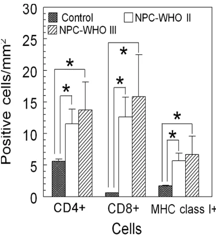
The positive staining for CD4+ and MHC class I+ cells were scattered in the stroma of NPC tissues (Figure 1). The distribution of these positive cells in NPC tissues with WHO II classiication was almost similar with that in NPC tissues with WHO III. Statistically, CD4+, CD8+ and MHC class I+ cells in cancerous tissues from NPC pa
-tients were higher than those from the healthy subject
(p<0.05) (Figure 2 and 3). No statistically differences in the number of positive cells between WHO II and WHO III of NPC could be found (p>0.05) (Figure 1 and 2). When NPC patients were divided into clinical stages, the num
-ber of positive cells between early and late stage were not also signiicantly different (p>0.05) (Figure 3). The number of CD4+ cells in each of pathological classiica
-tions and the clinical staging was not signiicantly differ
-ence with that of CD8+ cells (p>0.05) (Figure 2 and 3).
DISCUSSION
The present study showed that the number of iniltrating CD4+ cells in tissues from patients with NPC was signii
-cantly higher than the control, suggesting that iniltrat
-ing CD4+ cells may play a role in the progression of NPC as also previously demonstrated (4,5). Interestingly, the present study indicated that the number of CD4+T cells was independent upon the pathological classiication and clinical staging of NPC. The exact reason to explain Figure 1. CD4, CD8 and MHC class I protein expression in nasopharyngeal carcinoma (NPC). Panel A, C and E
are WHO Type II NPC, whereas panel B, D and F are WHO Type III. (A and B) = CD4 protein expression; (C and D) = CD8 protein expression; (E and F) = MHC class
I protein expression. Arrows indicate positive cells.
these results remains unclear. One of the possibilities is that CD4+T cells may be activated at the premalignant to malignant stage of EBV infection; so that, the num
-ber of iniltrating CD4+ cells remains stable throughout the progression of NPC as previously suggested (8,9).
CD8 cells can be activated by EBV-derived antigens in a MHC class I-dependent mechanism (1). Therefore, in
-creased number of iniltrating cells CD8+ cells in NPC seen in the present study is not surprising and is sup
-ported by previous studies (5,10). Interestingly, the number of CD8+T cells in NPC biopsies as seen in the present study was not associated with the pathological classiication and clinical staging of NPC. The exact rea
-son to explain these results is not clear, yet again. It is possible that the number CD8+T cells in the late stage of NPC as seen in the present study might relect a de
-fect of cell activation or functions in this clinical staging of this tumor (10,11), perhaps due to the action of IL-10 (6,12), thereby inhibiting further proliferation and dif
-ferentiation of this T cell subset in NPC. However, this notion needs to be further clariied.
Of interest, the CD4+ and CD8+ cell ratio in all patho
-logical classiications and clinical stages of NPC seen in
the present study was equal. In contrast, previous stud
-ies found that the number of iniltrating CD4+T cells in NPC is signiicantly higher that that of CD8+T cells (4,5). The exact reason to explain the discrepancy between the previous (5) and the present study is far from clear. Perhaps, this discrepancy may be due to different pa
-tient’s genetic background and/or EBV strains infected the NPC patients participated in the previous and the present study (1,2).
NPC cells posse normal expression of essential compo
-nents, such as TAPs and LMP, of MHC class I processing pathway and hence, normal MHC class I-antigen pro
-cessing functions (13). Therefore, the results of present study showing that MHC class I expression in NPC was in
-creased as compared with the healthy control indicates that EBV infection may stimulate the synthesis of MHC class I molecules and antigen-presentation functions of NPC cells. Furthermore, the present study also demon
-strated that the expression of MHC class I in NPC is inde
-pendent on the clinical staging and pathological classii
-cation of tumor and these results are in accordance with the previous study (5). One may assume, therefore, that MHC-class I-bearing NPC cells may present EBV-derived Figure 2. The number of CD4+, CD8+ and MHC class I+
cells in NPC tissues based on the WHO’s pathological classiication. Bars represent mean and standard devia
-tion. (*) = signiicant at p<0.05
Figure 3. The number of CD4+, CD8+ and MHC class I+ cells in NPC tissues based on the clinical staging. Bars
Budiani et al.
peptides to cytotoxic T cells at a similar magnitude in both early and late NPC stage or both non-keratinizing and undifferentiated NPC.
In conclusion, the results of the present study showed that the number of CD4+, CD8+ and MHC class I+ cells in the biopsies from NPC patients on Indonesia was higher than that in the healthy control. However, the number of these positive cells between WHO type II and III or between early and late clinical stage of NPC was not signiicantly differences. These results suggest, there
-fore, that the expression of CD4+, CD8+ and MHC class I may be increased in NPC but may not be associated with the pathological classiication and clinical staging of this carcinoma in Indonesia.
Acknowledgements
This work was supported by DCRG-URGE 2000/2001, the Ministry of National Education, the Indonesian Government. The authors gratefully thank to Drs. A. Haryadi and B. Hariwiyanto (Faculty of Medicine, Gadjah Mada University, Yogyakarta, Indonesia) for sharing the samples.
REFERENCES
1. Ohga S, Nomura A, Takada H, Hara T. The immunologi-cal aspects of Epstein-Barr virus infection. Crit Rev Oncol Hematol 2002;44:203-15.
2. Middeldorp JM, Brink AATP, van den Brule AJC, Meijer CJLM. Pathogenic roles for Epstein_/Barr virus (EBV) gene products in EBV-associated proliferative disorder. Crit Rev Oncol Hematol 2002;44:1-36.
3. Vasef M, Ferlito A, Weiss L. Nasopharyngeal carcinoma with emphasis on its relationship to Epstein-Barr virus. Ann Otol Rhinol Larynol 1997;106:348-56.
4. Hsu MM, Hsu HC, Liu LT. Local immune reaction in naso-pharyngeal carcinoma with special reference to its prog-nostic evaluation. Head Neck 1989;11:505-10.
5. Lai FM, Cheng PN, Tsao SY, Lai KN. Immunohistological characteristics of the iniltrating lymphoid cells and the expression of HLA class I and II antigens in nasopharyngeal carcinoma. Virchow Arch A Pathol Anat Histopathol 1990; 417:347-52.
6. Budiani DR, Hutahaean S, Haryana SM, Soesatyo MHNE, Sosroseno W. Interleukin-10 levels in Epstein-Barr vi-rus-associated nasopharyngeal carcinoma. J Microbiol Immunol Infect 2002;35:265-8.
7. Lu JJY, Chen CL, Hsu TY et al. Expression of Epstein-Barr virus latent membrane protein 1 and B-cell leukemia-lymphoma 2 gene in nasopharyngeal carcinoma tissues. J Microbiol Immunol Infect 2002;35:136-40.
8. Long HM, Haigh TA, Gudgeon NH et al. CD4+ T-cell re-sponses to Epstein-Barr virus (EBV) latent-cycle antigens and the recognition of EBV –transformed lymphoblastoid cell lines. J Virol 2005;79:4896-907.
9. Adhikary D, Behrends U, Moosmann A, Witter K, Bornkamm GW, Mautner J. Control of Epstein-Barr virus infection in vitro by T helper cells speciic for virion gly-coproteins. J Exp Med 2006;203:995-1006.
10. Zanussi S, Vaccher E, Caffau C et al. Interferon-gamma secretion and perforin expression are impaired in CD8+ T lymphocytes from patients with undifferentiated carcino-ma of nasopharyngeal type. Cancer Immunol Immunother 2003;52:28-32.
11. Ferradini L, Miescher S, Stoeck M et al. Cytotoxic poten-tial despite impaired activation pathways in T lympho-cytes iniltrating nasopharyngeal carcinoma. Int J Cancer 1991;47:362-70.
12. Yao M, Ohshima K, Suzumuya J, Kume T, Shiroshita T, Kikuchi M. Interleukin-10 expression and cytotoxic-T-cell response in Epstein-Barr-virus-associated nasopharyngeal carcinoma. Int J Cancer 1997;72:398-402.