1 23
Protoplasma
An International Journal of Cell Biology
ISSN 0033-183X
Protoplasma
DOI 10.1007/s00709-015-0869-3
development in
Panax ginseng
Meyer
Yu-Jin Kim, Moon-Gi Jang, Lu Zhu,
Jeniffer Silva, Xiaolei Zhu, Johan
1 23
ORIGINAL ARTICLE
Cytological characterization of anther development in
Panax
ginseng
Meyer
Yu-Jin Kim1,2&Moon-Gi Jang1&Lu Zhu2&Jeniffer Silva1&Xiaolei Zhu2& Johan Sukweenadhi1&Woo-Saeng Kwon1&Deok-Chun Yang1&Dabing Zhang2,3,4
Received: 15 March 2015 / Accepted: 5 August 2015 #Springer-Verlag Wien 2015
Abstract Ginseng (Panax ginseng), a valued medicinal herb, is a slow-growing plant that flowers after 3 years of growth with the formation of a solitary terminal umbel inflorescence. However, little is known about cytological events during gin-seng reproduction, such as the development of the male organ, the stamen. To better understand the mechanism controlling ginseng male reproductive development, here, we investigat-ed the inflorescence and flower structure of ginseng. Moreover, we performed cytological analysis of anther mor-phogenesis and showed the common and specialized
cytolog-ical events including the formation of four concentric cell layers surrounding male reproductive cells followed by sub-sequent cell differentiation and degeneration of tapetal cells, as well as the formation of mature pollen grains via meiosis and mitosis during ginseng anther development. Particularly, our transverse section and microscopic observations showed that the ginseng tapetal layer exhibits obvious nonsynchro-nous cell division evidenced by the observation of one or two tapetal layers frequently observed in one anther lobe, suggesting the unique control of cell division. To facilitate the future study on ginseng male reproduction, we grouped the anther development into 10 developmental stages accord-ing to the characterized cytological events.
Keywords Pollen . Microspore . Cell division . Ginseng . Reproductive development . Stages of anther development
Introduction
In higher plants, male reproductive development is a complex biological process that includes stamen identity specification from the floral meristem, anther morphogenesis, and the for-mation of pollen grains via meiosis and mitosis within the flower (Wilson and Zhang 2009). Microsporogenesis and male gametogenesis within the anther are critical for alteration between diploid sporophyte and haploid gametophyte in f l o w e r i n g p l a n t s . M a t u r e p o l l e n g r a i n s ( c a l l e d microgametophytes) release sperm cells to the female repro-ductive structures for double fertilization, leading to the for-mation of seeds and/or fruits, which are essential for species survivals and agricultural production. To ensure successful male reproduction, the development of the somatic tissue, anther wall layers, and reproductive cells is tightly
Handling Editor: Benedikt Kost
Yu-Jin Kim, Moon-Gi Jang and Lu Zhu contributed equally to this work.
Electronic supplementary materialThe online version of this article
(doi:10.1007/s00709-015-0869-3) contains supplementary material, which is available to authorized users.
* Yu-Jin Kim [email protected]
* Deok-Chun Yang [email protected]
1
Department of Oriental Medicine Biotechnology and Graduate School of Biotechnology, College of Life Science, Kyung Hee University, Youngin 446-701, South Korea
2
Joint International Research Laboratory of Metabolic and
Developmental Sciences, Shanghai Jiao Tong University–University of Adelaide Joint Centre for Agriculture and Health, School of Life Sciences and Biotechnology, Shanghai Jiao Tong University, Shanghai 20040, China
3
School of Agriculture, Food and Wine, University of Adelaide, Waite Campus, Urrbrae, South Australia 5064, Australia
4 Key Laboratory of Crop Marker-Assisted Breeding of Huaian
coordinated (Zhao2009; Feng and Dickinson2010; Chang et al.2011; Zhang et al. 2011; Kelliher et al.2014; Zhang and Yang2014).
Generally, the anther primordium that emerges from the floral meristem consists of three meristematic layers: the first layer forms the epidermis, the third layer forms the connective tissue, while the second layer differentiates into male repro-ductive cells (microsporocytes also called pollen mother cells, PMCs; or microspore mother cell, MMCs) and three inner somatic cell layers: the endothecium, the middle layer, and the tapetum (Zhang and Yang2014). After morphogenesis of the anther, the anther is composed of four somatic layers surrounding the PMCs (Heslop-Harrison1971; Bedinger
1992; Zhang et al.2011).
Development of male reproduction has been investigated extensively in rice (Oryza sativa) (Raghavan1988; Zhang and Wilson2009; Zhang et al.2011),Citrus sinensis(Koltunow et al.1995),Hibiscus syriacus(Kim and Kim1995),Nelumbo nucifera (Kreunen and Osborn 1999), Arabidopsis
(Arabidopsis thaliana) (Sanders et al.1999; Zhang et al.
2002; Zhao 2009; Feng and Dickinson2010; Chang et al.
2011), Bromeliaceae (Sajo et al. 2005), Onobrychis schahuensis(Chehregani et al. 2008),Carthamus tinctorius
(Yeung et al.2011), and maize (Zea maysL.) (Kelliher et al.
2014). By contrast, few studies on anther development have been performed in the familyAraliaceaeeven though there have been investigations on the morphological aspects of pol-len grains. For example, the polpol-len grains ofAcanthopanax
(Tseng et al.1983; Liu et al.1998),Aralia elata,Panaxgenus, andOplopanax elatus(Wen and Nowicke1999; Jeong2005; Reunova et al.2007; Reunov et al.2008), andTrevesia burckii
(Gabarayeva et al.2009a,b) on heteromorphism and meta-morphosis have been reported.
Panax ginsengis a slow-growing perennial plant in the genus PanaxL. in the family Araliaceae. Among the 17
Panaxspecies,P. ginseng, known as Korean/Asian ginseng, followed byP. quinquefolius, known as American ginseng, are widely used for medicinal purposes. Ginseng root has been widely used as a health food and traditional medicine for thousands of years. Before harvesting, ginseng plants typical-ly require cultivation for 4 to 6 years under shade conditions, which is challenging for efficient ginseng production because ginseng growth is susceptible to soil, climate, and shade per-turbations as well as pathogens and pests (Lee et al.2011; Oh et al.2014). Propagation of ginseng plants is largely depen-dent on seeds produced via self-fertilization. The third year after seed generation, ginseng plants initiate flowering, and at the fourth year, the plants undergo active reproductive devel-opment with formation of 30–50 flowers attached on an umbel-type inflorescence. Additionally, although the origin and development of inflorescence architecture have received much attention (Zhang and Yuan2014), little is known about reproductive development in Panax species. Therefore,
understanding the developmental events of ginseng plants, particularly their reproductive mechanisms, will help to im-prove ginseng production. In this study, we described the unique features on ginseng inflorescence and flower develop-ment. Particularly, we comprehensively investigated anther morphogenesis and pollen formation inP. ginseng together with morphological changes of the inflorescence in compari-son with the previous reports onPanax genus and its close generaAraliaandAcanthopanax(Tseng et al.1983; Wen and Nowicke 1999; Jeong2005; Reunova et al. 2007; Reunov et al.2008). This work will serve as the baseline knowledge for future investigations on ginseng reproduction.
Materials and methods
Plant growth
Fresh inflorescence and floral buds (also called as umbel) of 4-year-old cultivated Panax ginseng were collected from the ginseng field of Kyunghee University (South Korea) from the middle of April to end of May (15, 18, 22, 25 April and 2, 9, 13, 20 May), 2014. The length of stem, peduncle, and pedicle, the number and size of flowers were measured from 10 individual plants in each sampling time. The stage of anther and pollen development were determined by the length of anther measured by Olympus microscope BX61.
Light microscopy for semi-thin section
The ginseng inflorescences at various stages were fixed using formalin-acetic acid alcohol (FAA, 50 % ethanol, 5 % glacial acetic acid, 3.7 % formaldehyde). The fixed samples are dehydrated in a graded ethanol series (70, 80, 90, and 100 %) during 30 min for each steps, and then embedded in KULZER’s Technovit 7100 cold polymerizing resin (Heraeus Kulzer GmbH Philipp-Reis-Straße 8/13, D-61273 Wehrheim/ Ts) by three steps of preinfilteration, infiltration, and embed-ding at 45 °C (Igersheim and Cichocki1996; Beeckman and Viane 2000; Zhang et al. 2013). Embedding samples were sectioned as 3–4 μm in thickness using an Ultratome III
ul-tramicrotome (LKB) and stained with 0.25 % toluidine blue O (Chroma Gesellshaft Shaud) at 42 °C. Bright-field photo-graphs of the anther sections were taken using a Nikon ECLIPSE 80i microscope and a Nikon DXM1200 digital camera.
In situ hybridization
sections (4μm thick) were placed onto slide glasses. Ginseng
homogous ginseng gene of rice anther-specific gene
CYP703A3(Yang et al.2014) was analyzed using the geno-mic DNA sequence retrieved from ginseng genome database (http://im-crop.snu.ac.kr/new/index.php). The ginseng genome database was constructed as a part of on-going gin-seng genome project (Next-Generation BioGreen 21 program No. PJ008202) in Korea. The full-length PgCYP703A cDNA is prepared from RNA isolated from ginseng flowerbud then cloned to pJET clone vector. Transcripition in vitro under T7 or SP6 promoter with RNA polymerase using the DIG RNA labeling kit (Roche) was prepared for the DIG-labeled anti-sense (forward primer, 5′-ATG GAT TTC ACC CTC CTC CTA-3; reverse primer, 5′- AGC TCA TGA GTT ATG TGC AT-3′) or sense probes. In situ hybridization and immunolog-ical detection of the signals were performed as reported by Li et al. (2006).
Scanning electron microscopy
For scanning electron microscopy (SEM) examination, inflo-rescences with different size were collected, fixed, and washed as the same procedure for semi-thin section except the dehy-dration using 20, 30, 40, 50, 60, 70, 80, 90, and 100 % ethanol, respectively. The duration of each step was 3 min. After the dehydration, samples were thoroughly dried at critical point temperature (Leica EM CPD300). Leica EM SCD050 ion sputter was used for Aurum coating with 5-nm thickness. Aurum-coated samples were observed with Hitachi S3400N scanning electron microscope.
Transmission electron microscopy
For transmission electron microscopy (TEM) examination, anthers at different developmental stages were prefixed in 2.5 % glutaralhyde solution in 0.1 M sodium phosphate buffer (pH 7.2) and postfixed in 2 % osmium tetroxide (OsO4) in same buffer at room temperature (Kim and Kim1995; Li et al.
2006). The samples were then dehydrated through a graded series of ethanol and embedded in Epon 812 resins (Racich and Koutsky1976). Ultrathin sections (70 nm, Leica EM UC7, Germany) were double-stained with 2 % (w/v) uranyl acetate and 2.6 % (w/v) lead citrate aqueous solution and then examined with a 120-kV Biology Transmission Electron Microscope (Tecnai G2spirit Biotwin, FEI, Oregon, USA).
Results
Inflorescence development
Ginseng plants usually produce flowers in the third growth year when the stem develops three compound leaves and each
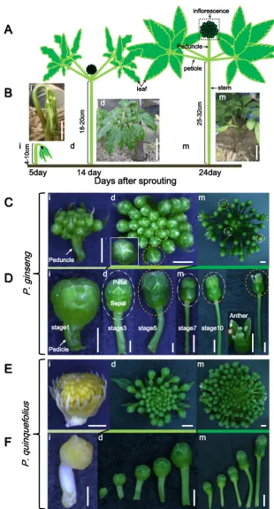
with five leaflets. In this study, we studied the inflorescence and flowers of 4-year-oldP. ginsengcv. K1 because of active reproductive development and abundant flowers at this growth stage (Fig.1). To correlate the developmental stages with the growth events, we measured the length of ginseng aerial parts, including the inflorescence and anther (Table1, Fig. 1a, b). At the initial stage, the aerial parts exhibit a parasol-like shape (Fig.1a(i), b(i); Kim et al.2014), showing unfolded leaves attached to the bending stem, and an inflores-cence primordia attached to a short peduncle (Fig.1c(i)). Each inflorescence contained about 8–15 flower buds (Fig. 1c(i), c(d), d(i)) and small immature flower buds (about 1 mm) with a short pedicle (1–5 mm) gradually increased in size with maturation. Two weeks after sprouting, active growth of the stem and peduncle occurred, and leaves partially expanded (Fig.1a(d), b(d)). During this developmental stage, the num-ber of flowers increased to 20–45 per inflorescence (Fig.1c(d)) and the pedicles of the outer flowers within the inflorescence were longer than those of the inner flowers (Fig. 1d(d)). At the mature stage, 3 weeks after sprouting, the height of the aerial parts was about 30 cm and leaves fully expanded (Fig. 1a(m), b(m)). Meanwhile, the peduncle (or inflorescence stalk) that arose from the shoot apical region kept a constant elongation and was finally located above the leaflets at the mature stage (Fig. 1c(m)). Furthermore, the multipedicelled umbel-shaped ginseng inflorescence had var-ious sized flowers at this stage, ranging from closed flower buds to fertilized flowers (Fig.1d(m)). About 1 month after sprouting, the pedicle linked with the calyx was as lengthen as the peduncle, and both pedicle and peduncle reached a max-imum height of about 10 and 20 cm, respectively (Table1).
Although the developmental events of theP. quinquefolius
inflorescence appear similar to those of P. ginseng,
P. quinquefoliushad about 50–80 flower buds and pedicles, about two-fold more thanP. ginseng(Fig.1e). Moreover, un-like the green color of theP. ginsenginflorescence at the initial developmental stage, the inflorescence primordium of
P. quinquefoliuswas pale yellow with a long involucre at this stage (Fig.1e(i)). In addition, the variation in developmental stages of flower buds was more obvious inP. ginseng com-pared withP. quinquefolius(Fig.1e(d), e(m), f(d), f(m)).
Floral morphology
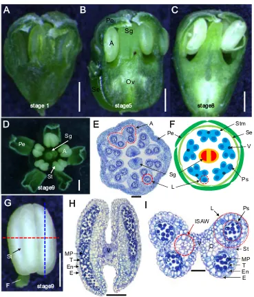
Each stamen consisted of a pollen-producing anther subtended by a filament (Fig.2g). During anther develop-ment, the color of the anthers changed from green at the initial stage to white during later stages (Fig.2a–c), and the stomium was well developed during anther maturation (Fig.2d, g, i). The anthers exhibited a butterfly shape in transverse sections
with four locules (Fig.2i). Anther locules were connected by flanking connective tissue and vascular elements continuous with the filament (Fig. 2h, i). The ginseng anther was com-posed of two theca linked with connective and vascular tis-sues, and each theca contained two locules, with the outer part of the locule longer and larger than the inner one. The two
ii
stage1 stage3 stage5 stage7 stage10
Fig. 1 Inflorescence morphology
at the different developmental stages of ginseng.aCartoon showingP. ginsengmorphology at the initial developing stage (i), the active developing stage (d), and the maturation stage (m) stage.bPictures ofP. ginseng
plants in a ginseng field.Bars
indicate 3 cm (i), 5 cm (d), and 10 cm (m), respectively.c Morphology of theP. ginseng
inflorescence at three different stages. Allbarsindicate 2 mm.d Morphological changes in a single flower ofP. ginsengwith an elongated pedicle during the three different stages.Bars
indicate 2 mm.fMorphology of a single inflorescence of
P. quinquefoliusduring three different developmental stages.
Barsindicate 500μm (i), 1 mm
locules were connected by the inner side of the anther wall (ISAW), a septum, and a stomium for anther dehiscence (Fig.2i). The anther was white till maturity, and the pollen sac was full of matured pollen grains and ready for release them when the flowers open (Fig.2g–i).
Anther and pollen development
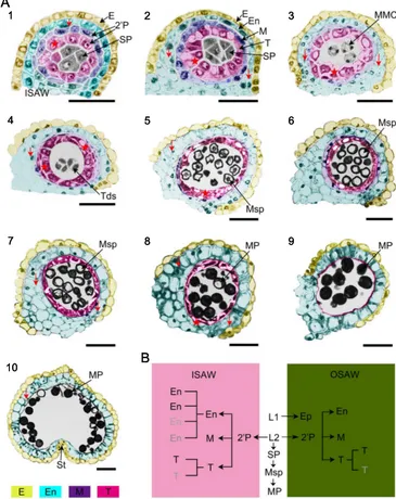
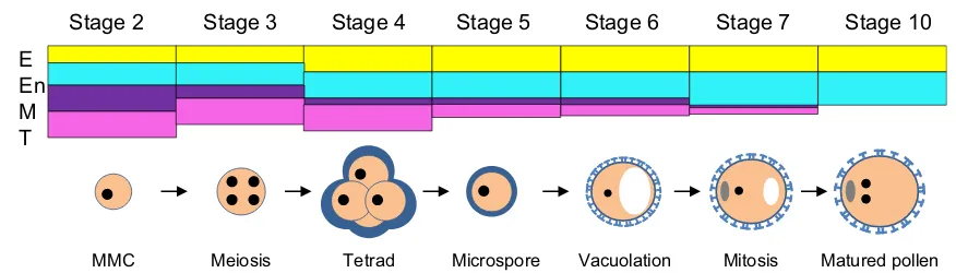
To understand cytological changes inP. ginsengduring anther development and the formation of pollen grains, we employed semi-thin section analysis. Generally, within a single ginseng flower bud, anther and pollen development varied in stages among individual stamens. Furthermore, pollen development varied along the length of the individual anther. We divided anther and pollen development ofP. ginsenginto 10 stages at which distinctive cellular events could be visualized at the level of a light microscope (Fig.3a). Table1summarizes the key events that occurred during the 10 stages of anther development.
After cell division and differentiation of the floral meri-stem, which contained layer 1 (L1) and L2, L1 cells within the anther primordium divided to form the epidermis and stomium, while two secondary parietal layers and sporoge-nous cells formed from L2 (Fig.3a(1)). We defined this stage as stage 1, during which anther length ranged from 0.2 to 0.25 mm. The outer secondary parietal layer generated the endothecium layer and the middle layer, and the inner second-ary parietal layer developed into the tapetal layer (Fig.3a(1)). At stage 2, four formatted concentric somatic layers surrounded the sporogenous cells: the epidermis, the endothe-cium, the middle layer, and the tapetal layer from outer to inner, and obvious nuclei were observed in these anther wall layers (Fig. 3a(2), (8)), suggesting active cell division. Remarkably, the endothecium and the tapetum of the anther wall opposite the central vascular tissues comprised a single cell layer each, respectively. In contrast, the endothecium of the ISAW had one to four cell layers while the tapetum com-prised one to two layers. Furthermore, the cell arrangement of the tapetal layer appeared uneven (indicated by stars and ar-rows in Fig.3a(2)), suggesting unsynchronized cell division of secondary parietal layers for the generation of endothecial and tapetal cells. In addition, at this stage, the middle layer only had one cell layer and appeared well developed, the spo-rogenous cells formed by cell division from the precursor cells within the central space of the anther had increased in number and had densely stained cytoplasm and obvious nuclei (Fig.3a(2)).
During morphological expansion of ginseng leaves at stage 3 (Fig. 1a(d), b(d)), there was further differentiation of the anther wall layers, epidermal cells appeared uneven, and the middle layer became thin, suggesting the initiation of cell degeneration (Fig.8). Furthermore, the tapetal layer appeared
larger and more irregular in cell shape, and MMCs formed from sporogenous cells initiate meiotic division (Fig.3a(3)).
At stage 4, when the flower bud had fully separated sepals (indicated by the box in Fig.1c(d)), the middle layer was almost fully degraded, and tapetal cells started the condensa-tion with dark-stained cytoplasm, an indicator of cell degen-eration triggered by programmed cell death (PCD). At this stage, MMCs continue the process of meiosis II with the for-mation of tetrads, consisting of four newly generated haploid microspores enclosed by a callose wall deposited on the primexine of the microspore (Fig.3a(4), (8); Supplementary Fig.S1B).
At stage 5, as the stem grew with fully expanded leaves and the peduncle elongated (Fig.1a(m), b(m)), free haploid young microspores were released from the tetrads, the endothecium and tapetal cells became vacuolated, and the tapetal layer con-tinued to degenerate as young microspores developed
(Fig.3a(5), (8)). At stage 6, the microspores increased in size through vacuolation, the endothecium became enlarged, and the tapetum continued to degenerate with much condensation (Fig.3a(6)). At stage 7, the anther appeared white, and vacu-olated microspores initiated the first mitosis with asymmetric cell division, giving rise to a much smaller generative cell and a larger vegetative cell. Meanwhile, the endothecium underwent secondary thickening (Fig. 3a(7)). At stage 8, when the pedicle and peduncle were the most elongated, the bicellular pollen grain underwent a second round of mitosis, and the endothecium contained expanded cells with thick cell walls, while tapetal cells formed a band-like structure (Fig.3a(8)).
At stage 9, mature spherical pollen grains full of storage starch formed. The middle layer and the tapetum had completely degenerated by this stage with tapetum-derived remnants, leaving the epidermis and the endothecium
Se
Fig. 2 Morphology of a single
flower ofP. ginsengshowing the floral organs and anther section.a Immature flower with unmatured green anthers at the initial developmental stage.bA flower with a clear ovary and anthers at the developing stage.cA mature flower with white anthers at the mature developing stage. Allbars
indicate 500μm. Opened flower
(d), transverse section (e), and cartoon (f) show the formation of five mature anthers and two stigmas.Barsindicate 500μm (d)
and 200μm (e). The mature
anther has a white appearance and is attached to the filament (g), longitudinal section (h), and transverse section (i).Bars
indicate 500μm (g) and 200μm
(H, I). Two sections of the anther at stage 7 showing the anther wall layers, microspores in the locule, as well as vascular tissues and connective tissues at the ISAW.A
anther,Stmstamens,Ovovary,Se
sepal,Pepetal,Sgstigma,Ps
pollen sacs,Vvascular bundle,T
tapetum,Enendothecium,MP
mature pollen,Eepidermis,F
filament,Llocule,Ststomium;
(Fig.3a(9)). At stage 10, the anther started to dehisce by breakage of the stomium, leaving behind the epidermis and endothecium layers. Tapetal cells were completely degraded and the two adjacent pollen sacs combined. The septum connecting the two pollen sacs broke, and anther walls at the stomium region located between the two locules started to break to release matured pollen grains by dehiscence (Fig.3a(10), (8)).
In situ analysis
The above light microscopic analysis revealed uneven cell division of the endothecium and tapetal layers. To further characterize the cell identity of these cell layers, we cloned a
DNA fragment calledPgCYP703identified from the ginseng genome database.PgCYP703shares 93 % identities with rice
CYP703A3 (Yang et al.2014) andArabidopsis CYP703A2
(Morant et al.2007), which are expressed in tapetal cells dur-ing pollen development. Further, the expression ofCYP703A3
is regulated by tapetum degeneration retardation (TDR) (Li et al. 2006; Yang et al. 2014) and GAMYB (Aya et al.
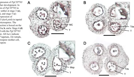
2009), two known regulators of tapetal PCD and pollen exine formation, respectively, in rice. Results of in situ hybridization usingPgCYP703showed high expression in one to two layers of tapetal cells of the anther during the formation of tetrads (Fig.4), confirming our assumption that ginseng anther un-dergoes uneven cell division during anther wall formation. Due to the lack of a marker gene for the endothecium, cells
1
1 2 3
4 5 6
7 8 9
10
A
B
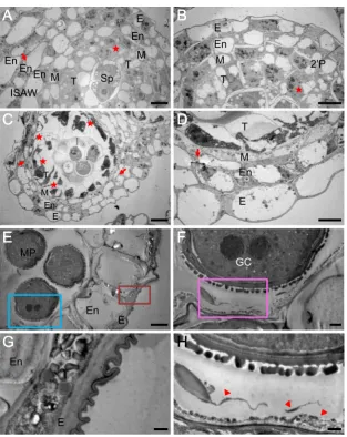
Fig. 3 Cytological observation
and diagrams ofP. ginsenganther development and pollen formation during the 10 developmental stages.aAnther sections were stained with toluidine blue and photographed by bright-field microscopy. Stages of anther development were observed in each locule. Anther cell layers differentiated into epidermis (E,yellow), endo-thecium (En,sky blue), middle layer (M,purple), and tapetum (T,
pink). Irregular cell division of endothecial and tapetum cells are indicted by thearrows and asterisks, respectively. Allbars
indicate 50μm.bDiagram of cell lineages during anther develop-ment in the inner sides of the an-ther wall (ISAW) and outer sides of the anther wall (OSAW). 2’P secondary parietal cell layer,E
epidermis,Enendothecium,T ta-petum,Mmiddle layer,Tds tet-rads,MMCmicrospore mother cell,Mspmicrospore,MPmature pollen,SPsporogenous cell,St
associated with the endothecium were not characterized at the molecular level.
TEM analysis of anther development
To precisely observe subcellular changes associated with an-ther development and pollen formation, we conducted TEM analysis (Fig.5). During the anther cell wall differentiation stage, the outer secondary parietal cells divided into the endo-thecium and middle layer, the inner secondary parietal cells developed into the tapetal layer (Fig.5a, b), and the central localized sporogenous cells differentiated into MMCs at stage 3 (Fig. 5c). MMCs contained numerous mitochondria and rough endoplasmic reticulum (ER) with expanded cisternae (Supplementary Fig.S3). Remarkably, during differentiation and development of the ginseng anther, tapetal cells became vacuolated and two-fold larger in size than the other anther wall cells. Consistent with the above observations based on examination of semi-thin sections and in situ analysis (Figs.3
and4), uneven cell division was also observed in the tapetum and the endothecium as evidenced by the presence of one or more layers of these cells in one anther (indicated by stars and arrows, respectively, in Fig.5a–d). At stage 3, the tapetal cells appeared well differentiated and could be clearly distin-guished from the other anther wall cell types by their dense cytoplasm (Fig.5c, d). The middle layer also became very thin (Fig.5d).
The mature anther wall consisted of dehydrated epidermal cells with a surface cuticle and expanded endothecium cells with secondary wall thickening (Fig.5e, g). Although the
tapetum degraded in the mature anther, tapetum-derived pol-len remnants were still present in the periphery of the locule, filling the cavities of a sporopollenin-based exine framework of tricellular pollen, completing pollen wall formation (indi-cated by the arrowheads in Fig. 5h). Ginseng tapetum ap-peared to be a secretory-type tapetum that produces granule structures (called orbicules) with dark-staining during TEM analysis (Fig.5f, h). The orbicules developed simultaneously from stage 7 to stage 9 during pollen exine development (Supplementary Fig.S4).
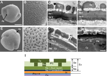
Each mature tricellular pollen grain possessed two gam-etes in the parietal position, and each had a prominent nucleus at stage 8 (Fig. 5f). During the maturation of a pollen grain, the outer pollen wall, called the exine, be-came well established as the synthesis, deposition, and as-sembly of sporopollenin precursors (Fig. 3a(5)). The for-mation of ginseng exine seems to be initiated from the tetrad stage (stage 4) through primexine formation and un-dulation of the plasma membrane. Young microspores re-leased from the tetrad started to develop baculum, which is the vertical element in the pollen wall, nexine, and tectum (Fig.7a–d) that gives mature pollen with the appearance of a fine reticulum (Fig. 7e–h). During maturation of pollen grains, there was differentiation between nexine I and nexine II (Fig.7c, d, g, h). After complementation of exine development, the intine, which is pecto-cellulosic in na-ture, developed as a thin layer after active development of high electron-dense organelles at the periphery of the pollen grain cytoplasm (Fig. 7d). Finally, mature pollen grains were characterized by a well-developed exine
A
B
C
D
MC
T
Tds
T
Msp
T
Tds
T
Fig. 4 Expression ofPgCYP703
during anther development. In situ analysis ofPgCYP703in
P. ginsenganther at stage 3 (a), stage 4 (b), and stage 5 (c) showing expression of
PgCYP703(dark pink) in tapetal cells and microspores. The enlarged section is boxed on the top right ofa,b, andc. Stage 4 (d) hybridized with thePgCYP703
sense probe. Allbarsindicate 500μm.Ttapetum,Tdstetrads,
MMCmicrospore mother cell,
comprising a uniformly thick nexine I (0.7 μm), thin
nexine II (0.1μm), and sexine (1μm) (Fig.7g). The sexine
consisted of a tectum (0.5 μm) linked by an irregular
bacula (0.1–0.5 μm) and pollen coat (Fig. 7g). Usually,
the thin layer of intine (0.4μm) between the exine and
cytoplasm plasma membrane in the non-apertural region of the pollen had a lower density than the exine (Fig.7g, i), but the intine was up to 3 μm thick in the apertural
region (Fig.7h).
Features of ginseng anther and pollen morphology
The anthers ofP. ginsengandP. quiquefoliushad obvious lobe boundaries (Fig.6a, c), and their anther cuticle displayed
striated patterns (Fig. 6b, d), whereas ginseng cells had a smoother appearance at early developmental stages (Supplementary Fig.S2). In addition, ginseng anthers contain various pollen grains with an average size of 20μm, ranging
from 16 to 27μm (Reunova et al. 2007) (Fig. 7e). Mature
pollen grains of ginseng are tricolpate and reticulate due to the external tectum structure (Fig. 7e, f). Even though
P. ginsengandP. quinquefoliusare the closest species in the same genus, minor differences in the pollen structure were evident between them: P. ginsengpollen grains had thick, almost continuous tecta and an infratectum comprising a short bacula and sparse granules, whereas pollen ofP. quinquefolius
had a thinner, discontinuous, and weakly striato-reticulate tec-tum, similar to previous report (Wen and Nowicke1999).
E
En
M
T E
En
M T Sp T M En
ISAW En En
A
A
B
E En T M
C
TM En
E
D
E En MP
GC
En
E
E
F
G
H
2’P
Fig. 5 Transmission electron micrographs of P. ginsenganthers at
different developmental stages. Somatic cells differentiated into six layers in the inner sides of the anther wall (ISAW) (a) and four layers in the outer sides of the anther wall (OSAW) (b) within a single anther locule at stage 1. Irregular cell division of endothecial and tapetum cells is indicted byarrows and asterisks, respectively.Barsindicate 10μm.
An-ther at stage 3 was characterized by four differentiated layers with irreg-ular division (c) and a high cell density (d) of tapetal cells.Barsindicate
20μm (c) and 10μm (d). Anther at stage 9 had a thin epidermis with the
formation of a cuticle, thickened endothecium (e,g), and one mature pollen grain with two generative cells (GC) (f). Pollen grain had an established exine in contact with orbicules (indicted asarrows) from complete degenerated tapetal cells (h).Barsindicate 10μm (e), 2μm (f), and 1μm (g,h).Eepidermis,Enendothecium,Mmiddle layer,T
Discussion
Despite the long history of the use of ginseng in medical applications, little is known about its reproduction. Development of the inflorescence and flower structure is es-sential for plant survival and agricultural yield. In this paper, we performed detailed observation on ginseng inflorescence and flower morphology and comprehensive cytological anal-ysis of ginseng male reproductive development. Furthermore, the anther developmental process is grouped into ten stages based on cytological events. In addition, the common and
specialized developmental features in ginseng are addressed in comparison with other model plants.
Characteristic morphology of ginseng inflorescence and flowers
Flower morphology of the family Araliaceae is extremely diverse, in contrast to low variation of mature flower structure of Umbelliferae, which is closely related family ofAraliaceae
(Nuraliev et al.2010).PanaxL. has a single terminal inflores-cence with an umbel shape, in contrast to other species in the
P. ginseng
(mature stage)P. quinquefolius
(mature stage)B
C
D
A
Fig. 6 Characterization of the
outer surface of ginseng anthers compared with those of
Arabidopsisand rice by scanning electron microscope.aP. ginseng
anther at stage 9,bar=200μm.b
Enlargement ofashowing the anther cuticle,bar=10μm.c
P. quinquefoliusanther at stage 9,
bar=200μm.dEnlargement ofc
showing the anther cuticle,
bar=10μm
Fig. 7 Pollen grain and pollen wall morphology analysis ofP. ginsengat
stage 5 (a–d) and stage 9 (e–h). Outer appearance of pollen grain (a), enlargement of pollen grain surface (b) by SEM, and pollen wall section by TEM (c,d) showing a less reticular structure than that ofArabidopsis. At this stage, bacula (Ba), tectum (Te), and nexine I (Ne I) were apparent, whereas the nexine II (Ne II), which was difficult to distinguish inc, further developed during development of pollen grain with highly electron-dense cells at the periphery of the cytoplasm (d). Mature pollen
grains had a rough outer appearance (e) and showed enlargement of the pollen grain surface (f) by SEM, while the pollen wall had a reticular structure as determined by TEM (g,h). Pollen grain had a thin intine at nonaperture regions (g) and a thick intine at the aperture region (h).Bars
indicate 5μm (a,e), 1μm (b–d,f–h).iDiagram of ginseng pollen exine
familyAraliaceae, whilePanax trifoliusand the nearest gen-eraAraliahave multiple inflorescences for each plant. Among
Panax L, two major important species, P. ginseng and
P. quinquefolius, are closely related and more advanced spe-cies (Wen and Nowicke1999). In ginseng plants, the transi-tion from vegetative growth into reproductive growth fre-quently occurs in the second growth year when the plant pro-duces at least two compound leaves with petioles in each stem. During third growth year, the indeterminate inflorescence at-tached by a single peduncle developed from the primordium forms from second-year growth plants. Unlike studies in an-nual plants which have multiple axillary meristems originated from the shoot apical meristem during inflorescence and flow-er development (Zhang and Yuan2014), ginseng plant usually produces a single inflorescence from the primordium contained within the underground bud of a rhizome formed during June to August of the previous year (Baranov1966; Thompson1987; Kim et al.1998). The feature of ginseng in producing the inflorescence primordium in the previous year and requiring winter dormancy for further development of inflorescence is a characteristic of perennial plants (Meloche and Diggle2003; Albani and Coupland2010). Furthermore, we observed the cell differentiation from sporogenous cells into pollen mother cells after dormancy in ginseng, which is similar to that in other woody perennial plants (Boss and Strauss1994; Julian et al. 2011). However, the mechanism underlying this switch still remains elusive. Furthermore, each ginseng flower has five green petals, and the upper region of the petals is white in color, with each petal surrounded by depressed sepals. The specialized flower structure of ginseng might be due to adaptation to the environmental factors and selection for pollination. The characteristic inflorescence and flower development imply tight regulation of genetic and en-vironmental factors during acquisition of meristem identity, which remain to be investigated in the future.
Nonsynchronous development of ginseng anther wall layers
To facilitate research on ginseng male reproduction, we grouped the development of the ginseng anther into 10 stages based on cytological observations and the length of the anther (Table1). Overall, the development of the ginseng anther ranging from anther primordium formation, anther wall, re-productive cell division, differentiation to pollen formation, maturation, as well as anther dehiscence, appears to be a well-conserved process in angiosperms, suggesting conserved control of male reproduction in higher plants. After morpho-genesis, each ginseng anther contained four anther wall layers, i.e., the epidermis, the endothecium, the middle layer, and the tapetum surrounding the male reproductive cells. After meio-sis, the middle layer and the tapetum degraded, likely promot-ed by PCD (Fig.8). Expansion and secondary wall thickening
in the endothecium layer and disappearance of the tapetum and middle layer, which were observed at stage 9, is essential for pollen dispersal by anther dehiscence along specialized cells of the epidermis (Mitsuda et al.2005; Yang et al.2007). Interestingly, unlike uniform anther wall layers such as the tapetal layer in rice and Arabidopsis (Sanders et al.1999; Zhang et al.2011; Quilichini et al.2014), we observed non-synchronous development of ginseng anther wall layers, par-ticularly the endothecium and the tapetum, during early anther development. Large number of middle layers also have been observed in other woody plants such asPopulus bolleanaand
Prunus armeniaca (Zhang et al. 2009; Julian et al.2011), which would contribute for thick anther walls. However, ta-petum is a usually uniseriate, rarely multiseriate in few plants fromPoaceaeandCucurbitaceaefamily andPeperonmia ge-nus (Fisher1914; Nakamura et al.2010; Pandey et al.2014). InAraliaceaefamily, it was also shown at least two layer of tapetum and uneven cell division during tetrad stage of
T. burckii microspore (Gabarayeva et al. 2009a). It will be interesting to investigate the mechanism underlying the for-mation of multiple tapetal layers.
From stages 1 to 4, anther parietal cells inP. ginseng dif-ferentiated into four anther cell walls, and the outer secondary parietal cell layer divided periclinally to form the middle layer and the endothecium, while the inner secondary parietal layer differentiated into tapetal cells (Supplementary Fig. S1A). Notably, nonsynchronous cell division occurred during for-mation of the tapetum and the endothecium, as evidenced by the observation of two to four layers in the ISAW, and one layer of tapetum and endothecium near the outside of the anther wall (OSAW), respectively (Figs. 3a, stages 1–3, b,
4a, b). The identity of tapetal cells was confirmed by in situ analysis usingCYP703homolog in ginseng as a probe (Fig.4) (Yang et al.2014). Due to the lack of a specific marker gene for the endothecium (Zhang et al. 2011; Zhang and Yang
2014), we were not able to determine the cell identity of the endothecium. Nonsynchronous cell division to generate an-ther wall layers, particularly of tapetal cells, has not been observed in Arabidopsis and rice (Sanders et al. 1999; Zhang et al.2011; Quilichini et al.2014). Recently, tapetum multilayer was observed as associated with male sterility in maize (Chaubal et al.2000), tomato (Sawhney and Bhadula
2011), andArabidopsis(Cecchetti et al.2015), confirming the important role of tapetum in normal pollen development reg-ulated by auxin transportation. This feature suggests a special-ized developmental program for ginseng anther wall develop-ment, which remains to be investigated.
degeneration exhibited a little delay compared with other re-ported species of angiosperms in which their tapetal cells be-gin to degeneration before the tetrad stage (Sanders et al.
1999; Parish and Li2010; Zhang et al.2011). The inner side of tapetal cell has the attached orbicule-like structures pending in the locular fluid (Fig.5h; Supplementary Fig.S4), which was also identified fromT. burckii in theAraliaceaefamily (Gabarayeva et al.2009b). These granule structures on the tapetum may play a key role in regulating the development of pollen exine, which is similar to that of Ubisch bodies/ orbicules on the inner surface of tapetal cells in cereal plants, such as rice and wheat (Huysmans et al.1998; Li and Zhang
2010), or tapetosomes in Arabidopsisand elaioplasts in
Brassicaceae, assumed to export tapetum-produced sporopol-lenin precursors across the hydrophilic cell wall to the locule (Wu et al.1997; Furness and Rudall 1998; Quilichini et al.
2014).
Pollen wall structures in ginseng
The pollen wall, surrounding the sperm cells, functions as a protective barrier for sperm cells and confers resistance to environmental stresses after anther dehiscence (Blackmore et al.2007; Li et al. 2010; Li and Zhang 2010; Shi et al.
2011). Development of the pollen wall in ginseng is obvious from the early tetrad stage (stage 4) and almost completes at the maturing pollen stage (stage 9), consistent with previous observations (Jeong2005). Compared with other plants, the
P. ginsengexine has a shorter bacula and thicker nexine, which are commonly characteristics of mostPanaxspecies, exceptPanax trifolius(Wen and Nowicke1999). It could be caused by high accumulation of pollen wall exine, comparable with less accumulation in close genus,Aralia(Reunov et al.
2008). The structural characteristics of pollen wall, reticulate cavities with an abundant pollen coat inside the pollen exine, may be associated with pollination habits, such as insect pol-lination (Zhang et al.2011). Moreover,P. ginsenghas thicker, almost continuous tecta (Fig.7), whereasP. quiqnefoliushas
thinner, and discontinuosus, and weakly striato-reticulate tec-tum (Wen and Nowicke1999).
In short, we described the characteristic development of the inflorescence and flower ofP. ginsengand performed com-prehensive cytological analysis of ginseng anther develop-ment and pollen formation. Furthermore, we categorized the developmental stages of the ginseng anther into 10 stages on the basis of biological events. Determination of the mecha-nism controlling nonsynchronous cell division within anther wall layers such as the tapetal layer in the same anther lobe is an interesting topic for future research. The information pro-vided in this study will benefit fundamental research on gin-seng reproductive development as well as plant breeding and male fertility manipulation.
Acknowledgments We express our thanks to Dr. Jakyung Lee,
Profes-sor Gynheung An (Kyung Hee Unviversity) for comments about flower sampling and fixation and Li Yang, Junping Yu, and Dr. Jie Xu (Shanghai Jiao Tong University) for comments about flower section. This work was supported by the funds from Ministry of Science and Tehnology (2015DFG32560), and the Programme of Introducing Talents of Disci-pline to Universities (111 Project, B14016), Young Scientist Exchange Program between Republic of Korea (NRF) and The People’s Republic of China (MOST) and NRF (2013R1A1A2064430) (YJ Kim); and Korea Institute of Planning and Evaluation for Technology in Food, Agriculture, Forestry and Fisheries (iPET; 112142-05-1-CG000), Republic of Korea (DC Yang).
Conflict of interest The authors declare that they have no conflict of
interest.
References
Albani MC, Coupland G (2010) Comparative analysis of flowering in annual and perennial plants. Curr Top Dev Biol 91:323–348 Aya K, Ueguchi‐Tanaka M, Kondo M, Hamada K, Yano K, Nishimura
M, Matsuoka M (2009) Gibberellin modulates anther development in rice via the transcriptional regulation of GAMYB. Plant Cell 21(5):1453–1472
MMC Meiosis Tetrad Microspore Vacuolation Mitosis Matured pollen
E En M T
Stage 2 Stage 3 Stage 4 Stage 5 Stage 6 Stage 7 Stage 10
Fig. 8 Schematic model of pollen and anther wall development in
P. ginseng. Anther somatic cell wall of P. ginsengat stage 2; four distinct cell layers were evident: epidermis (E,yellow), endothecium (En,sky blue), middle layer (M,purple), and tapetum (T,pink). Epidermis and endothecium became thicker at the tetrad stage of the
Baranov A (1966) Recent advances in our knowledge of the morphology, cultivation and used of ginseng (Panax ginsengC.A. Meyer). Econ Bot 20(4):403–406
Bedinger P (1992) The remarkable biology of pollen. Plant Cell 4(8): 879–887
Beeckman T, Viane R (2000) Embedding thin plant specimens for ori-ented sectioning. Biotech Histochem 75(1):23–26
Blackmore S, Wortley AH, Skvarla JJ, Rowley JR (2007) Pollen wall development in flowering plants. New Phytol 174(3):483–498 Boss TK, Strauss SH (1994) Floral phenology and morphology of black
cottonwood. Populus trichocarpa (Salicaceae) Am J Bot 81:562–
567
Cecchetti V, Brunetti P, Napoli N, Fattorini L, Altamura MM, Costantino P, Cardarelli M (2015) ABCB1 and ABCB19 auxin transporters have synergistic effects on early and late Arabidopsis anther devel-opment. J Integr Plant Biol. doi:10.1111/jipb.12332
Chang F, Wang Y, Wang S, Ma H (2011) Molecular control of microspo-rogenesis in Arabidopsis. Curr Opin Plant Biol 14(1):66–73 Chaubal R, Zanella C, Trimnell MR, Fox TW, Albersten MC, Bedinger P
(2000) Two male-sterile mutants ofZea mays(Poaceae) with an extra cell division in the anther wall. Am J Bot 87:1193–1201 Chehregani A, Tanaomi N, Ranjbar M (2008) Pollen and anther
devel-opment inOnobrychis schahuensisBornm. (Fabaceae). Int J Bot 4(2):241–244
Feng X, Dickinson HG (2010) Cell-cell interactions during patterning of the Arabidopsis anther. Biochem Soc Trans 38(2):571–576 Fisher GC (1914) Seed development in the genus Peperomia. Bull Torr
Bot Club 41:221–241
Furness CA, Rudall PJ (1998) The tapetum and systematics in monocot-yledons. Bot Rev 64(3):201–239
Gabarayeva N, Grigorjeva V, Rowley JR, Hemsley AR (2009a) Sporoderm development inTrevesia burckii(Araliaceae). I. Tetrad period: further evidence for the participation of self-assembly pro-cesses. Rev Palaeobot Palynol 156:211–232
Gabarayeva N, Grigorjeva V, Rowley JR, Hemsley AR (2009b) Sporoderm development inTrevesia burckii(Araliaceae) II. Post-tetrad period: further evidence for the participation of self-assembly processes. Rev Palaeobot Palynol 156:233–247 Heslop-Harrison J (1971) Wall pattern formation in angiosperm
micro-sporogenesis. Symp Soc Exp Biol 25:277–300
Huysmans S, El-Ghazaly G, Smets E (1998) Orbicules in Angiosperms: morphology, function, distribution, and relation with Tapetum Types. Bot Rev 64(3):240–272
Igersheim A, Cichocki O (1996) A simple method for microtome sec-tioning of prehistoric charcoal specimens, embedded in 2-hydroxyethyl methacrylate (HEMA). Rev Palaeobot Palynol 92: 389–393
Jeong BK (2005) Fine structural study of pollen wall development at late stage of microsporogenesis inPanax ginseng. Kor J Electron Microsc 35:1–10
Julian C, Rodrigo J, Herrero M (2011) Stamen development and winter dormancy in apricot (Prunus armeniaca). Ann Bot 108:617–625 Kelliher T, Egger RL, Zhang H, Walbot V (2014) Unresolved issues in
pre-meiotic anther development. Front Plant Sci 21:347–355 Kim MJ, Kim IS (1995) Microsporogenesis ofHibiscus syriacusL. and
its sporoderm differentiation. J Plant Biol 38(1):95–105
Kim YS, Lee HS, Lee MH, Yoo OJ, Liu JR (1998) A MADS Box gene Homologous to AG is expressed in seedlings as well as in flower of ginseng. Plant Cell Physiol 39(8):836–845
Kim YJ, Jeon JN, Jang MG, Oh JY, Kwon WS, Jung SK, Yang DC (2014) Ginsenoside profiles and related gene expression during fo-liation inPanax ginsengMeyer. J Ginseng Res 38(1):66–72 Koltunow AM, Soltys K, Nito N, McClure S (1995) Anther, ovule, seed,
and nucellar embryo development inCitrus sinensiscv. Valencia. Can J Bot 73(10):1567–1582
Kreunen SS, Osborn JM (1999) Pollen and anther development in
Nelumbo(Nelumbonaceae). Am J Bot 86(12):1662–1676 Lee OR, Sathiyaraj G, Kim YJ, In JG, Kwon WS, Kim JH, Yang DC
(2011) Defense genes induced by pathogens and abiotic stresses in
Panax ginsengC.A. Meyer. J Ginseng Res 35(1):21–30
Li H, Zhang D (2010) Biosynthesis of anther cuticle and pollen exine in rice. Plant Signal Behav 5(9):1121–1123
Li N, Zhang DS, Liu HS, Yin CS, Li XX, Liang WQ, Yuan Z, Xu B, Chu HW, Wang J, Wen TQ, Huang H, Luo D, Ma H, Zhang DB (2006) The rice tapetum degeneration retardation gene is required for tape-tum degradation and anther development. Plant Cell 18(11):2999–
3014
Li H, Pinot F, Sauveplane V, Werck-Reichhart D, Diehl P, Schreiber L, Franke R, Zhang P, Chen L, Gao Y, Liang W, Zhang D (2010) Cytochrome P450 family member CYP704B2 catalyzes the {ome-ga}-hydroxylation of fatty acids and is required for anther cutin biosynthesis and pollen exine formation in rice. Plant Cell 22(1): 173–190
Liu L-D, Wang Z-L, Guo-Wei T, Shen J-H (1998) Megasporogenesis, microsporogenesis and development of gametophytes in
Eleutherococcus senticosus(Araliaceae). Acta Phyto Sin 36(4): 289–297
Meloche CG, Diggle PK (2003) The pattern of carbon allocation supporting growth of preformed shoot primordia inAcomastylis rossii(Rosaceae). Am J Bot 90:1313–1320
Mitsuda N, Seki M, Shinozaki K, Ohme-Takagi M (2005) The NAC Transcription Factors NST1 and NST2 ofArabidopsisregulate sec-ondary wall thickenings and are required for anther dehiscence. Plant Cell 17(11):2993–3006
Morant M, Jorgensen K, Schaller H, Pinot F, Moller BL, Werck-Reichhart D, Bak S (2007) CYP703 Is an ancient cytochrome P450 in land plants catalyzing in-chain hydroxylation of lauric acid to provide building blocks for sporopollenin synthesis in pollen. Plant Cell 19(5):1473–1487
Nakamura AT, Longhi-Wagner HM, Scatena VL (2010) Anther and pol-len development in some species of Poaceae (Poales). Braz J Biol 70:351–360
Nuraliev MS, Oskolski AA, Sokoloff DD, Remizowa MV (2010) Flowers of Araliaceae: structural diversity, developmental and evo-lutionary aspects. Plant Div Evol 128:247–268
Oh JY, Kim YJ, Jang MG, Joo SD, Kwon WS, Kim SY, Jung SK, Yang DC (2014) Investigation of ginsenosides in different tissues after elicitor treatment inPanax ginseng. J Ginseng Res 38(4):270–277 Pandey AK, Dwivedi MD, Mathur RR (2014) Embryology of
Cucurbitaceae and circumscription of Cucurbitales: a review. Int J Plant Rep Biol 6:75–98
Parish RW, Li SF (2010) Death of a tapetum: a prgramme of develop-mental altruism. Plant Sci 178:73–89
Quilichini TD, Douglas CJ, Samuels AL (2014) New views of tapetum ultrastructure and pollen exine development inArabidopsis thaliana. Ann Bot 114(6):1189–1201
Racich JL, Koutsky JA (1976) Nodular structure in epoxy resins. J Appl Polym Sci 20(8):2111–2129
Raghavan V (1988) Anther and pollen development in rice (Oryza sativa). Am J Bot 75(2):183–196
Reunov AA, Reunova GD, Alexandrova YN, Muzarok TI, Zhuravlev YN (2008) The pollen metamorphosis phenomenon inPanax ginseng,Aralia elataandOplopanax elatus; an addition to discus-sion concerning thePanaxaffinity in Araliaceae. Zygote 17(1):1–17 Reunova GD, Reunov AA, Aleksandrova YN, Muzarok TI, Zhuravlev YN (2007) Pollen heteromorphism inPanax ginsengC. A. Meyer (Araliaceae) anthers. Dokl Biol Sci 412:76–78
Sajo MG, Furness CA, Prychid CJ, Rudall PJ (2005) Microsporogenesis and anther development in Bromeliaceae. Grana 44:65–74 Sanders PM, Bui AQ, Weterings K, McIntire KN, Hsu YC, Lee PY,
defects inArabidopsis thalianamale-sterile mutants. Sex Plant Reprod 11:297–322
Sawhney VK, Bhadula S (2011) Microsporogenesis in the normal and male-sterile stamenless-2 mutant of tomato (Lycopersicon esculentum). Can J Bot 66:2013–2021
Shi J, Tan H, Yu XH, Liu Y, Liang W, Ranathunge K, Franke RB, Schreiber L, Wang Y, Kai G, Shanklin J, Ma H, Zhang D (2011) Defective pollen wall of required for anther and microspore devel-opment in rice and encodes a fatty acyl carrier protein reductase. Plant Cell 23(6):2225–2246
Thompson GA (1987) Botanical characteristics of ginseng. In Herbs, Spices, and Medicinal Plants: Recent Advances in Botany, Horticulture, and Pharmacology, Edited by: Craker LE, Simon JE Vol. 2
Tseng CC, Shoup JR, Chuang TI, Hsieh WC (1983) Pollen morphology ofAcanthopanax(Araliaceae). Grana 22:11–17
Wen J, Nowicke JW (1999) Pollen ultrastructure ofPanax(The ginseng genus, Araliaceae), an eastern Asian and eastern north American disjunct genus. Am J Bot 86(11):1624–1636
Wilson ZA, Zhang DB (2009) From Arabidopsis to rice: pathways in pollen development. J Exp Bot 60(5):1479–1492
Wu SS, Platt KA, Ratnayake C, Wang TW, Ting JT, Huang AH (1997) Isolation and characterization of neutral-lipid-containing organelles and globuli-filled plastids fromBrassica napustapetum. Proc Natl Acad Sci U S A 94(23):12711–12716
Yang C, Vizcay-Barrena G, Conner K, Wilson ZA (2007) MALE STERILITY1 is required for tapetal development and pollen wall biosynthesis. Plant Cell 19(11):3530–3548
Yang X, Wu D, Shi J, He Y, Pinot F, Grausem B, Yin C, Zhu L, Chen M, Luo Z, Liang W, Zhang D (2014) Rice CYP703A3, a cytochrome P450 hydroxylase, is essential for development of anther cuticle and pollen exine. J Integr Plant Biol 56(10):979–994
Yeung EC, Oinam GS, Yeung SS, Harry I (2011) Anther pollen and tapetum development in safflower,Carthamus tinctoriusL. Sex Plant Reprod 24(4):307–317
Zhang D, Wilson ZA (2009) Stamen specification and anther develop-ment in rice. Chin Sci Bull 54(14):2342–2353
Zhang D, Yang L (2014) Specification of tapetum and microsporocyte cells within the anther. Curr Opin Plant Biol 17:49–55
Zhang D, Yuan Z (2014) Molecular control of grass inflorescence devel-opment. Annu Rev Plant Biol 65:553–578
Zhang C, Guinel FC, Moffatt BA (2002) A comparative ultrastructural study of pollen development inArabidopsis thalianaecotype Columbia and male-sterile mutantapt1-3. Protoplasma 219(1–2): 59–71
Zhang Z, Kang X, Wang S, Li D, Chen H (2009) Pollen development and multi-nucleate microspores ofPopulus bolleanaLauche. For Stud China 10:107–111
Zhang D, Luo X, Zhu L (2011) Cytological analysis and genetic control of rice anther development. J Genet Genomics 38(9):379–390 Zhang Y, Liang W, Shi J, Xu J, Zhang D (2013) MYB56 encoding a
R2R3 MYB transcription factor regulates seed size inArabidopsis thaliana. J Integr Plant Biol 55(11):1166–1178