20
Detection of Toxoplasmagondii infection by Polymerase Chain Reaction (PCR) and Histological Examination on Balb/c Mice
Muhammad Hanafiah1), Dwinna Aliza2), Erdiansyah Rahmi3), 4) Wisnu Nurcahyo
1The Laboratory of Parasitology, Veterinary Faculty, Syiah Kuala University, Darussalam, Banda Aceh
23111, Indonesia
2The Laboratory of Pathology, Veterinary Faculty, Syiah Kuala University, Darussalam, Banda Aceh 23111, Indonesia
3The Laboratory of Histology and Embryology, Veterinary Faculty, Syiah Kuala University, Darussalam, Banda Aceh 23111, Indonesia
4The Department of Parasitology, Veterinary Faculty, Gadjah Mada University, Sekip Unit II Yogyakarta
55281, Indonesia
Email for correspondence: [email protected]
Abstract
The purpose of this research was to compare the use of PCR method and histological examination to diagnose toxoplasmosis in tissues of Balb/c mice infected with sporulated oocysts through drinking water. A total of 20 male Balb/c mice aged approximately 2 months were used in this experiment. Each mouse was infected with 1x10³ Toxoplasmagondii tachyzoites intraperitoneally. Tissue samples (liver, lung, heart, kidney, and brain) were collected from 5 mice on day 1, day 5, day 7, and day 9 after infection. Samples were then examined by PCR and histological methods. The data collected were analyzed descriptively. The results showed that PCR method was more sensitive than histological examination. PCR examination using primer invitrogen gen can amplify DNA T. gondii at 436 bp of the samples from liver, lung, heart and brain on Day 7 and Day 9 after infection. The histological examination showed that the cyst of toxoplasma was found in the brain while mononuclear cells infiltration was found in other internal organs.
Key words: Toxoplasma gondii, PCR, Histological, Balb/c Mice
Background
Toxoplasma gondii is a worldwide parasite that can infect the central nervous system of warm-blooded animals, including humans (Montoya and Liesenfeld, 2004). Domestic cats and other felids have been shown as the major reservoir hosts of this parasite. It is generally assumed that cats play a major role in transmitting T. gondii through faecal contaminated soils, foods or water because they excrete millions of oocysts in a short period of time (1-2 weeks) (Dubey, 2008).
Toxoplasmosis caused by T. gondii infection has become a major public health
concern recently due to the
immunosuppressive effect of the ravaging HIV/AIDS pandemic (Lindstrom et al., 2006; Uneke et al., 2007). Several laboratory methods have been developed to detect antibody in the serum of infected cats such as PCR, ELISA, LAT (latex agglutination test), IHA (indirect hemagglutination assay), IFAT (indirect fluorescent antibody test) and immunochromatography (IC). Though these tests are more sensitive and specific, they are expensive and require a specialized Open Acces
Int. J. Trop. Vet. Biomed. Res. Vol. 1 (2) : 20-26; November 2016
www.jurnal.unsyiah.ac.id/IJTVBR
Res.2:20-26
21
laboratory (Zhang et al., 2009). IC is rapid, simple, sensitive, and specific. It will be a suitable diagnostic tool for detection of the specific antibodies in T. gondii infection in cats under field conditions (Xiaohong et al., 2004).
Bioassay in mice, although specific and relatively sensitive for the detection of T. gondii oocysts, relies on the presence of sporulated ocytests and thus infective oocysts in samples. Not only does this method introduces a possible biohazard element, it is also expensive, requires animal facilities, and is time consuming (Salant et al., 2007).
Faecal flotation technique is used to detect oocyst presence in faecal samples as well. Although flotation is a reference method for the detection of T. gondii oocysts, it has been suggested that an alternative test is also needed because microscopic examination is time consuming and needs an experienced microscopist (Greene, 2006).
In a routine histological examination, which is stained with hematoxylin and eosin (H&E), T. gondii antigen is generally invisible, especially when the number of the antigen is limited or dissolved (Lappin, 1994). Diagnosis using PCR from tissue samples (placenta, brain, liver and lung) has been conducted in many countries and shown satisfying result. Therefore, it can be assumed that PCR method has higher sensitivity and specificity to diagnose toxoplasmosis infection compared with conventional methods (Bell and Ranford-Cartwright, 2002; Contini et al., 2005; Calderaro et al., 2006; Bastien et al., 2007).
According to Edvinsson et al. (2006) PCR method can detect parasites in small amount or even only one with high specificity from various tissues, sputum, cerebrospinal fluid, blood, urine, and faeces. Based on the above explanations, this research was intended to study further the use of PCR method and histological examination on various tissue sampless (liver, lung, heart, kidney, and brain) for toxoplasmosis diagnosis using Balb/c mice infected with sporulated oocysts through drinking water as experimental animals
Materials and Methods
This research used 20 Balb/c mice. Oocysts were collected from faeces whereas T. gondii tachyzoites were obtained from some internal organs of the mice such as liver, lung, heart, and brain.
Toxoplasma gondii Oocysts Isolation from Cat’s Faeces
Isolation of T. gondii from faeces was done follows Lappin (1994). In brief, faeces was floated in a container, stored at 4 C and treated with water for 2 days till softened. Sulphuric acid 2% was added to change faecal emulsion into paste. The faeces was then resuspended in a 10-time volume of Sheather sugar solution (40% of sucrose in 0,8% of phenol as the preservatives). Suspension was filtered with 710 µm sieve (Endecott Ltd. England) to remove big particles. The filtrate was centrifuged at 3000 rpm for 10 minutes. Supernatant, 2 ml, was drawn using a pasteur pipette, inserted into a bottle and added with potassium bichromate 2%. The solution was stored in a sealed bottle at room temperature for 3-7 days so that the oocysts sporulated. The mixture was filtered through a 44 µm sieve. The remaining parts were sprayed with distilled water and then used for toxoplasmosis examination. Sporulated oocysts were given to 20 mice through drinking water.
Tachyzoite Isolation from Mouse Peritoneal Fluid
All infected mice, as marked by the formation of ascites liquid, were sacrified. Their stomaches were cleansed three times using 5 ml of physiological NaCl solution to obtain tachyzoites. Tachyzoites was used to infect 10 adult mice with dilution dosage of 1 X 10⁷ to obtain more tachyzoite using a technique similar to the previous ones. The cleansed stomach was centrifuged at 3000 rpm (Beckman), 4°C for 10 minutes. Pellets were washed 3 times using PBS solution pH 7.4 which contained tris-ammonium-chloride to lyse red blood cells contaminants. The exudate was incubated by being shaken at 37
Res.2:20-26
22 were repeated at least 4 times to eliminate
unwanted proteins. After the final centrifugation process, cells were resuspended in 100 ml of PBS. Using a 27-size needle, the suspension was drawn carefully so that the pellets containing peritoneal cells were not included. Free trophozoites suspension was then filtered with a 3 µm milipore sieve to remove cell debris (Salant et al, 2007).
Sample Collection
Tissue samples (liver, lung, heart, and brain) were collected from each of 5 mice on Day 1, 5, 7, and 9 after the infection for further process. These samples were used for DNA isolation and histological preparations.
Isolating DNA from tissues
A part of tissue samples (liver, lung, heart, kidney and brain) was used as histological preparation while the other part was scaled and grained in a mortar. These samples were then treated with liquid nitrogen and pounded till homogenized. Next, the sample was added with buffer lysis (b) with the ratio of 1 sample: 9 buffer lysis (b). Then the mixture was incubated at temperature of 370C for 15 minutes. After the incubation, the sample was centrifuged at the speed of 12.000 rpm for 15 minutes, and the supernatant was washed 3 times with sterile PBS with 7,2 pH. The pellet was added with buffer of digestion with the ratio of 500 mg sample: 1 ml buffer of digestion. The sample was then incubated in water bath at the temperature of 55oC for 2 hours and boiled at the temperature of 100oC for 8 minutes. It was then centrifuged at 12,000 rpm speed for 30 seconds. After that, the supernatant was taken to be amplified by PCR (Matsuura et al, 1990).
Amplifying DNA with PCR
Amplification of DNA samples was done by using PCR (Ready To Go TM PCR Beads from Amersham Pharmacia Biotech) commercial kit. The primers used, name 5-
GAC TCG GGC CCA GCT GCG -3
(forward) and 3- CCT CTC CTA CCC CTC
CTC - 3 (backward), were purchased Invitrogen, Berlin Buch GmbH Jerman. PCR reaction mixtures were prepared ifollwing the protocol provided. Here, 5 µl of DNA targets, 2 l of forward and reverse primers (final concentration of 2 pmol), and sterile aquadest up to 25 µl were added to a PCR tube containing a PCR bead. PCR reaction was done in the following condition: one cycle of preparation stage at 95C for 5 minutes, followed by 40 cycles for denaturation at 94
C for 1 minute, annealing at 60 oC for 30 seconds, elongation at 72 C for 1 minute and termination at 72 C for 10 minutes. PCR results were visualized using 1% of agarose electrophoresis; 0.5 x TBE (Tris borate buffer) using cDNA as the positive control.
Histological preparation
Tissue samples were poured in formalin 10%, cut into small pieces, inserted into formalin 4%, and then continued with dehydration process with graded alcohol (ethanol 80%, 95%, 100%), each for 2 hours. The next step was purification with xylol, blocked with paraffin, and cut using microtome. The cut tissues was mounted onto the surface of abject glasses, fixated and stained with haematoxylin-eosin (HE) staining. The occurrence of the parasite (in the form of bradyzoites cyst) inside the tissue was confirmed by miscroscopic examination using 100x Phase contrast, oil immersion objective lens with an Olympus binocular microscope.
Data analysis
Results obtained from PCR and histological examinations were analyzed descriptively.
Results
Res.2:20-26
23
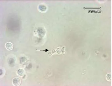
Figure 1. T. gondii tachyzoite which is found in mice peritoneum fluid. Note: arrows is directed to T. gondii tachyzoite
Isolation and Amplification of DNA from the Mice Organ using PCR
The result of PCR examination on organs of Balb/c mice to diagnose the toxoplamosis through infection by providing oocysts which had been sporulated can be seen in Table 1.
Day Sample Result Sample No Positive Negative
I
Liver Lung Heart Brain
- - - -
5 5 5 5
5 5 5 5
5
Liver Lung Heart Brain
- - - -
5 5 5 5
5 5 5 5
7
Liver Lung Heart Brain
2 1 1 2
3 4 4 3
5 5 5 5
9
Liver Lung Heart Brain
2 1 1 2
3 4 4 3
5 5 5 5
The result of examination on DNA in the organ of Balb/c mice which toxoplasma DNA was succesfully isolated by using primer invitrogen shows that the DNA is 436 bp. It can be seen in Figure 2.
Histological Preparation
Histological examinations
successfully found one cyst in the brain of infected mice as can be seen in Figure 3. This finding was somewhat different from what previously obtained by Hanafiah et al. (2013) in a research applying scouring method that a cysts was found in both brain and hearth
Figure 3. A cyst found in the brain tissue of the mouse containing bradyzoites with HE coloring (zoomed at 10 x 40)
Res.2:20-26
24 Figure 4. Black arrow shows focused
infiltration mononuclear cells in mice’s liver
stained with HE (zoomed 10 x 40)
Results of PCR and histological methods performed to particularly detect the occurance of 436 bp DNA sequence and bradyzoite cysts in the liver, lung, heart, kidney and brain of infected mice can be seen in Table 2.
Table 2. Examination parameter to diagnose the mouse tissue
Sample (Tissue)
PCR (sequence
436 bp)
Histological (the
presence/absence of bradyzoite
cysts)
+ - + -
Liver + -
Lung + -
Heart + -
Brain + +
Discussion
The result of PCR examinations found T. gondii DNAs detected in liver, lung, heart, and brain samples collected on day 7 and 9 after infection, but not from those collected on day 1 and 5 (Table 1). These might be caused by: until day-5 after infection the parasite had not yet grown or the amount of DNA extracted was insufficient for the amplification process. The damage of DNA molecules before being amplified due to some contaminants and the occurrence of porphyrin molecules in samples might also
decrease the sensitivity and specifity of PCR, as stated Long (1990). According to Grimwood and Smith (1992), porphyrin is biomolecule sensitive to light and has electron power that may interfere electrophoresis process. However, the result of this research is a bit different from what had been done by McLeod et al. (1991), in which the difference is found in the infection method and the stages used to infect the Balb/c mice and also the timing when the DNA is detected.
Positive results obtained by PCR examinations on Day-7 and Day-9 after infection from all samples (liver, lung, heart, kidney and brain) were in agreements with positive results of tachyzoits isolation from peritoneal samples at the same days showing parasitemia (Figure 2 and Table 2).
According to Cheng (1986), the selection of tissue samples (brain, liver, lung) for PCR examinations is suitable for evaluating the tissue on T. gondii infection. However, in this research, the result from examination of heart sample was also positive. It was probably due to all samples had already been infected since the mice treated with oocysts which had been sporulated in drinking water.
Table 2 shows results of examination on liver, lung, heart, and kidney using histological approach, showing that cysts was only found in brain tissue (Figure 3). Microscopic examinations on other organs, however, only show infiltration of mononuclear cells (Figure 4). This was contrast to results of PCR examinations where all organs showed positive results. It can be concluded that PCR method has higher sensitivity than histological examination.
Res.2:20-26
25
small or because they lacked attention during the cysts formation (Remington and Desmont, 1990). This is very obvious in the figure showing no inflammation in the cysts of the tissue found in each organ.
From Figure 1 above, T. gondii
tachyzoite in mice’s ascites can be seen. It explains that tachyzoite in ascites according to Frenkel (1990) at an acute infection grows rapidly. In host cell, tachyzoite is accumulated in parasitophorus vacuole. Parasites can grow at all types of eukaryotic cells and multiply in a non-eukaryotic cells. However, in a medium without cell, tachyzoite cannot multiply and die after 6 hours. Multiplication of tachyzoite occurs by endodyogeny, and it results in the accumulation of 8 or more parasites gathered in one host cell. When the host cell breaks, the tachyzoite will be out and infect the new cell (Long, 1990). According to (Grimwood and Smith, 1992), the tachyzoite stage is known as invasive stage. When the conoid emerges, tachyzoite rapidly invades the cell and enters parasitophorus vacuole. Anterior organ, micronema and rhoptries are naturally seen as sectrory organelle which produces substance to help penetrate into the host cell. It is proven by the state in which zoites are within the host cells. Parasitophorus vacuole is a safe place for the growth of the parasite since it does not create fusion with lysosom and acidification which can destroy the parasites.
Conclusion
From the results and discussion, this research it can be concluded that:
1. PCR examination developed
successfully amplified 436 bp DNA of T. gondii from some organs such as liver, lung, heart and brain on 7 and Day-9 post infection.
2. Using histological examination, cysts was only found in the brain while the other organs only showed mononuclear cells infiltration.
Acknowledgements
Sincere gratitude was sent to the project leader and all staffs of Batch II IMHERE that funded this research, the Dean
of the Faculty of Veterinary Medicine of Syiah Kuala University, and all staffs at Parasitology Laboratory of the Faculty of Veterinary Medicine, Unsyiah (Banda Aceh) and also Arsiah from Biotechnology Studies Program in UGM (Yogyakarta) for the big contributions to this research.
References
Bastien, P., Jumas-Bilak, E., Varlet-Marie, E., and Marty, P. 2007. Three years of multi-laboratory external quality control for the molecular detection of Toxoplasma gondii in amniotic fluid in France. Clin. Microbiol. Infect. 13 (4): 430-433.
Bell, A., and Ranford-Cartwright, L. 2002. Real-time quantitative PCR in parasitology. Trends Parasitol. 18 (8): 338.
Caldearo, A., Piccolo, G., Gorrini, C., Peruzzi, S., Zerbini, L., Bommezzadri, S., Dettori, G., and Chezzi, C. 2006. Comparison between two real-time PCR assays and a nested-PCR for the detection of Toxoplasma gondii. Acta Biomed. 77( 2): 75-80.
Cheng, T.C. 1986. General Parasitology. 2nd Ed. Academic Press College Division, London.
Contini, C., Seraceni, S., Cultrera, R., Incorvaia, C., Sebastiani, A., and Picot, S. 2005. Evaluation of a Real-time PCR-based assay using the light cycler system for detection of Toxoplasma gondii bradizoite genes in blood specimens from patients with toxoplasmic retinochoroiditis. Int. J. Parasitol. 35(3): 275-283.
Dubey, J.P. 2008. The history of Toxoplasma gondii – the first 100 years. J. Eukaryot. Microbiol. 55: 467-475.
Edvinsson, B., Lappalaine, M., and Evengard, B. 2006. Real-time PCR targeting a 529-bp repeat element for diagnosis of toxoplasmosis. Clin. Microbiol. Infect. 12(2): 131-136. Frenkel, J.K. 1990. Transmission of
Res.2:20-26
26 Greene, C.E. 2006. Infectious diseases of the
dog and cat. 3rd Ed. W.B. Saunders Co., Philadelphia
Grimwood, J. and Smith, J. 1992. Toxoplasma gondii: The role of a 30-kDa surface protein in host cell invasion. Exp. Parasitol. 74: 106-111.
Hanafiah, M., Nurcahyo, W. dan Sumartono. 2003. Studi eksperimental cysts tissue Toxoplasma gondii secara invivo. J. Sain Vet. (2): 27-32.
Lappin, M.R. 1994. Feline Toxoplasmosis. WALTHAM Focus 4(4): 2-8.
Lindstrom, I., Kaddu-Mulindwa, D.H., Kironde, F., and Lindh, J. 2006. Prevalence of latent and reactivated Toxoplasma gondii parasites in HIV-patients from Uganda. Acta Tropica 100: 218-222.
Long, P.L. 1990. Coccidiosis of Man and Domestic Animals. CRC Press. Inc., United States.
Matsuura, T., Tegoshi, T., Furuta-Matsuura, M., and Sugane, K. 1990. Epitope selected monospecific antibodies recombinant antigen from Toxoplasma gondii reacted with dense granules of Tachyoites. J. Histochem. Cytochem. 40: 1725-1730.
McLeod, R., Mack, D., Brown, C. 1991. Toxoplasma gondii-New Advances in Cellular and Molecular Biology. Exp. Parasitol. 63: 272-278.
Montoya, J.G., and Liesenfeld, O. 2004. Toxoplasmosis. Lancet. 363: 1965-1976. Remington, J. S., and Desmonts, G. 1990. Toxoplasmosis. In: Infectious diseases of the fetus and newborn infant. Remington, J.S. and Klein, J.O. (eds). 3rd
Ed. WB. Saunders Company,
Philadelphia.
Salant, H.A., Markovics, M.B., Spira, T., Hamburger, J. 2007. The development of a molecular approach for coprodiagnosis of Toxoplasma gondii. Israel Vet. Parasitol. 146: 214–220
Uneke, C.J., Duhlinska, D.D., Nqwu, B.A., and Njoku, M.O. 2007. Seroprevalence of Toxoplasma gondii infection in Kwal, a rural distraction of Plateau-Nigeria. Afr. J. Med. Sci. 36: 109-113.
Xiaohong, H., Xuenan, X., Haruyuki, H., Naoaki, Y., Longshan, X., Naoyoshi, S.,
and Ikuo, I. 2004. Rapid
immunochromatographic test using recombinant SAG2 for detection of antibodies against Toxoplasma gondii in cats. J. Clin. Microbiol. 42: 351-353. Zhang, H., Thekisoe, O.M., Aboge, G.O,