Tunability of gold-coated liposomes by changing the concentration of chloric acid in the formulation………36 3.3. Tunability of gold-coated liposomes with chitosan layer by the concentration of chloric acid in the.
Motivation
Gold nanoparticles (AuNPs) have been used to treat rheumatoid arthritis, while tremendous research is simultaneously being conducted to discover the potential antimicrobial, anti-cancer, and biodiagnostic applications of AuNPs for clinical settings.[6] AuNPs have important applications in the biomedical field due to (i) their chemical stability, making them less hazardous, (ii) their biocompatibility and lack of interference with other potentially labeled materials such as antibodies and other biomarkers, and (iii) direct their fabrication and synthesis process.[7] Another favorable characteristic of AuNPs is the ability to tailor their size, shape, and surface properties to suit specific biological applications.[8] For example, plasmonic AuNPs have the ability to be tuned to absorb near-infrared light, which penetrates tissue deeper than other wavelengths, and can efficiently convert this light into heat for the purpose of photothermal therapy (PTT).[9] The strong electromagnetic fields of these plasmonic AuNPs also enhance the Raman spectroscopy signal, allowing for sensitive molecular imaging and detection both in vitro and in vivo.[10] As such, AuNPs are useful for cancer therapy and imaging applications. To allow their biocompatibility and use in biodiagnostic applications, they are required to have further surface functionalization as well as proper characterization.
Cancer Biology
Thus, the expression and concentration of the proteins of interest will be the most important biological indicators to improve SERS detection. Cancer is known to have uncontrollable rate of cell cycles as well as protein synthesis, which translates to the overexpression of biomarkers/receptors of interest on the cell surface, and this can be used for the early detection of the disease.[17].
Plasmonic Properties of Gold Nanostars
Research advances over the past twenty years have contributed to the understanding of the mentioned features with the addition of two new features: reprogramming of energy metabolism and evasion of immune destruction.[15] The recognition of the widespread understanding of the above-mentioned characteristics will have a tremendous impact on the development of new methods of treating cancer. The energy difference between the incident light and the Raman scattered light is indicative of the energy of a molecular vibration. Raman scattering is a very inefficient process, resulting in low detection sensitivity.[20] To overcome the weak signals, plasmonic nanostructures such as Au and Ag have been used. Signal enhancement occurs when a molecule is in the near field of the LSPR nanostructures and can result in an enhancement of up to 1014 compared to traditional Raman scattering. SERS Raman reporters in combination with metal nanostructures have also been extensively used in biological applications.
Surface Enhanced Raman Spectroscopy, Photothermal Therapy, and Hyperthermia
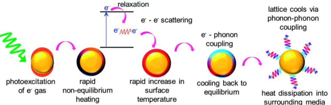
LSPR is the total oscillation of the electron cloud upon light stimulation at the characteristic plasmon resonance of AuNPs.[25] LSPR can create a strong enhancement of electromagnetic signals. After absorbing light, the plasmon energy must decay through a series of relaxation events and ultimately release energy either as light scattering (radiative relaxation) or as heat transfer (nonradiative relaxation) to the surroundings.[32] While the heating of AuNPs is rapid and in a non-equilibrium state, the cooling occurs through the quasi-equilibrium processes of electron–electron scattering, electron–phonon coupling, and phonon–phonon coupling, during which heat is dissipated and transferred to the surrounding medium. [33] The photothermal capability of AuNPs, or their efficient light-to-heat conversion, has been widely used in PTT, where heat derived from the particles can induce cell death and tumor shrinkage [25] .
Plasmonic SERS substrates for Biomolecular Detection
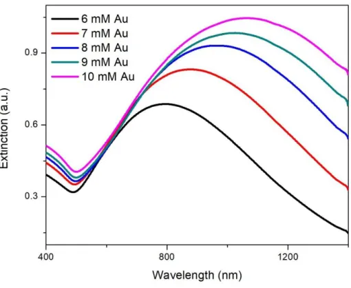
The higher the concentration of chloroauric acid used in the synthesis, the more gold is deposited on the surface of the liposomes. Zhang, X., et al., A simple microfluidic platform for rapid and efficient production of the radiotracer [(18)F]fallypride.
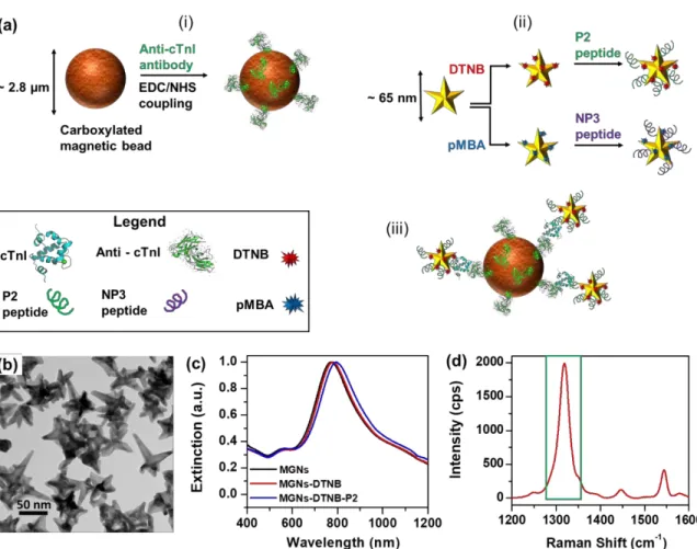
Gold Nanostars for Ultrasensitive and Multiplexed Biodiagnostics: PRADA, Portable
Summary
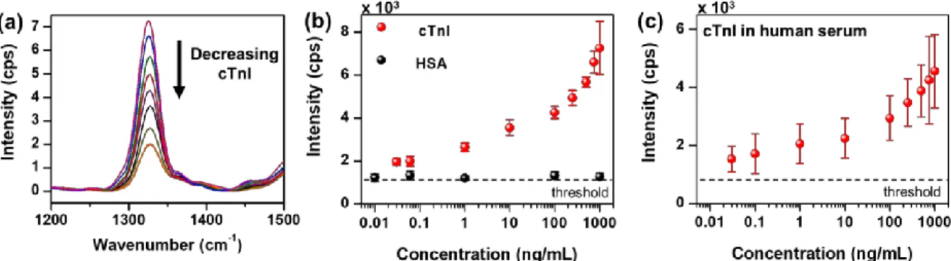
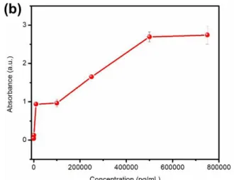
The nanostar/peptide conjugates are used to achieve high sensitivity, with a detection limit of 30 pg/ml cTnI. Furthermore, the narrow linewidths of SERS enabled multiplex detection of cTnI and neuropeptide Y (NPY), a biomarker of stress and cognition, in both buffer and human serum with high sensitivity and specificity.
Introduction
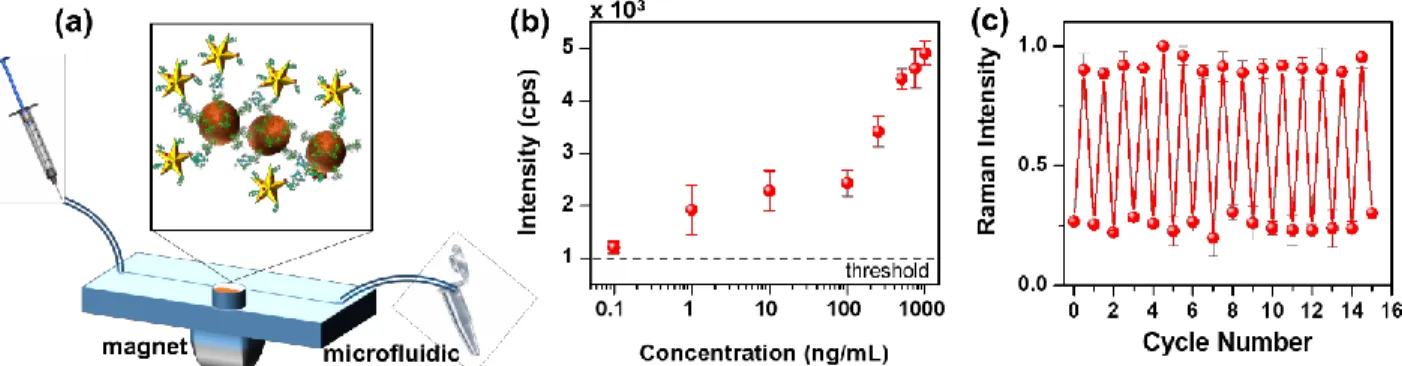
In this work, we have designed a new biodiagnostic sensor paradigm, PRADA, which synergistically integrates all these functionalities, enabling multiplexed detection of biomarkers in human serum at clinically relevant levels. We also demonstrated multiplexed detection of cTnI and neuropeptide Y (NPY) in human serum, achieving a highly sensitive detection range of 0.05 pg/ml for both biomarkers.
Results and Discussion
The sensitivity and specificity of PRADA is controlled by the controlled synthesis of the capture and detection probes as shown in Figure 2.2. The reusability of PRADA is made possible by the magnetic microbeads, as removing the magnet allows us to wash off.

Conclusions
There were several PRADA conditions that could be optimized and improved to achieve a higher detection limit and higher reproducibility of the results presented here. This is a crucial step and in the results presented in this thesis this step was missed or performed incorrectly, often resulting in high background and low signal-to-noise in the Raman signal. This step was not performed in the results presented here and should be performed in future work.
Therefore, the results presented here may differ significantly and can probably be improved if the proteins are treated in TBS buffer.
Experimental Methods
The reduction of gold on the surface of the liposome by using ascorbic acid as a reducing agent is given by the equation below (Figure 3.3). The gold particles reduced on the surface of the liposomes also had their zeta potential measured (Figure 3.8); the resulting surface charge of the gold particles was also negative. To observe the photothermal capacity of the gold-coated liposomes, ICP-OES was performed to quantify the concentration of gold that was reduced on the surface of the gold-coated liposomes.
The hollow cavity of the coated liposomes can potentially allow for the stable encapsulation of.
Liposome-Gold Hybrid Nanostructures, A Novel Approach to Combined Photothermal
Summary
Plasmon resonant gold nanoparticles of different sizes have been studied for their many applications in drug delivery, imaging, and photothermal therapy. Unfortunately, their capacity to degrade under physiological conditions after performing their function is limiting.[86] When used in combination with biodegradable liposomes, the gold nanostructures’ capacity to degrade improves. The ~100-120 nm sized liposome-gold constructs break down to form smaller ~5 nm sized particles that successfully achieve renal clearance.[87].
The resulting nanoparticles, or plasmonsomes (from plasmonic liposomes), have been analyzed for their resonances and their photothermal capabilities, as the liposome-gold constructs are produced within.
Introduction
The light-controlled content release would allow for precise and on-demand content delivery within individual cells in vitro or, when used in conjunction with endoscopic light delivery, could be used for precise medical intervention in vivo.[92] By coating the outer surface of a dipalmitoylphosphatidylcholine (DPPC) liposome, they produced a core-shell effect to provide an absorption in the near-infrared region that would be suitable for photothermally mediated drug release.[93] The use of near-infrared light promotes a greater depth of penetration with reduced tissue damage. Among cancer treatment methods, chemotherapy is particularly disappointing as it contributes about 2% to 5-year survival in all types of cancers.[97] While some produce lasting periods of remission in certain cancers such as acute lymphocytic leukemia and gestational choriocarcinoma, the use of most chemotherapeutic agents is through their narrow therapeutic window.[98] New formulations such as Doxil, doxorubicin encapsulated in thermosensitive liposomes, have been designed to limit the systemic exposure to such drugs.[99] The reduction of cardiotoxicity often associated with doxorubicin use is an advantage of drug encapsulation within the liposome.[100] This delivery system also allows for a more efficient and successful accumulation of the drug at the tumor site, due to the enhanced permeability and retention (EPR) effect, which is made possible by the leaky vasculature and reduced lymphatic drainage typical of the tumor environment.[101] Further improvement of the efficacy can be achieved by targeted delivery.[102] To carry out with the proposal of the implementation of controlled release, the substance,. The increase in temperature that causes the degradation of the liposomes indirectly kills the cancer cells, thus showing that the engineered liposome-gold constructs serve a dual purpose in delivering drugs to the site of interest as well as killing the cells make by providing heat.
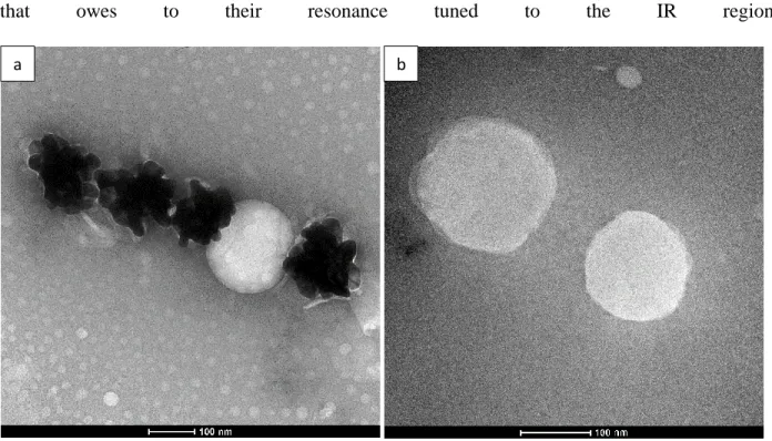
DPPC; MSPC; and PEG-2000, in a molar ratio of 90:10:4; DOX is also remotely loaded into these liposomes using a transmembrane pH gradient created when they are synthesized in buffer. The preparations were characterized by electron microscopy techniques to confirm the formation of the gold layer on the surface of the liposomes. The nanostructures have the potential to encapsulate therapeutic small molecules within the hydrophobic layer, allowing them to serve as a vehicle for additional drug delivery in addition to photothermal therapy.
Results and Discussion
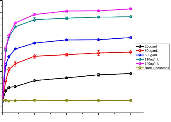
This demonstrates that the ability of gold-coated liposomes to convert light into heat is highly dependent on particle concentration; the higher the concentration, the higher the temperature reached after irradiation. The plasmon resonance of such shells is related to the interaction between plasmons supported on the inner and outer surfaces of the gold shell. This may be due to the excessive thickness of the gold shell (plasmonic hybridization).
As depicted in Figure 3.11, a rapid rise in temperature was observed in the case of gold-coated liposomes, where the temperature rose from room temperature to 46 ⁰C within 10 minutes.
Conclusions
Cytotoxicity/cell viability can be measured using the MTT assay to help determine the optimal concentration and power density. In addition, synergistic therapy, additive therapeutic efficacy, and independent photothermal treatment and chemotherapeutic treatment can be calculated and studied to demonstrate the advantages of this new combination platform. Proliferation and apoptosis assays can be performed to evaluate the therapeutic effect of the designed nanoparticles.
Possible experimental designs after optimizing reasonable ranges of parameters include the addition of immunotherapeutic agents while performing in vivo animal studies to study biodistribution, ablative ability and targeting capacity, but also to confirm the application of liposomes coated with gold as biocompatible and multifunctional agents. for effective cancer treatment, evaluating normal vs.
Experimental Methods
Smolsky, J., et al., Surface-Enhanced Raman Scattering-Based Immunoassay Technologies for Detection of Disease Biomarkers. Granger, J.H., et al., Prospects for point-of-care pathogen diagnostics using surface-enhanced Raman scattering (SERS). Ou, Y.C., et al., Diagnosis of immunomarkers in vivo via multiplexed surface-enhanced Raman spectroscopy with gold nanostars.
Yeh, E.C., et al., Zelfaangedreven geïntegreerde microfluïdische point-of-care low-cost enable (SIMPLE) chip.
Summary and Outlook