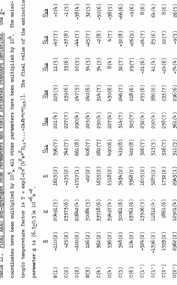
Harry Wood of the National Cancer Chemotherapy Center, who supplied several of the compounds I studied. 0 The absolute configuration of arabinose was determined using the anomalous scattering properties of P, O and Nin CuKo<.
7 ARACMP
DATA COLLECTION
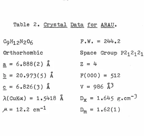
C9H12N2O6 Orthorhombic
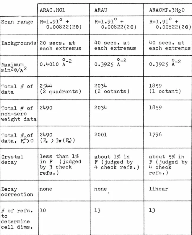
Backgrounds 20 secs. at each extremus
ARAU
13 STRUCTURAL SOLUTIONS
Now we can expand the expressions for IEI and obtain a relation between f', FH", .E, and E• Since.
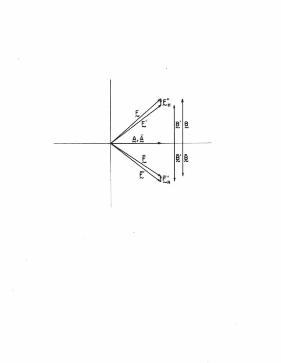
16 vector quantities. Likewise,
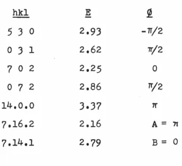
11) The only ambiguity remaining in the phase angle tan- 1 (B/A)
Due to the small anomalous effects observed in the intensities, only 7 of the 31 BRR phases were greater than 45°. Another important advantage of the BRR technique is that the model is right-handed.
20 ARAU
21 ARACMP.JH20
The position of the phosphorus atom was determined from a three-dimensional Patterson map to be approx. o.88). The four oxygen atoms in the phosphate group were sufficient to break the pseudosymmetry introduced by the phosphorus atom alone, and the resulting Fourier contained a molecular picture which was slightly stronger than the other.
30 REFINEMENT
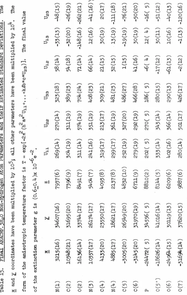
With the hydrogen atoms contributing to F 0 located, the anisotropic thermal parameters for the heavy atoms were introduced and refined for two cycles. Experimental details for the collection of this second set of data were provided (see EXPERIMENTAL-.
ARACMP),
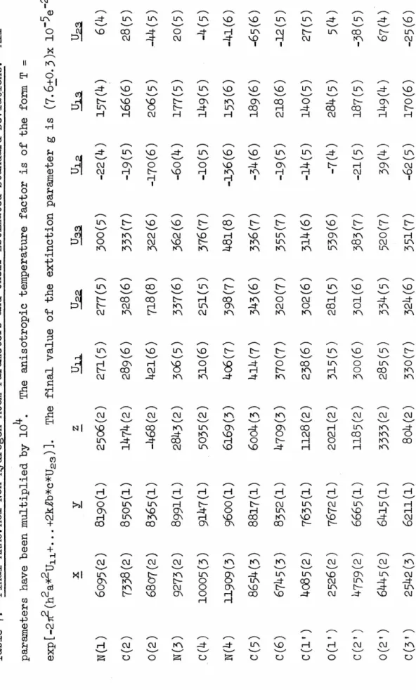
Initial least-squares refinement of the ARACMP trial model used a data set collected with molybdenum radiation. Isotropic refinement of all non-hydrogen atoms converges to an R index of 0.13 using all data collected to sin2 e/A 2 = 0.35. Two half-hydrogen atoms were used to represent the hydrogen bonding scheme here, and anisotropic refinement began.
Two cycles in which the six-parameter coordinates and temperature factors for each heavy atom were changed along with a scaling factor reduced the R index to. During this refinement phase it became increasingly clear that the quality of the fainter, high-angle data was poor enough to warrant re-collection of the data set using copper. Difference maps calculated after each refinement cycle failed to show anything but a disordered hydrogen-bonding pattern along the screw axis connecting 0(10) to 0(10), and so this pattern was retained through subsequent refinements.
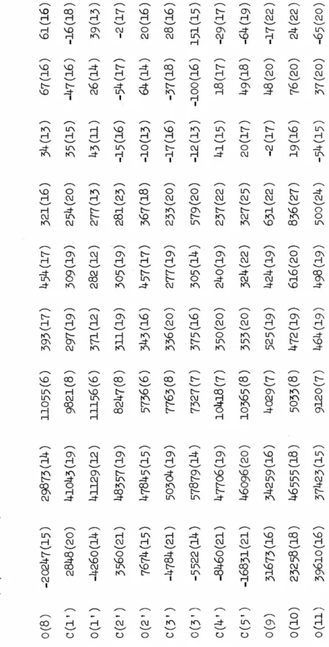
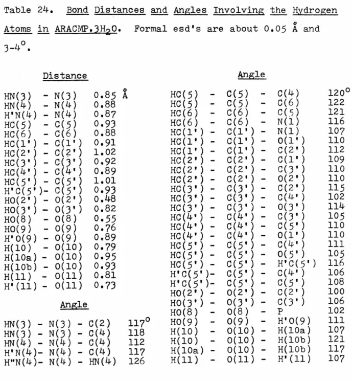
And although the methods of enantiomorph determination for both the ARAU and ARACMP cases were Hydrogen atoms associated with 0(10) were treated as follows: H(lO) was placed as accurately as possible from previous difference maps;.
57 STRUCTURE DESCRIPTIONS
A and 2
A possible manifestation of these dimension changes is that C(l') is now very close to the plane of the six-atom pyrimidine ring, as can be seen from Fig. Pseudo-translational relationships between symmetry-related uracil moieties are evident here, and were responsible for the relative ease of structure solution by Patterson techniques. There are no extremely short hydrogen bonds; in fact the interaction is the shortest observed in the structure.
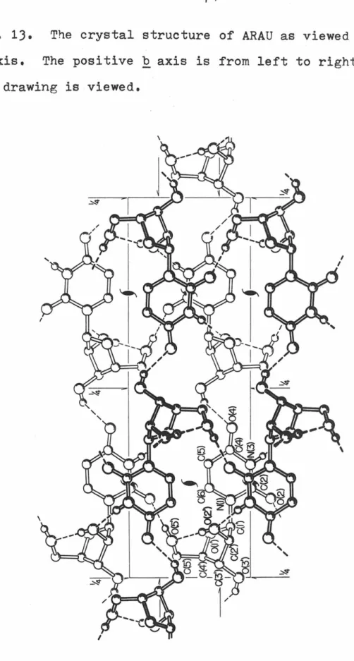
The crystal structure of ARAU as seen along the a-axis, The positive b-axis is from left to right.
78 ARACMP.JH2Q
The crystal structure of ARACMP,3H20 is shown in Figure 17. As mentioned in REFINEMENT:ARACMP.JH20, one hydrogen atom of the water molecule 0(10) was not unambiguously located and the disordered model (H(lOa), H(10b)) is was used to represent this hydrogen atom in the final refinement, but for the sake of clarity this. The view direction is parallel to -~, the ~ axis is from left to right, and the ~ axis is from top to bottom. In a manner similar to that found in the two structures of cvtidylic acid (54,55), exposed.
Again, as in all structures of cytosine compounds reported so far, atoms 0(2) and 0(1') do not participate in true hydrogen bonds.
DISCUSSION
In addition to the correlations discussed above, the length of the C(J')-C(4') bond distance appears to be related to the puckering of the sugar ring. The spread of values observed for this bond distance is quite large—the shorter lengths are associated with the C(J') endo conformation and the longer with the C(2') endo conformation. In the last six months, crystallographic studies of dinucleotides resembling the polynucleotide conformations suspected to be used in nucleic acid double helices have been published (70,71).
Arabinosyl nucleotides can also fit such descriptions, as shown by the results presented here. The correlations between ring-ring interactions, bond lengths, and out-of-plane displacements discussed above, while significant, are likely of minor importance compared to the correlations discussed above.
95 ADDENDUM
96 Footnotes
101 APPENDIX
- A feature common to the crystal structures of all hydrochloride salts of cytosine derivatives that have so
- In conjunction with the synthetic organic chemists at Caltech, and when time and funds permit, structural
- I propose to do the crystal structure of 5-methyl - isocytosine hydrochloride as the third of a series of
- I propose to carry out investigations as to the nature of the interaction of the antibiotic anthramycin with
- I propose to carry out a complete three -dimensional crystallographic analysis of a nucleic acid-protein complex
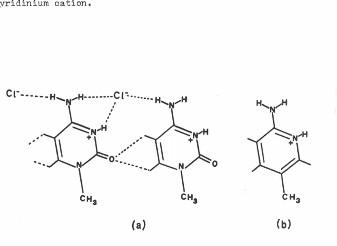
Although this arrangement must be primarily determined by the N-H ••• Cl hydrogen bonds, the C-H ••• o interactions may also be of some importance (6,7). A significant electrostatic attraction can contribute to the stability of the setup. To help determine the importance of these interactions, we examined the crystal structure of 2-amino-5-methylpyridine hydrochloride. derivative of 1-methylcytosine hydrochloride, and our intention was to compare the packing arrangements of the two crystal structures and determine the effect of removing the potential for C-H interactions. The volume of the new unit cell was half that of the original cell.
The 1,8-diiodonaphthalene molecule, due to the strong peri-interactions of the iodine atoms, is significant. The solution was done with Patterson-Fourier techniques, and the isotropic refinement of the structure stopped at an R-index of 0.14, although it was clear that a much better fit to the data was possible. 1 and J), in most cases the interactions in question appear to be the result of other, stronger packing forces and are not major contributors to the determination of the crystal structure.
Due to the uncertainty about the nature of the C-H ••• o contacts and possible hydrogen bonds involving a C-H group as a donor, it has been suggested that the crystal structure of the hydrochloride salt of 2-amino-5-methyl-pyridine by investigated by X-ray diffraction methods (11). I now propose to do the crystal structure of 5-methylisocytosine hydrochloride to further investigate the nature of the packing forces in this system of structures.
125 Proposition II
Based on the high stability of the anthramycin-DNA complex, it has been suggested that the interaction of anthramycin and DNA is at least partially a covalent one(5,6). The stability of the anthramycin-DNA complex in the presence of certain destabilizing agents was compared with that of the actinomycin-DNA complex. The observed behavior of the anthramycin-DNA complex is consistent with these mechanisms being the reaction rate.
A disadvantage of protein crystallography is that the resulting structure is that of the protein in a static structure. Attached to the other end of the tRNA is the amino acid appropriate for the specific anticodon. The function of the ribosome is to synchronize the mRNA codons and allow their specific interpretation.
The region of the mRNA around these two codons is protected by the ribosome and is not exposed to the environment. Regarding the problem of crystal growth of the native complex, it should also be taken.
HOH2C
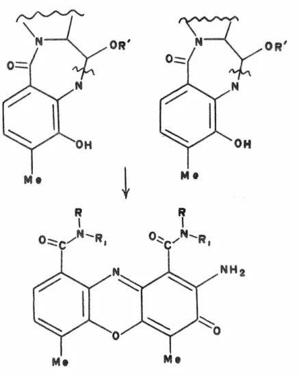
I suggest that the use of this new class of compounds be investigated as a possible new source of chemotherapeutic agents. Note that all of these nucleoside analogs are the. properties of the sulfur-substituted sugar and a purine base that by themselves have been shown to be effective in slowing cancer growth. All the analogues shown. except the adenine deriv.) were tested, and the results compared with the activities of the corresponding ribosyl derivs.
In some cases it was the ribosylpurine that was more effective, while in others it was the thioribosyl drug that was more potent. I believe that studies should be conducted to determine the effectiveness of these thioribosyl compounds, as antimetabolites, with both natural and synthetic bases attached. In addition to the adenine derivatives, other base substitutions should be tested; it is not uncommon that for a specific type of analog, one base derivative may be completely inactive, while another may be a potent antinetabolite (an example is arabinosylcytosine vs.
H HOH2C
That which adversely affects virus growth may very often not affect bacterial growth, and protozoan growth may be affected in a third way, all different from the effects on mammalian cell growth. Different cancer types should also be tested, as they often behave differently to various antimetabolites. Nucleotide analogs have been very useful in isolating a specific step or steps in biological reactions that were previously unrecognized (1).
To help determine the mechanism of action of these drugs, it is necessary to investigate which biochemical reactions they can and cannot undergo. If it is DNA, the drug must be introduced as the deoxy form, or it can be reduced enzymatically. If it is determined that any of these reactions are inhibited by the thioribosides (or deoxythioribosides), the inhibition can be reversed by the administration of a.
Answers to such questions would do much to define the mechanisms of thioribosides and could provide valuable insights into what we have about various metabolic reactions.
148 Proposition v
Each of the aforementioned components of a detector circuit has associated with it a dead time, which is In the same way a complete counting system can be assigned a value for a dead time, which is then an estimate of the count. This correction assumes that to a first approximation u 0 is of the same magnitude as ut• The validity of this assumption is.
If one reduces the lower discriminator level of the pulse height analyzer (FHA) to zero and records the number of counts reaching the scaler, the count rate is as indicated orders of magnitude greater than that due to the diffracted X-ray alone (4) ). Since derivation of the dead time correction factor given above assumes that only recorded counts contribute to dead time loss, not only is the functional form of the factor incorrect, but also the values commonly used for have no physical meaning. In any case, the source of the low energy pulses precedes the pulse height analyzer in the count.
This is important since no corrections now in use consider the existence of LEPs as. Second, a polynomial approximation for the true count rate would allow the effective dead time to be a function of the count rate and would not constrain it to a constant value.