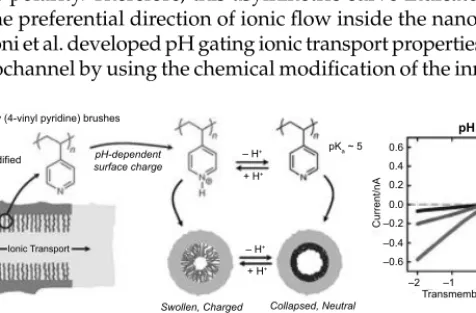
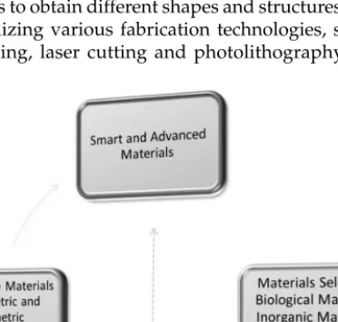
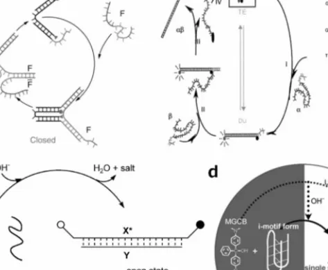
The bio-inspired research into the design and development of smart and advanced materials receives a lot of attention. Ionic current rectification of the single nanochannel before (lower left) and after (lower right) asymmetric chemical modification at 2 V.
Acknowledgements
The above bio-inspired nanochannel materials are intended as a general introduction to the design and fabrication of smart and advanced materials. The following chapters will be the systematic introduction and detailed discussion of the design, fabrication, properties and applications of various smart and advanced materials.
ABSTRACT
Introduction
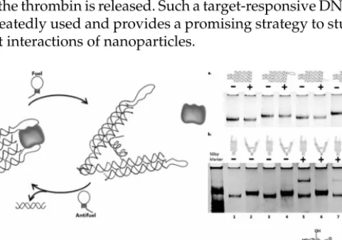
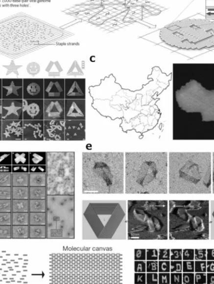
DNA origami will be highlighted in the fourth section with detailed descriptions about its invention, structural evolution and applications. Finally, we will try to give some perspectives on the development of DNA-based materials for nanoconstructions in the near future.
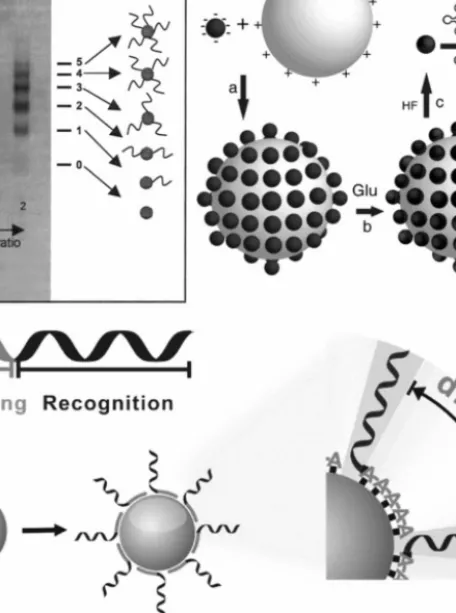
DNA-Nanoparticles Conjugates
In addition, monovalent DNA-QD conjugates were obtained using a steric exclusion strategy (Farlow et al. 2013). For the first strategy, the development of DNA conjugation with organic nanoparticles is attributed to DNA synthesis technology (Kedracki et al. 2013).
DNA Polyhedrons
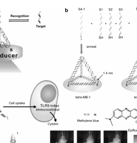
CpG can be recognized by Toll-like receptor 9 (TLR9) and induces immune responses (Hemmi et al. 2000). Second, apart from CpG and siRNAs, aptamers can also be delivered by DNA tetrahedra (Pei et al. 2012b).
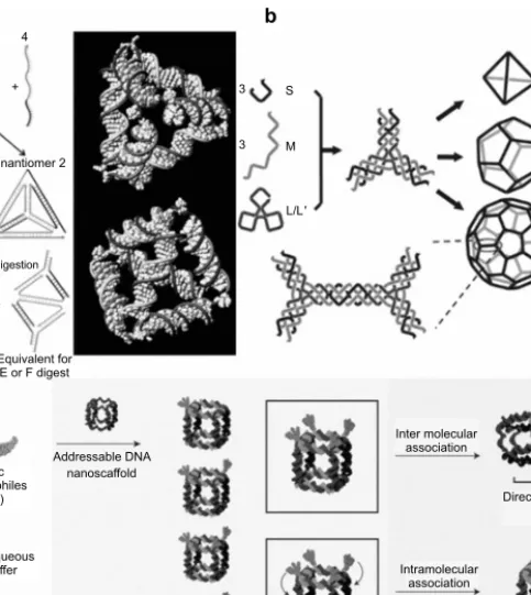
DNA Origami
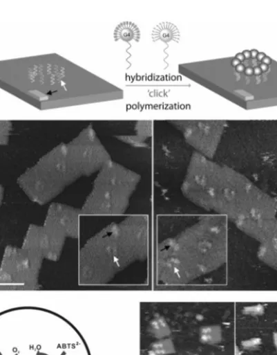
Yan's group reported an improved GOx-HRP cascade on rectangular DNA origami (Fu et al. 2012). Recently, Ding and colleagues reported a Dox delivery system based on DNA origami nanostructures (Jiang et al. 2012).
DNA Nanomachines
By controlling the alteration of DNA structures, the spatial arrangement of nanoparticles can be tuned accordingly (Sun et al. 2010, Chen et al. 2010). The yellow region represents the inner hydrophobic poly(aryl ether) and the blue regions represent the peripheral hydrophilic oligoethylene glycol (OEG) (Sun et al. 2010).
Perspectives
Precisely programmed and robust 2D streptavidin nanoarrays using nanometer-scale periodic wells embedded in DNA origami assembly. Modification of the quantum efficiency of organic fluorophores using gold nanoparticles on DNA origami scaffolds.
Self-Assembly of Proteins into Various Nanoscale Architecture
Another major component of the cytoskeleton is a globular multifunctional protein called actin, which assembles into a helical microfilament (Gunning et al. 2015). Intermediate filaments are the third major component of the cytoskeleton and are essential for maintaining proper tissue structure and function (Fuchs and Cleveland 1998). Elastic fibers represent interesting biomaterials that are a component of the extracellular matrix in tissues (Muiznieks et al. 2010).
Negative staining electron microscopy of elastin fibers revealed distinct filaments aligned parallel to the long axis of the fiber (Gotte et al.
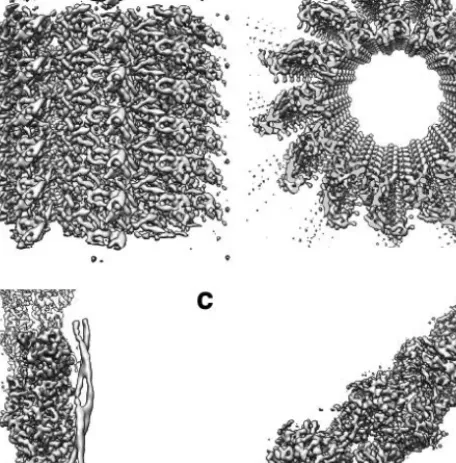
Proteins Assemble into Dynamic Nanomachines
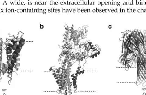
A crystal structure of the potassium channel from Streptomyces lividans with sequence similarity to all known K + channels, particularly in the pore region (PDB ID: 1BL8) (Doyle et al. 1998). In some bacteria, the ATP synthase works in the opposite direction, where the energy released by ATP hydrolysis drives the translocation of protons out of the cell, generating a proton gradient across the cytoplasmic membrane (Noji et al. 1997). The energy from ATP hydrolysis of the V1 domain is converted into rotation of the c-ring and proton transport.
The motor domain of kinesin (PDB ID: 1BG2) bears a striking structural similarity to the catalytic core domain of myosin, showing that it shares a similar force-generating strategy with myosin (Kull et al. 1996).
Engineering of Protein Nanomaterials 1 In Silico Design of Protein Nanomaterials
Recently, the principle of protein design was extended to predict the protein-protein interaction interfaces that can guide the self-assembly of de novo designed proteins in higher order architecture and biomaterials (King et al. 2012, King et al. 2014). This approach was later further generalized to the design of the co-assembly of multicomponent protein nanomaterials (King et al. 2014). Engineering the interface between protein nanomachines and inorganic nanomaterials represents another important avenue for the development of smart biomaterials (Ishii et al. 2003).
In addition, options remain to covalently bio-conjugate proteins to the surface of inorganic nanomaterials, integrating them into programmable hybrid building blocks (Marco et al. 2010).
Applications of Protein Nanomaterials 1 Energy
A protein fiber system that is joined by two peptides exhibits programmable assembly, stability and morphology (Papapostolou et al. 2007). Using antibody-coated nanoparticles, the detection sensitivity of the target protein can be increased down to the attomolar concentration (Nam et al. 2003). Fourth, protein nanomaterials can be used as part of tissue engineering to help regenerate or repair damaged tissues using suitable scaffolds based on nanomaterials and growth factors (Ellis-Behnke et al. 2006).
Protein hybrid with inorganic nanomaterials such as graphene, carbon nanotubes, molybdenum disulfide and tungsten disulfide can be used as reinforcing agents to produce mechanically strong biodegradable polymeric nanocomposites for bone tissue engineering applications (Lalwani et al. 2013).
Perspective
Crystal structure of Taq DNA polymerase in complex with an inhibitory Fab: The Fab is directed toward an intermediate in the enzyme's helix-coil dynamics. Three-dimensional structure of the flagellar rotor from a clockwise-locked mutant of Salmonella enterica serovar Typhimurium. Atomic Structure of the Extremophilic icosahedral 75 MDa Sulfolobus Virus Determined by CryoEM and X-ray Crystallography.
This chapter presents a comprehensive overview of the most promising smart materials for controlled drug release.
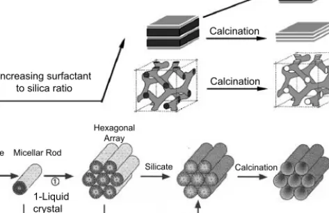
Smart Inorganic Nanomaterials for CDR 1 Mesoporous Silica Nanoparticles
Controlled release systems based on mesoporous silica nanomaterials have developed rapidly in the past decade (Ambrogio et al. 2011). MSNs with hyaluronic acid (HA) modified on the surface respond to Hyal-1 (Chen et al. 2013). Scheme of near-infrared light (NIR)-responsive MSNs for controlled drug release (Yang et al. 2012).
The results indicated that IONPs hold promise in the application of skin cancer treatment (Rao et al. 2015).
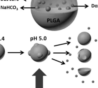
Smart Polymeric Materials for CDR 1 Polymeric Capsules
Scheme illustrating the capsules with an anti-cancer drug and NaHCO3, which could release drugs in an acidic state (Ke et al. 2011). The permeability of the hollow capsule shells can be easily photocontrolled at different irradiation times (Tao et al. 2004). The pH-cleavable property of the hydrazine bond would lead to a pH-triggered drug release (del Rosario et al. 2010).
In vitro studies also confirmed the possible reduction-induced release of DOX within tumor cells (Li et al. 2009).
Other Smart Materials for CDR 1 Albumins
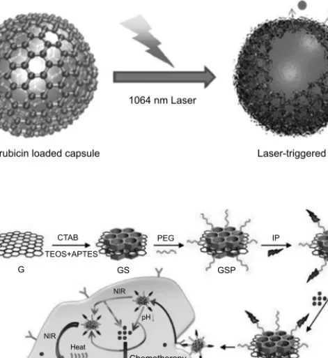
In the presence of 10 mM DTT, hydrolysis of the disulfide led to the release of DOX from the PMs particles. The GNR-incorporated liposome accumulated more efficiently at the tumor site and released the chemotherapeutic DOX upon NIR stimulation, leading to a significant increase in efficacy (Agarwal et al. 2011). A thiolated Bodipy dye (HSBOP) as cargo was loaded by covalent interaction, which could be released in the presence of GSH (Hong et al. 2006).
Upon NIR irradiation, the absorbance of AuNRs would transduce the light into heat, which denatured the DNA assembly, resulting in the release of DOX (Xiao et al. 2012).
Promise and Challenges
Stimuli-responsive conformational switching of peptide gatekeepers for controlled release of guests from mesoporous silica nanocontainers. Glucose- and pH-responsive controlled release of cargo from protein-gated carbohydrate-functionalized mesoporous silica nanocontainers. Lysozyme as a pH-responsive valve for the controlled release of gas molecules from mesoporous silica.
Mesoporous silica nanoparticle-based dual drug delivery system for glucose-responsive controlled release of insulin and cyclic enhancer.
Background and Basic Principles
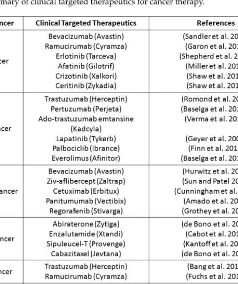
Considering tumor heterogeneity, we will review and discuss smart materials and relevant molecular targets for cancer based on cancer types, in order of cancer incidence.
Smart Materials for Cancer Diagnosis and Treatment 1 Lung Cancer
It works by disrupting both the HER2 and EGFR signaling pathways in HER2-positive breast cancers (Geyer et al. 2006). Currently, there is a common clinical targeted therapeutic for liver cancer, which is Sorafenib (Nexavar) (Cheng et al. 2009) (as seen in Table 1). The targeting ligand YC21 (an oligopeptide consisting of 21 amino acid units) was coupled to the nanovector composed of β-cyclodextrin and low molecular weight polyethyleneimine (PEI, Mw 600) to form the EGFR target gene vector (named YP ) (called YP) Liu et al. 2012).
FDA in 2012 for the treatment of a rare brain tumor called subependymal giant cell astrocytoma (Krueger et al. 2010).
Summary
A novel strategy for surface modification of superparamagnetic iron oxide nanoparticles for lung cancer imaging. Graphene quantum dots conjugated albumin nanoparticles for targeted drug delivery and imaging of pancreatic cancer. Single-chain epidermal growth factor receptor-conjugated nanoparticles for in vivo tumor targeting and imaging.
Lactosylated gramicidin-based lipid nanoparticles (Lac-GLN) for targeted delivery of anti-miR-155 to hepatocellular carcinoma.
Theory and Background
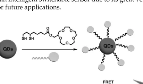
Therefore, it can be expected that electrostatic interaction between the donor and the acceptor moieties with different charge (i.e., positive charge or negative charge) will change the photophysical properties of the functionalized QDs. Fluorescence resonance energy transfer (FRET) is a distance-dependent physical process by which energy is transferred non-radiatively from an excited molecular fluorophore (the donor) to another fluorophore (the acceptor) via long-range intermolecular dipole— dipole coupling (Bagalkot et al. 2007). The photophysical properties of QDs can be controlled by their nanocrystal core sizes, the shell thickness and the composition of the semiconductor materials of cores and shell(s) and partially by their surface ligands (Green 2010).
The superior photophysical characteristics of semiconductor QDs (high fluorescence quantum yields and stability against photobleaching) are usually observed in organic solvents, and their introduction into aqueous media is usually accompanied by a drastic decrease in the luminescence yields of the QDs (Gao. 2005).
Quantum Dots-Based Smart Sensing Systems
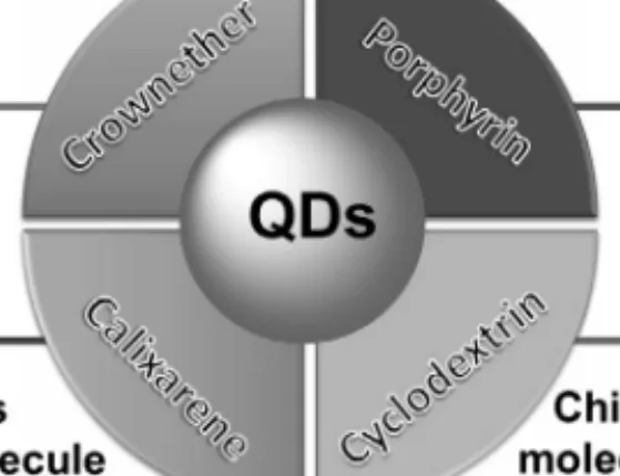
Through the strategy of supramolecular assembly, a quantum dot (QD) associated with palladium(II) porphyrins has been developed to detect oxygen (O2) in organic solvents (Lemon et al. 2013). This quenching was partly explained by FRET from CdSe/ZnS nanoparticles to porphyrins (Ellis et al. 1997). In addition, Patra's group has fabricated a fluorescence switch using an alloy (Cd1−xZnxS) quantum dot (QD) in the presence of porphyrin and cucurbituril (Mandal et al. 2013).
Increased stiffness of the system could suppress nonradiative pathways and protect the core (Wang et al. 2008).
Perspective of the Future Research
Structural changes of the host molecules around the QDs appeared to play an important role in the fluorescence changes. The calix[8]arene-capped CdSe/ZnS QDs showed intensive fluorescence emission with high quantum yield. After that, macrocyclic molecules functionalized QDs based on FRET mechanism will be one of the most promising methods for fluorescence-based biosensing.
After a multiplexed solid-phase nucleic acid hybridization test using quantum dots as donors in fluorescence resonance energy transfer.