Two new deep-reef basslets (Teleostei, Grammatidae, Lipogramma), with comments on the eco-evolutionary relationships of the genus
Carole C. Baldwin1, D. Ross Robertson2, Ai Nonaka1, Luke Tornabene1
1 Department of Vertebrate Zoology, National Museum of Natural History, Smithsonian Institution, Washing- ton, DC 20560 2 Smithsonian Tropical Research Institute, Balboa, Republic of Panama
Corresponding author: Carole C. Baldwin ([email protected])
Academic editor: D. Bloom | Received 9 September 2016 | Accepted 21 November 2016 | Published 7 December 2016 http://zoobank.org/B8ADA2DB-F7DF-41F7-977D-9EB79DDDC34A
Citation: Baldwin CC, Robertson RD, Nonaka A, Tornabene L (2016) Two new deep-reef basslets (Teleostei, Grammatidae, Lipogramma), with comments on the eco-evolutionary relationships of the genus. ZooKeys 638: 45–82.
https://doi.org/10.3897/zookeys.638.10455
Abstract
The banded basslet, Lipogramma evides Robins & Colin, 1979, is shown to comprise two species: L. evides, which inhabits depths of 133–302 m, and a new species described here as Lipogramma levinsoni, which in- habits depths of 108–154 m and previously was considered to represent the juvenile of L. evides. A second new species of banded basslet, described here as Lipogramma haberi, inhabits depths of 152–233 m and was previously not reported in the literature. Morphologically, the three species differ in color patterns and modal numbers of gill rakers, whereas various other morphological features distinguish L. levinsonsi from L. evides and L. haberi. DNA barcode data and multilocus, coalescent-based, species-delimitation analysis support the recognition of the three species. Phylogenetic analysis of mitochondrial and nuclear genetic data supports a sister-group relationship between the two deepest-living of the three species, L. evides and L. haberi, and suggests that the shallower L. levinsoni is more closely related to L. anabantoides Böhlke 1960, which inhabits depths < 120 m. Evolutionary relationships within Lipogramma thus appear to be correlated with species depth ranges, an eco-evolutionary pattern that has been observed in other Carib- bean marine teleosts and that warrants further investigation. The new species represent the eleventh and twelfth new fish species described in recent years from exploratory submersible diving in the Caribbean in the globally poorly studied depth zone of 50–300 m. This study suggests that there are at least two ad- ditional cryptic species of Lipogramma, which are being analyzed in ongoing investigations of Caribbean deep-reef ecosystems.
Copyright Carole C. Baldwin et al. This is an open access article distributed under the terms of the Creative Commons Attribution License (CC BY 4.0), which permits unrestricted use, distribution, and reproduction in any medium, provided the original author and source are credited.
Keywords
Manned submersible, cryptic species, integrative taxonomy, phylogeny, ocean exploration, Smithsonian Deep Reef Observation Project (DROP)
Introduction
The western Atlantic family Grammatidae comprises small, usually brightly colored fishes in two genera, Gramma with four species and Lipogramma with eight (Robertson and Van Tassell 2015). Among other characters, the two genera are distinguished by the absence of a lateral line and presence of thickened, spinous, outer procurrent rays in Lipogramma (Mooi & Gill, 2002). The Banded Basslet, Lipogramma evides Robins
& Colin, 1979, was described based on six specimens collected from Barbuda, Jamaica, Mexico, and Nicaragua. The original description also included observations of the spe- cies from Belize by Colin (1974). Subsequently, six additional specimens from the Bahamas were reported by Gilmore and Jones (1988). Robins and Colin (1979) noted differences in pigment pattern between adults and what they thought was a juvenile L. evides (Fig. 1A, B), in particular the presence of broader and more intense dark bands on the juvenile that completely encircle the body. Gilmore and Jones (1988) further commented on the presumed color differentiation between ontogenetic stages and noted that heavily banded “juveniles” (Fig. 1C) inhabit shallower waters (< 200 m) than adults (as deep as 250 m).
Exploratory submersible diving to 300 m in the southern and eastern Caribbe- an over the past several years by the Smithsonian Institution’s Deep Reef Observa- tion Project (DROP) has resulted in the collection of over 50 specimens of “banded basslets” assignable to Lipogramma based on the absence of a lateral line and presence of spinous procurrent caudal-fin rays. That material includes individuals with both pigment patterns observed by previous authors and a third pigment pattern not previ- ously described. Genetic and morphological analyses of individuals with those three pigment patterns suggested three distinct species and show that the heavily banded pattern is not an ontogenetic feature but diagnostic of a separate species. That species reaches a smaller maximum size than L. evides and has a shallower depth range. The other new species is similar in size and depth of occurrence to L. evides. Here we de- scribe those two new species of Lipogramma, morphologically and genetically compare them with L. evides, and discuss depth distributions and evolutionary relationships of species of the genus.
Methods and materials
Collecting and morphology. Basslets were collected using Substation Curaçao’s manned submersible Curasub (http://www.substation-curacao.com). The sub has two flexible, hy draulic arms, one of which is equipped with a quinaldine/ethanol-ejection
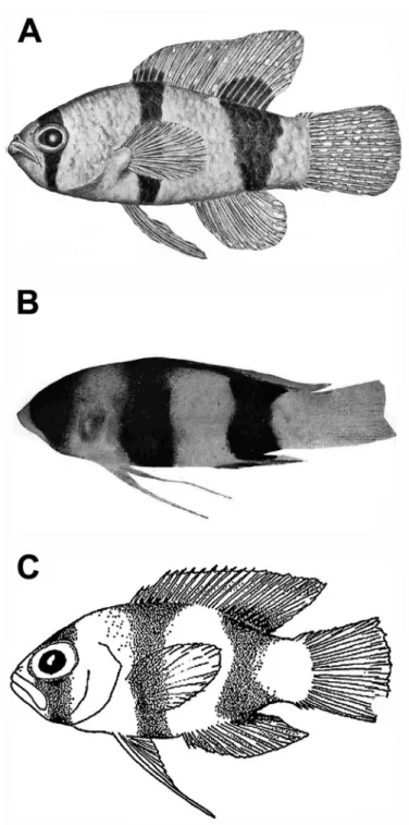
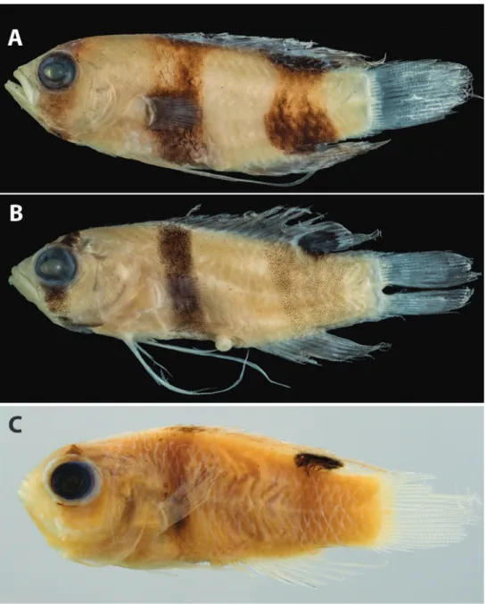
Figure 1. Previously published images of A Lipogramma evides, 34.4 mm SL, ANSP 134329, holotype, from Robins and Colin (1979: fig. 1) B Lipogramma levinsoni sp. n., 12.6 mm SL, ANSP 134332, as juvenile paratype of L. evides in Robins and Colin (1979: fig. 2) C Lipogramma levinsoni sp. n., 14.1 mm SL, IRCZM 107: 07660, as juvenile of L. evides in Gilmore and Jones (1988: fig. 3). Images reproduced with permission from Bulletin of Marine Science.
system and the other with a suction hose. Anesthetized fish specimens were captured with the suc tion hose, which empties into a vented plexiglass cylinder attached to the outside of the sub. At the surface, the specimens were photographed, tissue sampled, and fixed in 10% formalin. Measurements were made weeks to months after fixa- tion and subsequent preservation in 75% ethanol and were taken to the nearest 0.1 mm with dial calipers or an ocular micrometer fitted into a Wild stereomicroscope.
Selected preserved specimens were later photographed to document preserved pig- ment pat tern and X-rayed with a digital radiography system. Images of supraorbital pores and tooth-like structures on gill rakers were made using a Zeiss Axiocam on a Zeiss Discovery V12 SteREO microscope. Counts and measurements follow Hubbs and Lagler (1947). Specimens were cleared and stained following the protocol of Dingerkus and Uhler (1977). Symbolism for configuration of supraneural bones, anterior neural spines, and anterior dorsal pterygiophores follows Ahlstrom et al.
(1976). USNM = Smithsonian Institution, National Museum of Natural History;
ANSP = Academy of Natural Sciences, Philadelphia; IRCZM = Indian River Coastal Zone Museum, Harbor Branch Foundation, Fort Pierce, Florida; UF = University of Florida, Gainesville.
Molecular analyses. Tissue samples for 97 specimens assignable to eight species of Lipogramma were used for molecular analyses (Appendix 1). Tissues of L. rosea Gilbert, 1979 (in Robins and Colin 1979), L. regia Robins & Colin, 1979, and L.
flavescens Gilmore & Jones, 1988 were not available. Tissues were stored in saturated salt-DMSO (dimethyl sulfoxide) buffer (Seutin et al. 1991). DNA extraction and cy- tochrome c oxidase subunit I (COI) DNA barcoding were performed for 96 specimens (i.e., for all available specimens except one L. anabantoides – Appendix 1) as outlined by Weigt et al. (2012). Four nuclear markers were amplified and sequenced—TMO- 4C4, Rag1, Rhodopsin, and Histone H3—for 18 specimens of Lipogramma, and one or more of those genes was sequenced for an additional three specimens (Appendix 1). Primers and PCR conditions for the nuclear markers followed Lin and Hastings (2011, 2013). Sequences were assembled and aligned using Geneious v. 9 (Biomatters, Ltd., Aukland). A neighbor-joining (NJ) network was generated for the COI data using the K2P substitution model (Kimura 1980) in the tree-builder application in Geneious. Mean within- and between-species K2P genetic distances were calculated from the COI data in MEGA v. 7 (Kumar et al. 2015). Genetic distances were con- sidered as corroborating morphology-based species delineation if the distances be- tween species were ten or more times the intraspecific differences (Hebert et al. 2004).
The alignments of COI and nuclear genes were concatenated and phylogeny was inferred using Bayesian Inference (BI) and Maximum Likelihood (ML), partitioning by gene. For the Bayesian analysis, substitution models and partitioning scheme were chosen using PartitionFinder (Lanfear et al. 2012) according to Bayesian Informa- tion Criterion scores. The chosen scheme had the following partitions and models:
COI, HKY+I+G; Histone H3 plus Rhodospin, HKY+G; TMO-4C4, K80+G; Rag1,
K80+G. All partitions in the ML analysis received a GTR-GAMMA substitution model. The BI phylogeny was inferred in the program MrBayes v. 3.2 (Ronquist et al. 2012) using two Metropolis-coupled Markov Chain Monte Carlo (MCMC) runs, each with four chains. The analysis ran for 10 million generations sampling trees and parameters every 1000 generations. Burn-in, convergence and mixing were assessed using Tracer (Rambaut and Drummond 2007) and by visually inspecting consensus trees from both runs. The ML analysis was done in the program RAxML v.8.2.9 (Stamatakis, 2014), using 20 initial random searches, and topological support was assessed using 1000 bootstrap replicates. Outgroups for the phylogenetic analysis included two species of Gramma and several other genera from the Ovalentaria sensu Wainwright et al. (2012): Acanthemblemaria (Labrisomidae), Helcogramma (Trip- terygiidae), Blenniella (Blenniidae), and Tomicodon (Gobiesocidae).
To corroborate the morphologically diagnosed species using our molecular data, we conducted a coalescent-based, Bayesian species-delimitation analysis (Yang and Rannala 2010, 2014). We used the computer program BP&P ver. 3.2 (Bayesian Phy- logenetics and Phylogeography – Yang and Rannala [2010], Yang [2015]), which ana- lyzes multi-locus DNA sequence alignments under the multispecies coalescent model (Rannala and Yang 2003). We used the five DNA alignments for the 21 Lipogramma specimens in BP&P, with each sequence in the alignments being assigned to one of eight groups a priori, based on the diagnostic morphological and coloration charac- ters discussed in the ‘Morphological Comparisons’ section below. BP&P was then used to jointly infer a species tree and calculate posterior probabilities of different species-delimitation models containing either eight species, fewer than eight species (i.e. lumping multiple ‘morphological species’), or more than eight species (i.e. split- ting ‘morphological species’ into multiple cryptic species).
Depth distributions. To evaluate depth distributions we searched FishNet2 (www.fishnet2.net) for all Lipogramma specimens that were identified to species and that included data on the depth of capture. For some specimens, capture depth was given as a range of possible depths, and in instances where this range was 50 m or nar- rower, we took the mean depth as a proxy for a point estimate of the exact depth of capture. Broader depth ranges of capture were excluded. Depth records for L. evides were only included for specimens whose identifications we confirmed to avoid possible confusion with one of the two new species described here. When combined with depth data from specimens from DROP collections, this search resulted in depth records for 278 identified specimens of Lipogramma. We also included depth records from 83 visual observations from DROP submersible dives, excluding those observations where there was uncertainty regarding identification of the three morphologically similar spe- cies (L. evides and the two species described here).
Accession numbers. GenSeq nomenclature (Chakrabarty et al. 2013) and Gen- Bank accession numbers for DNA sequences derived in this study are presented along with museum catalog numbers for voucher specimens in Appendix 1.
Taxonomy Hourglass Basslet
Lipogramma levinsoni Baldwin, Nonaka & Robertson, sp. n.
http://zoobank.org/C12172C1-B3BF-48B8-B267-61D845EDCC63 Figure 2
Lipogramma evides Robins & Colin, 1979: 43, fig. 2, table 1, ANSP 134332, paratype from Jamaica (photograph, counts, measurements).
Lipogramma evides Robins & Colin, 1979, fig. 3 in Gilmore and Jones (1988: 441), IRCZM 107:07660 from San Salvador, Bahama Islands (illustration, habitat in- formation).
Type locality. Curaçao, southern Caribbean.
Holotype. USNM 406139, 28.3 mm SL, tissue no. CUR11139, Curasub submers- ible, sta. CURASUB11-02, Curaçao, off Substation Curaçao, 12.083197 N, 68.899058 W, 137–146 m depth, 23 May 2011, C. Baldwin, D. Robertson & B. Van Bebber.
Paratypes. BONAIRE: USNM 426784, 24.2 mm SL, tissue no. CUR13183, Cu- rasub submersible, Bonaire, Bonaire City Dock, Kralendijk, Dive 2, 12.15 N, 68.2829 W, 121-137 m depth, 30 May 2013, B. Van Bebber, A. Schrier, C. Baldwin, T. Chris- tiaan; CURAÇAO: ANSP 201863, 24.0 mm SL, Curasub submersible, Curaçao, off Substation Curaçao, 12.083197 N, 68.899058 W, no depth data available; UF 238589, 25.0 mm SL, tissue no. CUR11018, Curasub submersible, sta. CURASUB11-22, Curaçao, off of Substation Curaçao downline, 12.083197 N, 68.899058 W, no depth data available, 27 February 2011, C. Baldwin & L. Weigt; USNM 406393, 25.7 mm SL, tissue no. CUR11393, Curasub submersible, sta. CURASUB11-06, Curaçao, 132 m depth, 31 May 2011, C. Baldwin, A. Driskell, A. Schrier & B. Van Bebber; USNM 414877, 25.3 mm SL, cleared and stained, tissue no. CUR12159, Curasub submers- ible, sta. CURASUB12-15, Curaçao, off of Substation Curaçao downline, 12.083197 N, 68.899058 W, 128 m depth, 10 August 2012, A. Schrier, B. Brandt, C. Baldwin, A. Driskell & P. Mace; USNM 440229, 12.7 mm SL, Curasub submersible, sta. CU- RASUB14-07, Curaçao, in between Porto Marie and Daaibooi beaches, 12.202842 N, 69.089507 W, 123 m depth, 21 March 2014, C. Baldwin et al.; USNM 440230, 13.4 mm SL, Curasub submersible, sta. CUR13-18, Curaçao, Playa Forti, Westpoint, 12.3679 N, 69.1553 W, 127 m, 15 August 2013, C. Baldwin, B. Brandt, A. Schrier, K. Johnson & C. DeForest; USNM 406140, 19.5 mm SL, tissue no. CUR11140, Cu- rasub submersible, sta. CURASUB11-02, Curaçao, 137-146 m depth, 23 May 2011, C. Baldwin, D. Robertson & B. Van Bebber. DOMINICA: USNM 440231, 17.0 mm SL, tissue no. DOM16229, Curasub submersible, off northwest Dominica, no specific collection data available, March 2016, R/V Chapman Crew.
Non-type specimens. BONAIRE: USNM 426754, 21.2 mm SL, tissue no.
CUR13184, Curasub submersible, Bonaire, Bonaire City Dock, Kralendijk, Dive 2,
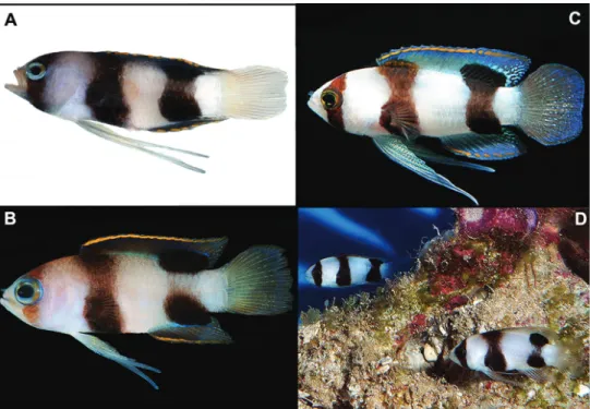
Figure 2. Lipogramma levinsoni sp. n. A USNM 406139, holotype, 28.3 mm SL, photographed prior to preservation, photo by D. R. Robertson and C. C. Baldwin B USNM 406394, 22.2 mm SL , photo- graphed prior to preservation, photo by D. R. Robertson and C. C. Baldwin C and D Aquarium photos, Curaçao Sea Aquarium, photos by D. Ross Robertson.
12.15 N, 68.2829 W, 121–137 m depth, 30 May 2013, B. Van Bebber, A. Schrier, C.
Baldwin, T. Christiaan; USNM 426802, 9.4 and 18.3 mm SL, Curasub submersible, Bonaire, Bonaire City Dock, Kralendijk, 12.15 N, 68.2829 W, 114–137 m depth, 30 May 2013, B. Van Bebber, A. Schrier, C. Baldwin, T. Christiaan. CURAÇAO: USNM 426774, 17.6 mm SL, tissue no. CUR13267, Curasub submersible, sta. CURAS- UB13-18, Curaçao, Playa Forti, Westpoint, 12.3679 N, 69.1553 W, 118 m depth, 15 August 2013, C. Baldwin, B. Brandt, A. Schrier, K. Johnson & C. DeForest; USNM 426730, 12.3 mm SL, tissue no. CUR13268, Curasub submersible, sta. CURAS- UB13-18, Curaçao, Playa Forti, Westpoint, 12.3679 N, 69.1553 W, 118 m depth, 15 August 2013, C. Baldwin, B. Brandt, A. Schrier, K. Johnson & C. DeForest; USNM 406011, 20.9 mm SL, tissue no. CUR11011, Curasub submersible, sta. CURAS- UB11-22, Curaçao, off of Substation Curaçao downline, 12.083197 N, 68.899058 W, no depth data available, 27 February 2011, C. Baldwin & L. Weigt; USNM 406012, 18.0 mm SL, tissue no. CUR11012, Curasub submersible, sta. CURASUB11-22, Curaçao, off of Substation Curaçao downline, 12.083197 N, 68.899058 W, no depth data available, 27 February 2011, C. Baldwin & L. Weigt; USNM 406019, 14.0 mm SL, tissue no. CUR11019, Curasub submersible, sta. CURASUB11-22, Curaçao, off of Substation Curaçao downline, 12.083197 N, 68.899058 W, no depth data avail-
able, 27 February 2011, C. Baldwin & L. Weigt; USNM 406394, 22.2 mm SL, tissue no. CUR11394, Curasub submersible, sta. CURASUB11-06, Curaçao, 132 m depth, 31 May 2011, C. Baldwin, A. Driskell, A. Schrier & B. Van Bebber. DOMINICA:
USNM 438703, 19.0 mm SL, tissue no. DOM16052, Curasub submersible, sta. CU- RASUB16-07, Toucari Bay, Toucari, Dominica, NW corner of island, 15.608047 N, 61.471788 W, no depth data available, 2 March 2016, A. Schrier, R. Bakmeijer, B.
Van Bebber & F. van der Hoeven; JAMAICA: ANSP 134332, 12.6 mm SL, Nekton Gamma dive 141, collection 151-2, Jamaica, Discovery Bay, 145 m depth, 15 August 1972, L. Land & S. Hastings.
Diagnosis. A species of Lipogramma distinguishable from congeners by the fol- lowing combination of characters: pectoral-fin rays 16–18 (modally 17), gill rakers 17–20 (modally 19); three supraorbital pores present along dorsal margin of orbit, no pore present between pore at mid orbit and one at posterodorsal corner of orbit;
caudal fin truncate, tips of lobes rounded; body with three broad blackish bars (one on head, two on trunk) on white background, width of bar on head sufficient to en- compass entire eye, width just ventral to eye averaging 26.4% head length; trunk bars sometimes hourglass shaped, with narrower and less intensely colored central regions;
anterior trunk bar covering pectoral-fin base; posterior trunk bar extending onto dorsal and anal fins as large oval blotches bordered in part by white or blue pigment to form partial ocelli; dorsal and anal fins with thin orange sub-marginal stripe. The new spe- cies is further differentiated from congeners for which molecular data are available in mitochondrial COI and nuclear Histone 3, Rhodopsin, TMO-4C4, and RAG1.
Description. Counts and measurements of type specimens given in Table 1.
Frequency distributions of pectoral-fin rays and gill rakers on the first arch are given in Table 2. Twenty specimens examined, 9.4 to 28.3 mm SL. Dorsal-fin rays XII, 9 (last ray composite); anal-fin rays III, 8 (last ray composite); pectoral-fin rays 16–18, modally 17, 17 on both sides in holotype; pelvic-fin rays I,5; total caudal-fin rays 25 (13 + 12), principal rays 17 (9 + 8), spinous procurrent rays 6 (III + III), and 2 addi- tional rays (i + i) between principal and procurrent rays that are neither spinous nor typically segmented; vertebrae 25 (10 + 15); pattern of supraneural bones, anterior dorsal-fin pterygiophores and dorsal-fin spines 0/0/0+2/1+1/1/; ribs on vertebrae 3–10; epineural bones present on vertebrae 1-16 in holotype and cleared and stained paratype (difficult to assess in radiographs of most other specimens); gill rakers on first arch 17–20 (5-6 + 12–14), modally 19 (6 + 13), 19 (6 + 13) in holotype; up- permost four and lowermost one or two rakers very small or present only as nubs, all other gill rakers elongate and slender with tooth-like secondary rakers as in L. evides (Fig. 3); pseudobranchial filaments 5–7 (7 in holotype), filaments fat and fluffy;
branchiostegals 6.
Spinous and soft dorsal fins confluent, several soft rays at rear of fin forming el- evated lobe that extends posteriorly beyond base of caudal fin. Pelvic fin, when de- pressed, extending posteriorly to point between anterior base of anal fin and beyond base of caudal fin, elongate first pelvic-fin ray broken in most preserved specimens.
Dorsal profile from snout to origin of dorsal fin convex. Diameter of eye of holotype
Table 1. Counts and measurements of type specimens of Lipogramma levinsoni sp. n.. Measurements are in percent SL except width of bar ventral to eye, which is in percent head length. C&S = cleared and stained; CP = caudal peduncle; PFO = pelvic-fin origin; P1 = pectoral fin; P2 = pelvic fin; DXII = twelfth dorsal-fin spine. “Other Caudal” rays include “i” – a slender, flexible, non-spinous, and typically non-segmented ray and “I” – a spinous procurrent ray.
USNM 406139 USNM 406393 USNM 406140 USNM 414877 USNM 426784 USNM 440230 USNM 440229 USNM 440231 ANSP 201863
UF 238589 HolotypeParatypeParatypeParatype (C&S)ParatypeParatypeParatypeParatypeParatypeParatype SL28.325.719.525.3 24.213.426.317.024.025.0 Dorsal-fin raysXII, 9XII, 9XII, 9XII, 9XII, 9XII, 9XII, 9XII, 9XII, 9XII, 9 Anal-fin raysIII, 8III, 8III, 8III, 8III, 8III, 8III, 8III, 8III, 8III, 8 Principal caudal9+89+89+89+89+89+89+8Broken9+89+8 Other caudalIIIi+iIIIIIIi+iIIIIIIi+iIIIIIIi+iIIIIIIi+iIIIIIIi+iIIIIIIi+iIIIBrokenIIIi+iIIIIIIi+iIII Pectoral-fin rays17, 1717, 1617, 1717, 1717*, 1717, 1717, 1816, -17, 1717, 17 Gill rakers 19201919191918181918 Head length33.236.235.4-38.037.334.639.435.837.0 Eye diameter12.012.114.9-11.615.711.413.512.113.0 Snout length7.17.46.2-7.46.75.75.97.15.5 Depth at CP20.118.717.4-19.818.716.317.617.117.2 Depth at PFO33.936.634.4-35.535.135.431.240.033.6 Length P1 25.824.523.1-21.926.124.722.428.825.6 Length P2 72.445.5Broken-47.142.562.7Broken83.3Broken Length DXII18.720.220.5-16.517.219.015.922.221.0 Width of bar ventral to eye25.528.021.5-26.126.023.125.425.628.4 *6th and 7th rays (counting from dorsalmost ray) separate proximally but joined distally within same sheath and appearing as a single fat ray.
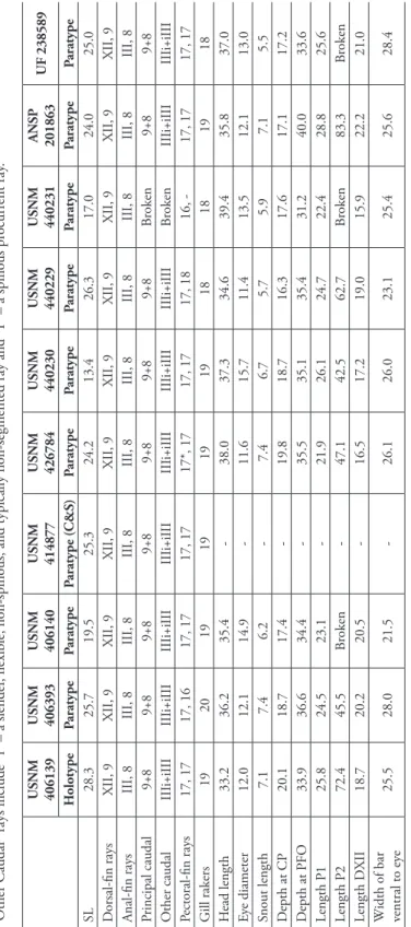
Table 2. Frequency distributions of gill rakers on first arch and left and right pectoral-fin rays in Lipo- gramma levinsoni sp. n., L. evides, and L. haberi sp. n. Counts for the holotype and three paratypes of L.
evides are included from Robins and Colins (1979). Counts of gill rakers and pectoral-fin rays for a fourth paratype of L. evides, ANSP 134330, were not given in the original description. The fifth and smallest paratype, ANSP 134332, is a specimen of L. levinsoni, and counts of that specimen made in this study are included. An asterisk indicates count of gill rakers or left pectoral-fin rays in holotype.
Gill Rakers Pectoral-fin Rays
15 16 17 18 19 20 21 22 15 16 17 18 19
L. levinsoni 1 5 9* 1 5 26* 3
L. evides 3 14* 11 1 1 45* 9
L. haberi 1* 2 1 5*
Figure 3. Tooth-like, secondary rakers on the first gill arch in Lipogramma evides, USNM 34771, cleared and stained paratype. Photo by L. Tornabene.
contained 2.8 times in head length. Pupil slightly tear shaped, with small aphakic space anteriorly. Scales extending anteriorly onto posterior portion of head, ending short of coronal pore. Scales present on cheeks, opercle, preopercle, interopercle, and isthmus.
Scales lacking on top of head, snout, jaws, and branchiostegals. Scales large and de- ciduous, too many scales missing in most specimens to make accurate scale counts. In holotype, approximately 23 lateral scales between shoulder and base of caudal fin, ap- proximately 4 scale rows on cheek, and approximately 9 scale rows across body above
anal-fin origin. Scales on head and nape without cteni, scales on rest of body ctenoid.
Fins naked except small scales present at bases of soft dorsal and anal fins.
Margins of bones of opercular series smooth, opercle without spines. Single row of teeth on premaxilla posteriorly, broadening to 2-3 rows anteriorly, teeth in innermost row smallest, some teeth in outer row enlarged into small canines. Dentary similar, holotype with 3 enlarged teeth in outer row near symphysis. Vomer with chevron- shaped patch of teeth, palatine with long series of small teeth. Several canals and pores visible on head, but most pores inconspicuous. Conspicuous pores present in infraor- bital canal (2 pores) and portion of supraorbital canal bordering dorsal portion of orbit (3); less conspicuous pores present on top of head (1 median coronal pore), pre- opercle (7), and lateral-line canal in the posttemporal region (3). Anteriormost of the 3 supraorbital pores situated at anterodorsal corner of orbit, middle supraorbital pore situated above mid orbit, and posteriormost supraorbital pore situated at posterodorsal corner of orbit (Fig. 4). This pore with fleshy rim in holotype, and mid-orbit supraor- bital pore with smaller fleshy rim. Posterior nostril situated just ventral to anteriormost supraorbital pore, nostril a single large opening with ventral portion of rim slightly elevated. Anterior nostril in tube with anterior flap and situated just posterior to upper lip. No lateral line present on body.
Coloration: In life (Fig. 2), ground color of head and trunk white to tan dorsally grading to white below. Head: dark brown to black bar encompassing orbit and extend- ing ventrally to ventral midline; above orbit, bar narrowing across dorsal midline; eye with dark brown outer ring, yellowish to bluish iris. Trunk: two broad, dark brown to blackish bars present beneath dorsal fin, bars sometimes hourglass shaped, with nar- rower and less intensely colored central regions (central regions losing almost all dark color in some freshly dead specimens); anterior bar extending ventrally from anterior third of spinous dorsal fin to ventral midline, its anterior border extending forward to encompass base of pectoral fin; posterior bar extending ventrally from base of soft dorsal fin to posterior half of anal fin. Dorsal fin: dark trunk bars extending onto base of fin
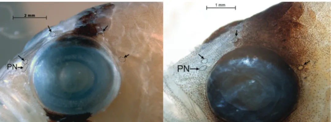
Figure 4. Supraorbital pore patterns in Lipogramma evides, UF 238591, 34.5 mm SL (left) and L. levin- soni sp. n., UF 238589, 25.0 mm SL (right). Arrows point to pores, which have been outlined with tiny dots for emphasis. PN – posterior nostril.
as two blotches, anterior blotch short, low, less conspicuous (than posterior blotch) and sometimes with faint orange upper border. Posterior blotch an intense, dark, longitudi- nal oval spanning lower half of soft dorsal and bordered posteriorly by white to bluish- white pigment. Base of fin between trunk bars whitish, central portion of fin brown to grey, and distal third of fin with bluish tint and thin, orange, submarginal stripe; this stripe breaking into spots along the rear third of fin. Anal fin: posterior trunk bar ex- tending onto proximal portion of posterior half of fin as a strong, horizontally elongate, black blotch edged distally with bluish white line; base of fin with thin, white stripe, fin color grading into blackish to bluish-black distally. A thin, orange, sub-marginal stripe breaks into spots along posterior portion of fin. Caudal fin: basal half translucent pale orange, grading into translucent bluish distally, sometimes with indistinct, very narrow, submarginal orange band around entire edge. Pectoral fins: base blackish, fin trans- lucent, rays translucent or tinted with orange. Pelvic fins: translucent white to bluish white, with orange tint medially on basal half of fin. In preservative (Fig. 5A), barred color pattern retained, but orange, yellow, and bluish pigments absent.
Distribution. Known from specimens collected from the Bahamas, Bonaire, Curaçao, Dominica, and Jamaica. This species was also clearly observed in October 2016 by DRR and LT from the mini-submarine “Idabel” at 140 m depth adjacent to Half Moon Bay, Roatan, Honduras.
Habitat. Lives in or hovers above small rocky rubble on gradual slopes at depths of 108-154 m. When approached by the submersible, L. levinsoni disappears into the rubble. We observed them often in pairs.
Etymology. Named Lipogramma levinsoni in recognition of the generous, contin- uing support of research on neotropical biology at the Smithsonian Tropical Research Institute (Panamá) made by Frank Levinson.
Common name. We propose “Hourglass basslet” (Cabrilleta hierba-horaria as the Spanish equivalent) to differentiate this species from the Banded Basslet, Lipogramma evides, and the Yellow-banded Basslet, L. haberi (see description below), both of which have narrower, straight-sided bars on the trunk.
Genetic comparisons. Table 3 shows average inter- and intraspecific divergences in COI among species of Lipogramma analyzed genetically in this study. With the exception of a single substitution in one specimen, the 15 specimens of Lipogramma levinsoni exhibit no intraspecific genetic variation at this locus and differ from other Lipogramma species by 15.4–26.0%. Lipogramma levinsoni differs from L. evides by 17.1% and L. haberi by 19.0%.
Comments. The smallest paratype of L. evides, ANSP 134332 (Fig. 1B), 12.6 mm SL, is a specimen of L. levinsoni. Although Robins and Colin (1979) indicated 15 pec- toral-fin rays on both sides of this specimen, we count 17 on the right and find the left side too bent to make an accurate count. Lipogramma levinsoni typically has 17–18 pec- toral-fin rays, modally 17. The gill-raker count of 19 given by Robins and Colin (1979) was confirmed by our examination, and is the typical count for L. levinsoni. Counts of pectoral-fin rays (15–16, usually 16) and gill rakers on the first arch (19–21, usually 20 or 21) given by Robins and Colin (1979) for the remaining paratypes of L. evides
Figure 5. Preserved specimens of A Lipogramma levinsoni sp. n., holotype, USNM 406139, 28.3 mm SL B Lipogramma haberi sp. n., holotype, USNM 422679, 40.1 mm SL C L. evides, paratype, ANSP 134330, 30.5 mm SL Photos A and B by Sandra Raredon, C by Mark Sabaj.
support their identification as specimens of L. evides. As noted, previous authors have mistakenly identified the broad-banded L. levinsoni as the juvenile form of the more narrow-banded L. evides. Our material includes juvenile specimens of both L. levinsoni and L. evides, which in each case have the adult configuration of dark bands (Fig. 6).
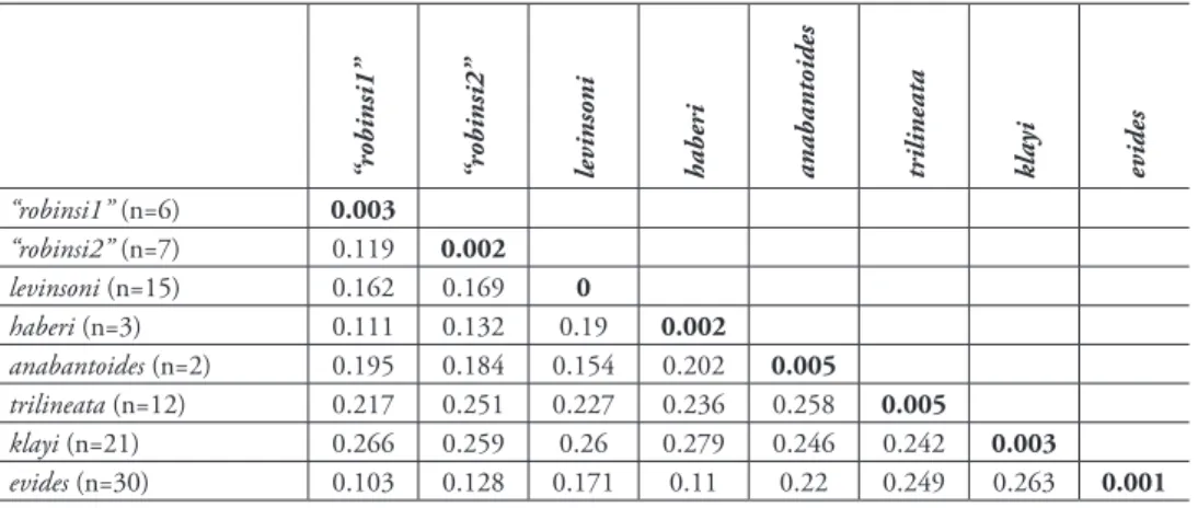
Table 3. Average Kimura two-parameter distance summary for species of Lipogramma based on cy- tochrome c oxidase I (COI) sequences analyzed in this study. Intraspecific averages are in bold.
“robinsi1” “robinsi2” levinsoni haberi anabantoides trilineata klayi evides
“robinsi1” (n=6) 0.003
“robinsi2” (n=7) 0.119 0.002
levinsoni (n=15) 0.162 0.169 0
haberi (n=3) 0.111 0.132 0.19 0.002
anabantoides (n=2) 0.195 0.184 0.154 0.202 0.005
trilineata (n=12) 0.217 0.251 0.227 0.236 0.258 0.005
klayi (n=21) 0.266 0.259 0.26 0.279 0.246 0.242 0.003
evides (n=30) 0.103 0.128 0.171 0.11 0.22 0.249 0.263 0.001
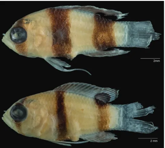
Figure 6. Comparison of juveniles of A Lipogramma levinsoni sp. n., USNM 440230, paratype, 13.4 mm SL and B L. evides, USNM 431410, 12.7 mm SL.
Yellow-banded Basslet
Lipogramma haberi Baldwin, Nonaka & Robertson, sp. n.
http://zoobank.org/4A8447E9-205C-4639-9209-428D8DCDAC1F Figure 7
Type locality. Curaçao, southern Caribbean
Holotype. USNM 422679, 40.1 mm SL, tissue no. CUR13171, Curasub sub- mersible, sta. CURASUB13-09, Curaçao, southwest tip of Klein Curaçao, 11.975783 N, 68.646192 W, 152 m depth, 27 May 2013, M. Harasewych, L. Weigt, B. Van Bebber & A. Schrier.
Paratypes. USNM 434772, 26.4 mm SL, tissue no. CUR15092, Curasub sub- mersible, sta. CURASUB15-12, northwest corner of Klein Curaçao, 11.998453 N, 68.651308 W, 187 m depth, 27 August 2015, B. Brandt, A. Schrier, S. Haber &
T. Haber; USNM 422670, 23.0 mm SL, tissue no. CUR13158, Curasub submers- ible, sta. CURASUB13-08, Curaçao, southwest tip of Klein Curaçao, 11.975783 N, 68.646192 W, 233 m depth, 27 May 2013, C. Baldwin, D. Robertson, B. Brandt, A.
Schrier & L. Weigt.
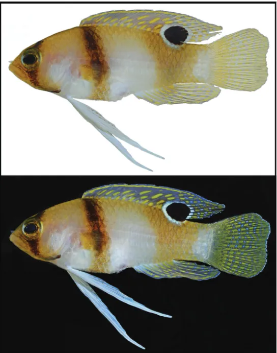
Diagnosis. A species of Lipogramma distinguishable from congeners by the fol- lowing combination of characters: pectoral-fin rays 15–16 (modally 16), gill rakers 15–16 (modally 16); four supraorbital pores along dorsal portion of orbit, a pore pre- sent between pore at mid orbit and one at posterodorsal corner or orbit; caudal fin truncate, tips of lobes rounded; body with three dusky bars (one on head, two on trunk) on yellow/white background; width of bar on head sufficient to encompass pu- pil but not entire eye, width just ventral to eye averaging 17.6% head length; anterior trunk bar narrow and not extending forward to cover pectoral-fin base, bar lighter and less conspicuous ventrally; posterior trunk bar a broad, yellow/tan triangle that is wider dorsally than ventrally; this triangle extending onto soft dorsal fin as large, round, well- defined ocellus; posterior trunk bar not extending onto anal fin; dorsal fin with thin yellow sub-marginal stripe; no yellow submarginal stripe on anal fin; dorsal, anal, and caudal fins with numerous yellow spots. The new species is further differentiated from congeners for which molecular data are available in COI and RAG1.
Description. Counts and measurements of type specimens given in Table 4. Fre- quency distributions of pectoral-fin rays and gill rakers on the first arch are given in Table 2. Three specimens examined, 23.0–40.1 mm SL. Dorsal-fin rays XII, 9 (last ray composite); anal-fin rays III, 8 (last ray composite); pectoral-fin rays 15–16, modally 16, 16 on both sides in holotype; pelvic-fin rays I,5; total caudal-fin rays 25 (13 + 12), principal rays 17 (9 + 8), spinous procurrent rays 6 (III + III), and 2 additional rays (i + i) between principal and procurrent rays that are neither spinous nor typically segmented; vertebrae 25 (10 + 15); pattern of supraneural bones, anterior dorsal-fin pterygiophores and dorsal-fin spines 0/0/0+2/1+1/1/; ribs on vertebrae 3-10; epineural bones present on vertebrae 1–15 in one paratype, difficult to assess in other specimens;
gill rakers on first arch 15–16 (4-5 + 11), 15 (4 + 11) in holotype, both paratypes with
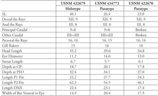
Figure 7. Lipogramma haberi sp. n., USNM 422679, holotype, 40.1 mm SL, photographed prior to preservation against white (top) and black (bottom) backgrounds. Photos by D. R. Robertson and C. C.
Baldwin.
16 (5 + 11); lowermost two rakers very small, all other gill rakers elongate and slender with tooth-like secondary rakers as in L. evides (Fig. 3); pseudobranchial filaments 6, filaments fat and fluffy; branchiostegals 6.
Table 4. Counts and measurements of type specimens of Lipogramma haberi sp. n. Measurements are in percent SL except width of bar ventral to eye, which is in percent head length. CP = caudal peduncle;
PFO = pelvic-fin origin; P1 = pectoral fin; P2 = pelvic fin; DXII = twelfth dorsal-fin spine. “Other Caudal”
rays include “i” – a slender, flexible, non-spinous, and typically non-segmented ray and “I” – a spinous procurrent ray.
USNM 422679 USNM 434772 USNM 422670
Holotype Paratype Paratype
SL 40.1 26.4 23.0
Dorsal-fin Rays XII, 9 XII, 9 XII, 9
Anal-fin Rays III, 8 III, 8 III, 8
Principal Caudal 9+8 9+8 Broken
Other Caudal IIIi+iIII IIIi+iIII Broken
Pectoral-fin Rays 16, 16 16, 15 16, 16
Gill Rakers 15 16 16
Head Length 35.2 39.0 34.8
Eye Diameter 11.2 14.0 13.0
Snout Length 6.7 5.7 6.1
Depth at CP 18.7 20.1 17.8
Depth at PFO 32.4 34.1 27.0
Length P1 Fin 22.2 27.7 24.3
Length P2 Fin 62.3 54.5 46.1
Length DXII 22.4 23.1 17.4
Width of Bar Ventral to Eye 14.9 20.4 17.5
Spinous and soft dorsal fins confluent, several soft rays in posterior portion of fin forming elevated lobe that extends posteriorly beyond base of caudal fin. Pelvic fin extending posteriorly to anterior third of caudal peduncle in holotype when depressed, longest pelvic-fin rays broken in preserved specimens. Dorsal profile from snout to origin of dorsal fin convex. Diameter of eye of holotype contained 2.7 times in head length. Pupil slightly tear shaped, with small aphakic space anteriorly. Scales extending anteriorly onto top of head, ending short of coronal pore. Scales present on cheeks, opercle, preopercle, interopercle, and isthmus. Scales lacking on frontal region, snout, jaws, and branchiostegals. Scales large and deciduous, too many missing in paratypes to make counts, holotype with approximately 24 lateral scales between shoulder and base of caudal fin, 5 cheek rows, and 11 rows across body above anal-fin origin. Scales on head and nape without cteni, scales on rest of body ctenoid. Fins naked except small scales present at bases of soft dorsal and anal fins.
Margins of bones of opercular series smooth, opercle without spines. Premaxilla with band of small conical teeth, band widest at symphysis, outer row with largest teeth, 3 or 4 near symphysis enlarged into canines. Dentary similar except 4-6 anterior teeth enlarged into canines. Vomer with chevron-shaped patch of teeth, palatine with long series of small teeth. Several canals and pores visible on head, but most pores inconspicuous. Conspicuous pores present in infraorbital canal (2) and in supraorbital
canal bordering dorsal portion of orbit (4); less conspicuous pores present on top of head (1 median coronal pore), preopercle (8), and lateral-line canal in posttemporal region (3). An additional 4 tiny pores present beneath orbit in holotype in infraorbital canal. Supraorbital pore pattern as in L. evides (Fig. 4): anteriormost of 4 supraorbital pores situated at anterodorsal corner of orbit, second supraorbital pore situated above mid orbit, and posteriormost supraorbital pore situated at posterodorsal corner of or- bit. Between second and posteriormost supraorbital pores, another pore present and situated closer to latter. Posterior nostril situated just ventral to anteriormost supraor- bital pore, nostril a single large opening with ventral portion of rim slightly elevated.
Anterior nostril in tube with anterior flap and situated just posterior to upper lip. No lateral line present on body.
Coloration: In life, ground color of head and trunk pale yellow to tan dorsally, white ventrally. Head: mostly pale yellow-tan with white blotch on operculum; a brown to black C-shaped bar with yellow-brown edges originating on top of head, widening ventrally above orbit to width of pupil and passing over orbit at that width, then narrowing ventrally and continuing as dark line along lower edge of operculum;
iris dark brown above and below where bar passes through, yellowish-white anteriorly and posteriorly, a thin gold ring circling pupil. Trunk: two dark bars beneath dorsal fin, anterior one brown to blackish (edged with yellow-brown) originating below anterior dorsal spines and descending obliquely behind pectoral-fin base to ventral midline; bar fading below pectoral-fin base; posterior bar much broader than anterior bar but paler and less conspicuous, bar spanning dorsal and ventral body margins and covering anterior half of caudal peduncle; bar narrowing ventrally. Dorsal fin: grey with a bluish tint (when photographed against black background – Fig. 7, bottom), with thin, submarginal yellow stripe; spinous dorsal fin with row of round to oblong yellow spots along base, 1–2 rows of obliquely oriented, oval, yellow spots above that;
soft dorsal with large, conspicuous, circular, black ocellus covering lower half of fin and extending onto dorsal portion of trunk; thin, white, outer ring surrounding ocel- lus on both fin and trunk complete in holotype (Fig. 7), absent along underside of ocellus in both paratypes; above ocellus, soft dorsal fin with approximately three rows of rounded yellow spots; grey spaces between yellow spots appearing as well-defined grey to blue spots posteriorly. Anal fin: grey with bluish tint (when photographed against black background), each ray with 3-6 elongate yellow spots from base to fin edge; grey spaces between yellow spots appearing as well-defined grey to blue spots posteriorly. Caudal fin: base of fin mostly yellow, remainder of fin with rows of yel- low spots along fin rays; grey spaces between yellow spots appearing as well-defined grey or blue spots. Pectoral fins: base yellowish with black dots, fin translucent.
Pelvic fins: bright white, inner 2-3 rays with series of small yellow-brown dots. In preservative (Fig. 5B), barred color pattern retained, posterior trunk bar faint, and yellow and bluish pigments absent.
Distribution. Known only from Klein Curaçao, a 1.7 km2 island 11 km southeast of Curaçao.
Habitat. No specific habitat information recorded.
Etymology. Named in honor of Spencer and Tomoko Haber, who funded and participated in a submersible dive by the Smithsonian’s Deep Reef Observation Project (DROP) that resulted in the collection of USNM 434772, a paratype of the new species.
Common name. We propose “Yellow Banded Basslet” (“Cabrilleta cinta-amaril- la” as the spanish equivalent) to distinguish L. haberi from L. evides and L. levinsoni.
Although L. evides has a submarginal yellow stripe along the dorsal and anal fins, it lacks the overall yellow body color of L. haberi.
Genetic comparisons. Table 3 shows average inter- and intraspecific divergences in COI among species of Lipogramma analyzed genetically in this study. Lipogramma ha- beri exhibits 0.2% intraspecific genetic variation and 11.0–27.9% divergence from other Lipogramma species. It differs from L. evides by 11.0% and from levinsoni by 19.0%.
Comments. Relative to L. levinsoni and L. evides, which are known from multiple localities within the Caribbean Sea, L. haberi is an uncommon species on deep reefs and may have a more restricted geographic distribution. Although both L. levinsoni and L. evides are frequently observed and collected off the southern coast of Curaçao, in more than one hundred submersible dives there we have not collected L. haberi.
Rather, we have only collected L. haberi on infrequent trips to Klein Curaçao, a small island, as noted above, 11 km southeast of Curaçao.
Discussion
Comments on Lipogramma evides. The type series of L. evides includes the holotype and five paratypes (Robins and Colin 1979). We examined specimens or photographs of specimens of the type series from ANSP and FMNH and conclude that all except one, ANSP 134332, 12.6 mm SL, represent L. evides. We also examined 31 specimens of Lipogramma evides that we recently collected at Curaçao and that range in size from 12.7–45.4 mm SL. Frequency distributions of pectoral-fin rays and gill rakers on the first arch are given in Table 2, an illustration of the holotype that was included in the original description of the species is shown in Fig. 1A, color patterns of live and recent- ly deceased individuals are shown in Fig. 8, a photographed of a preserved paratype (ANSP 134330) is provided in Fig. 5C, secondary spines on gill rakers of the first arch are shown in Fig. 3, supraorbital pore pattern is shown in Fig. 4, and a photograph of a preserved juvenile is featured in Fig. 6.
The illustration of the holotype (Fig. 1A) shows a triangular-shaped bar on the posterior portion of the trunk that more closely resembles the shape of that bar in L. haberi (Fig. 7) than L. evides (Fig. 8). The pigment is too faded in the preserved holotype now to determine the shape of that bar, but we note that in specimens or photographs of the four paratypes of the type series that are actually specimens of L.
evides, the posterior trunk bar is narrow (e.g., Fig. 5C). This suggests that the shape of the posterior trunk bar illustrated in the holotype of L. evides is either incorrectly drawn or represents an anomaly for the species. Lipogramma evides and L. haberi are easily distinguished by numbers of gill rakers on the first arch—15 or 16 in L. haberi,
19-22 (modally 20) in L. evides (Table 2). Our examination of the holotype confirms the count by Robins and Colin (1979) of 20 gill rakers in the holotype of L. evides.
Furthermore, the triangular-shaped posterior trunk bar in L. haberi is very pale rela- tive to the anterior trunk and head bars. Robins and Colin’s (1979) illustration of the holotype shows three body bars of equal intensity.
Colin’s (1974) observation of “Lipogramma sp.” at Glover’s Reef, Belize, was cited as L. evides by Robins and Colin (1979), and based on the recorded depths of observa- tions, 165–180 m, we tentatively agree with this identification, as Belize is between Arrowsmith Bank and Nicaragua, where L. evides does occur. Lipogramma evides in- habits depths of 133-302 m, whereas L. levinsoni occurs from 103 to 154 m. However, Colin’s (1974) observed fish could have been L. haberi, which occurs from 152–233 m. Polanco et al. (2012) recorded several specimens of L. evides from the Coralinos Ar- chipelago off Colombia, and based on the stated counts of 15–16 pectoral-fin rays and 20–21 gill rakers, those specimens are correctly identified. In addition to Colombia and the tentative Belize location, L. evides is known definitively from the type locality of Arrowsmith Bank in the Yucatan Peninsula, Nicaragua, southeast of Barbuda, and Curaçao (including Klein Curaçao). It was also observed but not collected in October 2016 by DRR and LT from the mini-submarine “Idabel” at 232–250 m depth adja- cent to Half Moon Bay, Roatan, Honduras. It was not collected or observed on DROP submersible dives at Bonaire or Dominica. A list of L. evides material examined in this study is given in Appendix 2.
Morphological comparisons. Lipogramma levinsoni, L. evides, and L. haberi can be readily distinguished from all congeners in having three dark bars (one on the head, two on the trunk) on a white background vs. a brown body with a reddish head in L.
anabantoides; a yellow body with one black bar on the head in L. flavescens; a purple head and yellow trunk in L. klayi Randall, 1963; a brown body with one broad white bar and multiple narrow orange bars in L. regia; a brownish body with about 12 thin dark bars in L. robinsi Gilmore, 1997; a pink head and trunk with a yellow stripe along the dorsal profile of the head in L. rosea; and a yellow head and trunk with three long iridescent blue stripes on the head in L. trilineata Randall, 1963. The major differences among L. levinsoni, L. evides, and L. haberi are summarized in Table 5. Lipogramma evides and L. haberi are morphologically similar and reach a similar maximum size (45.5 and 40.1 in our material, respectively). They are easy to distinguish from one another on the basis of number of gill rakers on the first arch (usually 20-21 in L. evides, 15-16 in L.
haberi – Table 2) and by live and preserved color pattern (Figs 5, 7, 8). In life, L. haberi has a considerable amount of yellow as ground color and associated with the dark bars, whereas the ground color of L. evides is mostly white. In fresh and preserved specimens, the posterior trunk bar in L. haberi is broad and much wider at the top than the bottom, whereas in L. evides it is narrower and of uniform width. There is also a difference in the shape of the caudal fin of the two species, with L. haberi having a truncate fin with rounded lobe tips and L. evides having a slightly emarginate fin with pointed lobe tips.
Lipogramma levinsoni reaches a smaller maximum size than L. haberi and L. evides (largest specimen examined 28.3 mm SL) and differs in modal numbers of gill rakers
Figure 8. Lipogramma evides A Aquarium photograph by Barry Brown, Substation Curacao B USNM 276560, 45.3 mm SL, illustration by Grant Gilmore in Gilmore and Jones (1988: fig. 1) C and D USNM 414885, 24.4 mm SL, photos by D. R. Robertson and C. C. Baldwin against black (C) and white (D) backgrounds.
on first arch and pectoral-fin rays (Table 2), supraorbital pore pattern (Fig. 4), and numerous aspects of color pattern (Figs 2, 5, 7, 8). In life, L. levinsoni is easily distin- guished from L. haberi and L. evides by having an orange submarginal stripe on the dorsal fin (vs. yellow) and an orange submarginal stripe on the anal fin (vs. no stripe in L. haberi, a yellow submarginal stripe in L. evides). In preservative, L. levinsoni is easily distinguished from L. haberi and L. evides by the shape, size, and configuration of the dark body bars (Table 5).
Species delimitation and phylogeny. The neighbor-joining network (Suppl. ma- terial 1) shows eight distinct genetic lineages with an average within-lineage genetic distance of 0.002 substitutions/site and an average between-lineage genetic distance of 0.20 substitutions/site (Table 3). Considering between-lineage distances that are 10 or more times greater than within-lineage distances as indicative of distinct species (Hebert et al. 2014), genetic distances corroborate the recognition of the L. levinsoni and L. haberi lineages as species. Average between-lineage divergence for L. levinsoni is 19% (18% between L. levinsoni and the other two banded basslets, L. haberi and L. evides), whereas average within-lineage divergence is 0%. For L. haberi, average between-lineage divergence is 18% (11% between L. haberi and L. levinsoni/L. evides), whereas average within-lineage divergence is 0.2%.
Table 5. Comparisons among Lipogramma levinsoni sp. n., L. haberi sp. n., and L. evides.
L. levinsoni L. haberi L. evides
Standard length (mm) 9.4–28.3 23.0–40.1 13.7–45.5
Gill rakers on 1st arch Usually 19 15–16 Usually 20–21
Pectoral-fin rays Usually 17 Usually 16 Usually 16
Supraorbital pores/pore present between pore at mid orbit and one at posterodorsal corner of orbit (Fig. 4)
3/Absent 4/Present 4/Present
Ground color White, grey on nape &
snout Yellow above, white below White
Dark bar on head
Black; relatively wide, widens to encompasses
entire eye No rearward extension
along lower edge of opercle
Brown, edged with yellow;
relatively narrow, widens to encompass pupil Narrow rearward extension along lower edge of opercle
Black; relatively narrow, widens to encompass pupil Narrow rearward extension along lower edge of opercle Width of dark bar on head
(measured immediately
ventral to eye) in % HL) 21.5–34.8 (x‒ = 26.4) 14.9–20.4 (x‒ = 17.6) 8.7–19.4 (x‒ = 14.7)
Anterior trunk bar
Black, wide, vertical, center often narrower
& paler Covers pectoral base Extension onto dorsal fin
large, intense
Brown, edged with yellow;
narrow, slightly oblique, uniform width, paler on
belly Behind pectoral base No extension onto dorsal fin
Black; narrow, slightly oblique, uniform width,
paler on belly Behind pectoral base Extension onto dorsal fin
small, weak
Posterior trunk bar
Same form and color as anterior bar Extension onto dorsal fin
= oval partial ocellus Extension onto anal fin =
elongate, partial ocellus
Yellow-brown; broad dorsally, thinning ventrally,
triangular in shape Extension onto dorsal fin = round, well defined ocellus No extension onto anal fin
Same form and color as anterior bar but usually paler than anterior bar Extension onto dorsal fin = round, well defined ocellus
Extension onto anal fin absent or a small,
weak smudge
Dorsal-fin pigment
Submarginal stripe orange Remainder of fin without
pale spots
Submarginal stripe yellow Remainder of fin with 2–3
rows of yellow spots
Submarginal stripe yellow Remainder of fin with 1–2
rows of yellow spots
Anal-fin pigment
Submarginal stripe orange No pale spots on remainder of fin
No pale submarginal stripe Remainder of fin with 1–6
rows of yellow spots
Submarginal stripe yellow Remainder of fin with 1–3 rows of yellow spots
near base Caudal-fin shape Truncate, lobe tips
rounded Truncate, lobe tips rounded Slightly emarginate, lobe tips pointed
Depth range (m) 108–154 152–233 133–302
Geographical distribution Bahamas, Bonaire, Curaçao, Dominica, and
Jamaica Klein Curaçao
Barbuda, Belize(?),Colombia, Curaçao, Klein Curaçao, Mexico (Caribbean), and
Nicaragua
The ML and BI analyses resulted in identical topologies, with most relationships sup- ported by 1.0 posterior probability and 100% bootstrap support (Fig. 9). The BP&P anal- ysis inferred a coalescent-based species-tree that was identical in topology to the ML and BI trees. In addition, the BP&P analysis provided overwhelming support for the presence of eight species of Liprogramma in our phylogeny (posterior probability 0.99981), includ- ing three distinct species of banded basslets (L. evides, L. haberi and L. levinsoni), indicat- ing perfect congruence between molecular and morphology-based species delimitation criteria. Lipogramma trilineata and L. klayi, which have two of the shallowest depth ranges among Lipogramma species (Fig. 10), were recovered as sister species. This pair is sister to a larger clade comprising L. anabantoides + L. levinsoni (as sister species) and L. evides + L.
haberi + two putative new species superficially resembling L. robinsi. Not surprising given their morphological similarity, L. evides, and L. haberi resolve as sister species. There is strong support for a clade comprising the four deepest-known species in our phylogeny—
L. evides, L. haberi, and the two “L. robinsi” species (Figs 9, 10). Baldwin and Robertson (2014) found a similar evolutionary grouping of deep-water species in the serranid genus Liopropoma, and Tornabene et al. (2016a) found repeated invasions of deep-reef depths in the family Gobiidae with subsequent species radiations entirely within the deep-reef zone.
Lipogramma flavescens, which also inhabits deep water (200–300 m, Fig. 10), may be part of this clade. A dark ocellus on the base of the soft dorsal fin appears to be a synapomor- phy of the clade comprising L. anabantoides, L. levinsoni, L. evides, L. haberi, and the two
“L. robinsi” species. Presence of this ocellus in L. flavescens and L. regia suggests that they may also belong to this group, but genetic samples of both are needed for phylogenetic analysis. Lipogramma flavescens may be closely related to L. haberi, as they share a narrow dark bar through the eye, yellow coloration, and low gill-raker count (15–16), and they inhabit similar deep-reef depths (152–233 m for L. haberi, 200–300 m for L. flavescens). If the evolutionary relationships of Lipogramma species are correlated with depth as our data suggest, and if Lipogramma regia, which is known only from depths < 100 m is a mem- ber of the clade diagnosed by a dark ocellus on the soft dorsal fin, it may be most closely related to L. anabantoides and L. levinsoni, which are known only from depths < 120 m (L. anabantoides) and < 154 m (L. levinsoni). Those three are the only known Lipogramma species with a modal pectoral-fin count of 17 (Gilmore and Jones 1988: Table 2, this study). We note that the addition to our molecular phylogeny of the four known species of Lipogramma that are currently unavailable for analysis (L. flavescens, L. regia, L. rosea, and L. robinsi) could change our hypotheses of relationships within the genus.
The two “L. robinsi” included here (Table 3, Figs 9, 10) are genetically distinct and superficially different from one another and from L. robinsi Gilmore, 1997. A more thorough investigation of those three taxa is in progress, after which a key to all Lipo- gramma species will be constructed.
Lipogramma is currently classified along with Gramma in the family Grammatidae based on a single synapomorphy in the arrangement of cheek musculature (Gill and Mooi 1993). Molecular data have failed to corroborate the monophyly of the Grammatidae (Betancur-R et al. 2013, Mirande 2016); rather, those data suggest that Lipogramma and
Figure 9. Bayesian Inference molecular phylogeny of Lipogramma based on combined mitochondrial and nuclear genes. Numbers of individuals analyzed for each species are given in Appendix 1, along with the genes sequenced for each individual. Topology is identical to that from Maximum Likelihood analysis. Support values are Bayesian posterior probabilities (above) and bootstrap values (below). Nodes without labels have 1.0 posterior probability and 100 bootstrap values. Photos or illustrations by C. C.
Baldwin, D. R. Robertson, R. G. Gilmore, and C. R. Robins.
Figure 10. Depth distributions for species of Lipogramma. Photos or illustrations by C. C. Baldwin, D.
R. Robertson, R. G. Gilmore, and Mooi and Gill (2002: fig. 9).
Gramma are each related to different taxa within the diverse Ovalentaria. Relationships within the Ovalentaria have proven difficult to resolve with traditional molecular markers (Betancur-R et al. 2013), molecular markers plus some morphological characters (Mirande 2016), and phylogenomic data (Eytan et al. 2015). Potential close relatives of Lipogramma based on molecular data include blennioids, cichlids, plesiopids, pseudochromids, and Pholidichthys. Some of these groups have been previously linked to either Lipogramma, Gramma, or both, based on shared morphological characters, but the homologies of many of these characters have been questioned (Gill and Mooi 1993). At present, the phyloge- netic position of Lipogramma is unresolved.
Ecology and life history. Little is known about community structure on deep reefs, including food networks. Although an analysis of the diet of banded basslets based on stomach contents is beyond the scope of this study, the gastrointestinal tract of the cleared and stained L. evides (USNM 434771) contained numerous individuals of a planktonic foraminiferan that was tentatively identified by Smithsonian Curator of Planktic Fo-
Figure 11. Items from stomachs of deep-reef Lipogramma: A Planktonic foraminiferan, possibly Glob- orotalia manardii, from L. evides, USNM 434771, collected at 174 m B Diatom, possibly Coscinodiscus eccentricus, from L. levinsoni sp. n., USNM 406140, collected between 137 and 146 m C Parasitic Nema- toda from same specimen as B. Photos by A. Nonaka and L. Tornabene.