Academy of Natural Sciences
Two New Species of Acanthoclininae (Pisces: Plesiopidae) with a Synopsis and Phylogeny of the Subfamily
Author(s): William F. Smith-Vaniz and G. David Johnson
Source: Proceedings of the Academy of Natural Sciences of Philadelphia, Vol. 142 (1990), pp.
211-260
Published by: Academy of Natural Sciences Stable URL: http://www.jstor.org/stable/4064977 Accessed: 02/06/2010 12:06
Your use of the JSTOR archive indicates your acceptance of JSTOR's Terms and Conditions of Use, available at
http://www.jstor.org/page/info/about/policies/terms.jsp. JSTOR's Terms and Conditions of Use provides, in part, that unless you have obtained prior permission, you may not download an entire issue of a journal or multiple copies of articles, and you may use content in the JSTOR archive only for your personal, non-commercial use.
Please contact the publisher regarding any further use of this work. Publisher contact information may be obtained at http://www .jstor.org/action/showPublisher ?publisherCode=ans.
Each copy of any part of a JSTOR transmission must contain the same copyright notice that appears on the screen or printed page of such transmission.
JSTOR is a not-for-profit service that helps scholars, researchers, and students discover, use, and build upon a wide range of content in a trusted digital archive. We use information technology and tools to increase productivity and facilitate new forms of scholarship. For more information about JSTOR, please contact [email protected].
Academy of Natural Sciences is collaborating with JSTOR to digitize, preserve and extend access to Proceedings of the Academy of Natural Sciences of Philadelphia.
STOR
http: //www .j stor.org
Two New Species of Acanthoclininae
(Pisces: Plesiopidae) with a Synopsis and Phylogeny of the Subfamily
WILLIAM F. SMITH-VANIZ Department of Ichthyology Academy of Natural Sciences
19th and the Parkway, Philadelphia, PA 19103 G. DAVID JOHNSON
Division of Fishes
National Museum of Natural History Smithsonian Institution Washington, DC 20560
ABSTRACT.—Osteology and soft anatomy were studied to advance a hypothesis of phylogenetic relationships within the previously recognized family Acanthoclinidae in order to determine the correct generic placement of two new species. The following genera are recognized, with the number of their included species given in parentheses: Acanthoclinus Jenyns (5), Belonepterygion McCulloch (1), Beliops Hardy (2) and Acanthoplesiops Regan (4). A cladistic analysis based on 33 adult characters resulted in a highly corroborated hypothesis indicating that each of the four genera comprise monophyletic clades ordered in the above phyletic sequence. Taumakoides Whitley is a synonym of Acanthoclinus.
Beliops batanensis n. sp., known only from the Batan Islands, northern Philippines, shares three unique apomorphies with its allopatric congener. It exhibits a surprisingly large number of derived character states not present in its plesiomorphic sister-species, including short and robust dorsal-fin spines and distinctive body scales with membranous flaps. Acanthoplesiops echinatus n. sp., known only from Jolo, Sulu Archipelago and Ambon, Indonesia, is most closely related to the allopatric A. psilogaster, and is readily distinguished from its congeners by having most scales on the posterior half of body with one or two membranous flaps supported by elongate ctenii and 6 (versus 3-5) segmented anal-fin rays; the occurrence of minute, pointed papillae on the head of the two Ambon specimens (inexplicably absent in the single Jolo specimen) is unique within the subfamily. [Acanthoclinidae, biogcography, cladistic analysis, osteology, Perciformes, Plesiopidae, scale morphology, systematics]
In April-May of 1987 the authors spent all served to inhibit biological exploration three weeks collecting fishes in the in this poorly collected region. The Batanes Province, northern Philippines Batanes Province lies at the northern tip of under the auspices of a Smithsonian Insti- the Philippine Archipelago, between north- tution sponsored project to sample the ern Luzon and Taiwan. Most of its dozen biota. Limited transportation facilities and islands are of volcanic origin and much of frequent bad weather and sea conditions the shoreline consists of rocky ledges and combined with geographic isolation have boulders, with coral development relatively
211
212 W. F. SMITH-VANIZ AND D. W. JOHNSON
sparse.
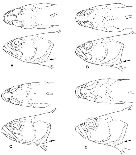
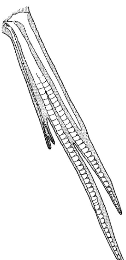
Among the fishes collected were two specimens of an enigmatic acanthoclinid that seemingly did not agree with any pre- viously described genus. Subsequently, we discovered an undescribed species of Acanthoplesiops in an unsorted collection of fishes from Ambon, Indonesia deposited at the Bernice P. Bishop Museum. Our efforts to place these new taxa led us to re-evaluate the phylogenetic relationships of the Acanthoclinidae as outlined by Hardy (1985). The phylogenetic relationships of the family Acanthoclinidae have been uncertain since it was first established by Gunther (1861:297). Although most recent authors have recognized the family as valid, both Regan (1913) and McCulloch (1915) allocated acanthoclinid genera to the Plesiopidae. Mok et al. (1990) treated the Acanthoclinidae and Plesiopidae as sister taxa, although the characters they used to support such a relationship are invalid or were misinterpreted. In his recent revision of the Acanthoclinidae, Hardy (1985) did not consider the possibility of a close rela- tionship between these two groups. Both nominal families share one external feature that we have seen in no other fishes. The third branchiostegal ray (counting postero- dorsally) is positioned so that it extends farther posteriorly than adjacent rays result- ing in a slight to pronounced notch or rounded projection on the posterolateral margin of the exposed branchiostegal membranes (Fig. 1). This condition can be seen clearly in previously published illus- trations of Plesiops (Inger 1955:fig. 3a), Paraplesiops (Hoese and Kuiter 1984:figs.
3-4), and Steeneichthys (Allen & Randall 1985: figs. 1-2). The branchiostegal mem- branes of Trachinops, unlike those of other
plesiopids, usually are not exposed in lat- eral view, but when the opercle is raised the characteristic membrane configuration is readily apparent. The monotypic plesiopid genus Calloplesiops is exceptional in lack- ing notched branchiostegal membranes.
As noted by Mooi (1990), the Plesiop- idae has never been satisfactorily defined although three of the seven genera tradi- tionally assigned to the family (Trachinops, Paraplesiops, and Fraudella) have eggs with similar and unique chorionic struc- tures. Furthermore, and of more relevance to this study, no synapomorphy has been found that supports a monophyletic Plesi- opidae exclusive of the Acanthoclinidae.
The most recent assessment (results of which were presented at the annual meet- ing of the American Society of Ichthy- ologists and Herpetologists, Mooi 1990 abstract) of plesiopid relationships has re- vealed a number of putative synapo- morphies that define various monophyletic clades of plesiopid genera + the Acantho- clinidae. We defer discussion of these characters to a future paper by Mooi that is in an advanced state of preparation.
Although the monophyly of the fishes treated herein has never been questioned seriously, we believe their cladistic rela- tionships are best expressed by recognizing the Acanthoclinidae as a subfamily within an expanded Plesiopidae1.
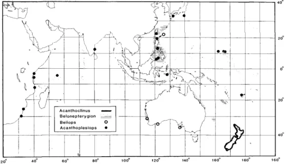
The four genera and 12 species of acanthoclinine fishes, the so-called spiny basslets (Smith & Heemstra 1986), that we recognize occur in depths of 2-73 meters (one trawl collection of Acanthoclinus marilynae in 73-91 m) on reefs and along rocky shores in tropical and temperate waters of the Indo-west Pacific (Fig. 3).
Our study of these fishes benefited greatly
'The Plesiopidae dates from Gunther (1861:362) who first recognized the family as the supra- generic taxon Plesiopina; the Acanthoclinidae dates from the same work (p. 297). Past workers either were unaware that both family-group names date from the same source or did not consider it nomenclaturally relevant because they recognized both families as distinct phylogenetic units of equal taxonomic rank. In accordance with article 24 of the International Code of Zoological Nomenclature (ICZN 1985), we hereby act as first revisers in selecting the Plesiopidae as taking precedence over the Acanthoclinidae. This action preserves the more commonly cited family name and best serves nomenclatural stability.
Fig. 1. General physiognomy and cephalic pore patterns in selected species of Acanthoclin- inae: A, Acanthoclinus fuscus, ANSP 165085, 57.4 mm SL; B, A. littoreus, ANSP 165089, 73.3 mm; C, A. rua, ANSP 165086, 44.8 mm; D, Belonepterygion fasciolatum, USNM 257883, 35.5 mm. (Small arrows indicate location of rounded projection on posterolateral margin of exposed branchiostegal membrane.)
from Hardy's (1985) revision, and readers herein. We follow his taxonomic nomencla- should refer to that work for complete ture, except for our synonymization of synonymies, expanded descriptions, meristic Taumakoides. In the species accounts all frequency tables and good photographs of primary synonyms are given but secondary all except the two new species described synonymies are selective.
214 W. F. SMITH-VANE AND D. W. JOHNSON
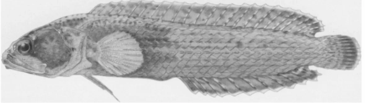
Fig. 1. (continued): E, Beliops xanlhokrossos, ANSP 165557, 25.7 mm SL; F, B. batanensis, USNM 288976, holotype, 21.0 mm; G, Acanlhoplesiops hiatti, USNM 257631, 18.5 mm; H, A.
echinatus, BPBM 44177, holotype, 19.8 mm.
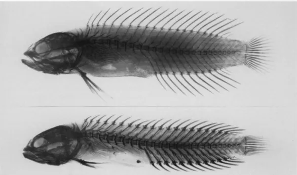
METHODS AND MATERIALS Counts.—Counts of dorsal-, anal- and caudal-fin rays and vertebrae were taken from radiographs. The last two elements of the dorsal and anal fins have the "split to
base" condition and were counted as one, in accord with the general practice of most authors. The distinction between precaudal and caudal vertebrae, as observed from radiographs, was occasionally difficult to determine with confidence. Except for the
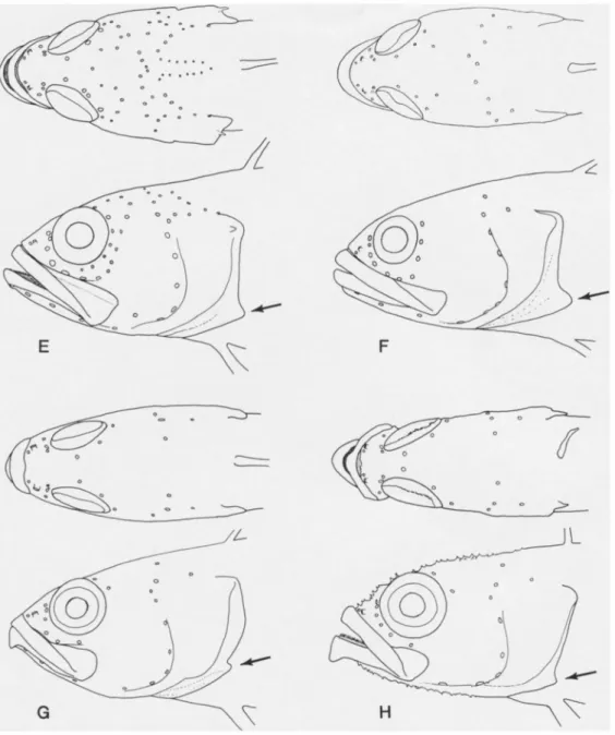
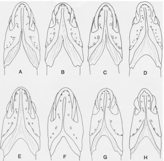
Fig. 2. Ventral views of heads showing general physiognomy and cephalic pore patterns in selected species of Acanthoclininae (data as in Fig. 1.): A, Acanthoclinus fuscus; B, A. littoreus;
C, A. rua; D, Belonepterygion fasciolatum; E, Beliops xanthokrossos; F, B. batanensis; G, Acanthoplesiops hiatti; H, A. echinatus. (Small arrows indicate location of posteriormost dentary pore position.)
two new species and Acanthoclinus matti, for which no cleared and stained whole specimens were available, the number of precaudal and caudal vertebrae was con- firmed by examination of C & S speci- mens. Caudal-fin ray counts include the spinelike dorsal and ventral procurrent rays and the segmented rays. In species of Acanthoclinus the posteriormost dorsal and ventral "procurrent" rays are often difficult to distinguish from segmented rays; often a ray will have only one or two segmental
joints which can easily be overlooked. For enumeration purposes only rays with at least three segments were counted as
"segmented rays." The most important con- sideration is that the other acanthoclinine genera have fewer total fin elements (18- 23, rarely 23 versus 24).
Measurements and illustrations.—All measurements were taken to the nearest 0.1 mm using needle-point dial calipers.
Methods of taking measurements were con- ventional. Except for the two whole fish
216 W. F. SMITH-VANIZ AND D. W. JOHNSON
Fig. 3. Distribution of genera of Acanthoclininae; see Fig. 9 for distributions of individual species of Acanthoplesiops.
drawings, all specimen illustrations were made by Smith-Vaniz using a microscope and camera lucida. Pore diameters in Figs.
1-2 have been slightly enlarged for clarity.
Material examined.—In the listings of material, C&S designates cleared and stained specimens; the number of speci- mens and their standard lengths (SL) in mm are given in parentheses. In the spe- cies accounts for Acanthoplesiops we list all known collections, but do not give lengths unless we have personally exam- ined specimens. Institutional abbreviations for specimen repositories are as follows:
Auckland Institute and Museum, Auckland (AIM); Australian Museum, Sydney (AMS); Academy of Natural Sciences of Philadelphia (ANSP); The Natural History Museum, London (BMNH); Bernice P.
Bishop Museum, Honolulu (BPBM); Na- tional Museum of New Zealand, Welling- ton (NMNZ); Queensland Museum, Bris- bane (QM); J.L.B. Smith Institute of Ich- thyology, Grahamstown (RUSI); Kyoto University, Seto Marine Biology Laboratory (SMBL); National Museum of Natural His- tory, Smithsonian Institution, Washington,
D.C. (USNM).
The following cleared and stained spe- cimens are the basis for the osteological illustrations and comparative observations noted in the text. If not given below, lo- cality data are given under "material" in individual species accounts.
Plesiopidae
Acanthoclinus fuscus Jenyns, ANSP 165085 (2:44.6-46.5).
Acanthoclinus littoreus (Forster), ANSP 165089 (2:53-69).
Acanthoclinus rua (Hardy), ANSP 165087 (1:46.3).
Acanthoclinus marilynae (Hardy), ANSP 134947 (2:62.8-66.6).
Acanthoclinus matti (Hardy), ANSP 165088 (1:51.0), jaws, suspensorium, gill arches and infraorbitals of right side only.
Acanthoplesiops indicus (Day), ANSP 165570 (1:23.7); ANSP 122483 (1:26.5).
Acanthoplesiops hiatti Schultz, ANSP 165421 (1:18.8); USNM 257874 (2:16.8-21.4).
Acanthoplesiops psilogaster Hardy, USNM 257871 (1:18.8).
Acanthoplesiops echinatus Smith-Vaniz &
Johnson, ANSP 166316 (1:21.0), jaws, suspensorium, gill arches, hyoid arch and infraorbitals of right side only.
Assessor macneilli Whitley, ANSP 141752 (4:36.2-36.7) Australia, Great Barrier Reef, One Tree Is.
Belonepterygion fasciolatum (Ogilby), ANSP 142690 (1:30.6); USNM 257875 (3:14.3-28.7).
Beliops batanensis Smith-Vaniz & Johnson, USNM 309905 (1:21.1), jaws, sus- pensorium, gill arches, hyoid arch and infraorbitals of right side only.
Beliops xanthokrossos Hardy, ANSP 165557 (1:25.9).
Fraudella carassiops Whitley, QM 1.19760 (1:42.0), Australia, Great Barrier Reef.
Plesiops coeruleolineatus Riippell, ANSP 106612 (2:32.7-49), Seychelles, Beacon Is.
Trachinops taeniatus Giinther, ANSP 135461 (2:44.7-51.5), Australia, Sydney.
Notograptidae
Notograptus guttatus Giinther, ANSP 109653 (4:67.5-81.0), Australia, Queensland, Little Hope Is.
Io infraorbital Max maxilla Met metapterygoid Nsp neural spine Op opercle Pal palatine
Pb pharyngobranchial (=infrapharyngobranchial) Pmax premaxilla
Pop preopercle Pp parapophysis Ptemp posttemporal Quat quadrate Rad radial
Retart retro-articular Scap scapula Sclei supracleithrum Subop subopercle Sym symplectic Up uncinate process
Phylogenetic procedures.—Phylogenetic analyses (grouping taxa on the basis of shared derived characters; Wiley 1981) were performed using Hennig-86, version 1.5 (Farris 1988). The outgroup method (Maddison et al. 1984) was used to assign character polarities. Refer to "Character Descriptions and Analysis" and "Phylo- genetic Analysis" for specific details.
SYSTEMATIC ACCOUNTS Anatomical abbreviations.—The follow-
ing abbreviations are used in the text and figures.
Al, A2 separate sections of adductor mandibulae
Af articular facet Angart angulo-articular Cor coracoid Clei cleithrum Dent dentary Ect ectopterygoid Eb epibranchial End endopterygoid Fl flange
Hpu haemal spine of preural centrum Hyom hyomandibular
Hypop hypurapophysis Intop interopercle
Acanthoclininae Giinther
Description (see also Table 1).—Indo- Pacific marine shorefishes ranging in size from 20-140 mm SL (except 210 mm SL maximum in Acanthoclinus fuscus). Dorsal fin XVII-XXVI, 2-6; anal fin VII-XVI, 2- 6. Supraneural bones 0-2. Anteriormost proximal pterygiophore (first two in Acanthoplesiops psilogaster) of dorsal fin inserted between second and third neural spines. Pterygiophores supporting dorsal- and anal-fin spines with proximal, middle and distal radials fused (distal radials not absent as reported by Mok et al. 1990);
distal radials of pterygiophores of dorsal- and anal-fin segmented rays autogenous, middle and proximal radials autogenous or
218 W. F. SMITH-VANE AND D. W. JOHNSON Table 1. Comparison of selected characters in genera of Acanthoclininae. "N" indicates character states that logically do not apply; number of species in parentheses.
Genus
Size of adults (maximum mm SL)
Acanthoclinus Belonepterygion (5) (1)
64-210 50
Beliops (2) 21-26
Acanthoplesiops (4) 21-26.5
Number of lateral lines Lateral-line scales in
upper series 47-97 36-46 24-35 0-13
Infraorbital bones 6 5 5 1
Suborbital shelf no yes yes N
Vertebrae:
precaudal caudal total
11-12 17-23 28-35
10-11 17-18 27-29
10 16-17 26-27
12-14 14-17 27-30 Dorsal-fin:
spines rays
18-26 3-5
17-20 4-5
18-20 2-4
19-21 3-6 Anal-fin:
spines rays
9-16 3-5
10-12 3-5
10-11 2-3
7-10 3-6
Gill membranes separate united united united
Basihyal teeth yes or no no yes no
Supramaxilla yes no yes or no no
Primary opercular spine platelike platelike pungent pungent
Preopercular canal open open tubular tubular
Neural spine association with 1st precaudal centrum
autogenous autogenous fused fused
2nd and 3rd epurals separate separate fused fused
Interarcual cartilage long long short short
Metaptergoid-quadate joint smooth smooth interdigitated smooth
Secondary opercular spine no no no yes
Bilobed mid-lateral scales no no no yes
fused in various species. Ultimate dorsal- and anal-fin pterygiophores each serially supporting two fin elements (last ray split to base). Tips of dorsal- and anal-fin spines with thickened, fleshy pads (spine tips with fleshy tassels in Beliops xantho- krossos) that are always pale and contrast conspicuously with heavily pigmented spine shaft and interradial fin membranes (see species account for description of fins in yellow color form of above species). Ori- gin of pelvic fin in advance of vertical from anteriormost insertion of pectoral fin;
pelvic fin I, 2; the outermost segmented ray robust basally and deeply bifurcated, the two branches undivided, or very slight- ly, and with the innermost ray very slen- der and unbranched or weakly branched distally (Fig. 4). Pectoral fin 15-21. Gill membranes either separate or united across ventral midline; the third branchiostegal ray and immediately adjacent rays positioned so as to produce the distinctive posterola- terally projecting membranous margin (see Fig. 1 and discussion in introduction). At least posterior body scales ctenoid in rep- resentatives of all genera but only cycloid scales present in some species (Acantho- clinus 2 spp.; Acanthoplesiops 1 sp.); head naked except for 1-5 cycloid scales in ant- erodorsal angle of opercle in three species of Acanthoclinus; fins naked except for caudal-fin base. Lateral lines on body in three basic patterns: three essentially com- plete series (Acanthoclinus & Beloneptery- gion), except middle lateral line scarcely if at all extending beneath appressed pectoral fin; two incomplete series (Beliops), the first from upper angle of opercle to rear of spinous dorsal fin, and the middle from slightly to well behind pectoral fin to caudal-fin base; or one incomplete dorsal series (Acanthoplesiops) extending posteri- orly to below verticals between origin and about middle of spinous dorsal fin, but with area of the body that is occupied by the middle lateral line in the other genera with a row of scales containing superficial neuromasts (refer to discussion of character 7 in "Character Descriptions and Analy- sis").
Fig. 4. Ventral view of left pelvic fin of Acanthoclinus littoreus, ANSP 165089, 53 mm SL.
Vertebrae 10-14 + 14-23 (total 26-35).
Haemal spine of penultimate (second pre- ural) vertebra fused to vertebral centrum (except autogenous in Acanthoclinus 3 spp.). Hypurals 1 and 2 and parhypural fused together as autogenous plate; hyp- urals 3 and 4 fused to each other and to urostylar complex; hypural 5 present and independent. No autogenous uroneurals; 3 epurals, 2nd & 3rd partially fused in Beliops & Acanthoplesiops). Branched caudal-fin rays 12 or 14, 6-7 + 6-7; total fin elements 18-24. No procurrent spur.
Interarcual cartilage present, very small in Beliops & Acanthoplesiops; infrapharyngo- branchial 1 present (toothless) but may be entirely cartilaginous in Acanthoplesiops hiatti. Basibranchials 1-3 ossified, 4th pre- sent as cartilage. Basibranchial 1 anterior and ventral to basihyal. Urohyal articulating
220 W. R SMITH-VANIZ AND D. W. JOHNSON
with ventral surface of basibranchials 1 and 2. Six branchiostegals, anterior 4 on ceratohyal. No ligament connecting cerato- hyal and dentary. Dorsal and ventral hypo- hyals present. Uppermost pectoral-fin ray articulating with scapula. Scapular foramen complete. Dorsal and ventral postcleithra present and usually attached to each other.
Basisphenoid present. Vomer toothed;
palatine toothed (except in Belone- pterygiori), broadly articulating with ante- riorly elongate process of ectopterygoid.
Infraorbital bones 1-6; suborbital shelf pre- sent or absent. Each eye with one pair of concave sclerotics. One extrascapular (lateral) on each side (supratemporal canal passing only through skin medially). Ecto- pterygoid and endopterygoid anteriorly elongate, closely associated with palatine.
Supramaxilla present or absent. Sesamoid articular (coronomeckelian) present.
At least five readily observed character states (derived) distinguish the Acanthoclin- inae as a group from all or most of the seven other plesiopid genera:
1. Lower lip with continuous free ventral margin across front of lower jaw (also present in Calloplesiops & Steeneich- thys) versus ventral margin free only later- ally, interrupted by isthmus.
2. Head naked (except 2-5 cycloid scales on anterodorsal angle of opercle in Acanthoclinus 3 spp.) versus preopercle (scales embedded in some spp.), opercle, and often dorsum of head mostly to com- pletely scaled.
3. Dorsal and anal fins with higher numbers of spines (16-26 and 7-16 versus
11-15 and 3, respectively) and concomitant lower numbers of segmented rays (2-6 and 2-6 versus 6-21 and 7-23, respectively).
4. Fewer pelvic-fin rays, 1,2 (versus 1,4).
5. Fewer branched caudal-fin rays in adults, 14 (12 in Beliops batanensis) (versus 15-17, rarely 14 in Steeneichthys).
Additional characters that bear on the relationship of the Acanthoclininae to other plesiopid genera will be presented else- where by Randall Mooi.
The Notograptidae, consisting of two or three species known only from Australia and southern Papua-New Guinea, shares with the Acanthoclininae the combination of pterygiophores supporting dorsal- and anal-fin spines with proximal, middle and distal radials fused, high numbers of dorsal- and anal-fin spines (66-68 and 39- 43, respectively), 1,2 pelvic-fin rays, scale- less head, and lower lip with continuous free ventral margin (although produced into a barbel at symphysis of lower jaw). Noto- graptids differ from plesiopids (sensu lato) most notably in absence of the charac- teristic branchiostegal membrane configura- tion discussed in the introduction, absence of uncinate process on epibranchial 1 (and associated loss of the interarcual cartilage), absence of infrapharyngobranchial 1, and in having the anterior half of the suspensor- ium only weakly connected to the posterior half (Gosline 1968:fig. 8d). The precise phylogenetic relationships of the Notograpt- idae are uncertain and require further in- vestigation, which is beyond the scope of the present study.
Key to the Species of Acanthoclininae
la. Body with 3 separate lateral lines 2 lb. Body with 1 or 2 lateral lines 7 2a. Gill membranes fused in ventral midline to form a broad free fold across isthmus (Fig. 2D); adults with 9-16 narrow, dark bands on body; palatine teeth absent; 5 infraorbital bones, the 3rd with a well developed subor- bital shelf (Fig. 13C) (Western Australia and western Pacific Ocean, excluding New Zealand) Belonepterygion fasciolatwn
Fig. 5. Lateral line configuration in Acaruhoclinus fuscus.
2b. Gill membranes separate ventrally, not fused as a continuous fold across isthmus (Figs. 2A-C); adults without narrow, dark bands on body; palatine teeth present; 6 infraorbital bones, the 3rd without a suborbital shelf (Figs. 13A-B) (endemic to New Zealand) Acanthoclinus ... 3 3a. Ventral lateral line with branch at anal-fin origin that continues anteriorly on either side of ventral midline (Fig. 5); nape scaled; dorsal- and anal- fin spines 20 and 9, respectively; haemal spine of PU2 fused to centrum
Acanthoclinus fuscus
3b. Ventral lateral line unbranched; nape naked; dorsal- and anal-fin spines 17-26 and 9-16, respectively; haemal spine of PU2 autogenous 4 4a. Body scales all cycloid; innermost pelvic-fin ray branched; dorsal- and anal-fin spines 24-26 and 14-16, respectively; first anal-fin pterygiophore with 1 supernumerary spine; total vertebrae 34-36
Acanthoclinus littoreus
4b. Body scales ctenoid, at least posteriorly; innermost pelvic-fin ray unbran- ched; dorsal- and anal-fin spines 18-26 and 9-15, respectively; first anal- fin pterygiophore with 2 supernumerary spines; total vertebrae 28-33 . .
5 5a. Lower jaws anteriorly with a single pair of sensory pores (each dentary
with 4 pores, Fig. 2C); dorsal- and anal-fin spines 22-24 and 14-15, respectively Acanthoclinus rua 5b. Lower jaws anteriorly with two pairs of sensory pores (each dentary with
5 pores); dorsal- and anal-fin spines 18-20 and 9-12, respectively ... 6 6a. Dark blotch on opercle, head without conspicuous white spots; body without narrow pale stripes; dorsal- and anal-fin spines 19 or 20 and 11 or 12, respectively Acanthoclinus marilynae 6b. No dark blotch on opercle, head with conspicuous white spots; body with
narrow white stripes; dorsal- and anal-fin spines 18 and 9 or 10, respec- tively Acanthoclinus matti
222 W. F. SMITH-VANIZ AND D. W. JOHNSON
7a. Opercle with 1 or 2 secondary spines (Figs. 16F-H); single lateral line extending from upper angle of opercle to no more than half-way along dorsal-fin base; pectoral-fin base with a large pale spot; single infraorbital (lacrimal) bone (Fig. 13F); first anal-fin pterygiophore with 2 super- numerary spines Acanthoplesiops ... 9 7b. Opercle without secondary spines; two lateral lines, dorsalmost extending from upper angle of opercle to base of last dorsal-fin ray, the other extending forward a variable distance from middle of caudal-fin base;
pectoral-fin base without a large pale spot; 5 infraorbital bones (including lacrimal); first anal-fin pterygiophore with 1 supernumerary spine ....
Beliops ... 8 8a. Scales elliptical (Fig. 20F) ; dorsal- and anal-fin spines relatively long
and slender (Fig. 8), and with interradial membranes of anterior spines incised about 1/2 to 2/3 spine length; dorsal- and anal-fin spines 18 or 19 and 10, respectively; supram axilla present (Western and South Australia) Beliops xanthokrossos 8b. Scales on posterior half of body strongly lanceolate, with membranous
flaps (Fig. 20G,); dorsal- and anal-fin spines relatively short and stout, and with interradial membranes of spines very weakly incised; dorsal- and anal-fin spines 22 and 11, respectively; supram axilla absent (Batan Islands) Beliops batanensis, n.sp.
9a. Terminal dorsal- and anal-fin rays with broad membranous attachment to caudal fin (Fig. 10); broad white band on base of caudal fin, caudal peduncle and posteriormost rays of dorsal and anal fins; supraneural bones absent (western Indian Ocean and India)
Acanthoplesiops indicus 9b. Terminal dorsal- and anal-fin rays completely free or scarcely mem-
branously attached to caudal peduncle; no broad white band on caudal peduncle and caudal-fin base; 1 or 2 supraneural bones 10 10a. Anterior two thirds of belly naked; first two pterygiophores of dorsal fin inserted between 2nd and 3rd neural spines; 1 supraneural (Japan, Taiwan and Batan Islands) Acanthoplesiops psilogaster 10b. Belly completely scaled; only first pterygiophore of dorsal fin inserted
between 2nd and 3rd neural spines; 2 supraneural bones 11 11a. Symphysis of lower jaws with terminal pair of pores (each dentary with
4 pores, Fig. 2G); segmented anal-fin rays 3-5; posterior profile of scales ovate or bluntly rounded; preopercle occasionally with 2 secondary spines (Sulu Archipelago, Banda Sea, Fiji and Marshall Is.)
Acanthoplesiops hiatti lib. Symphysis of lower jaws without terminal pair of pores (each dentary
with 3 pores, Fig. 2H); segmented anal-fin rays 6; posterior profile of scales lanceolate with membranous flap (Fig. 20J); preopercle with 1 secondary spine; (Sulu Archipelago and Banda Sea)
Acanthoplesiops echinatus, n.sp.
Genus Acanthoclinus Jenyns Acanthoclinus Jenyns, 1842:91 (type
species Acanthoclinus fuscus Jenyns, 1842, by monotypy, a second species questionably included).
Taumakoides (as a subgenus of Acantho- clinus) Whitley, 1955:111 (type species Acanthoclinus trilineatus Griffin, 1933,
= Acanthoclinus littoreus (Forster) in Bloch & Schneider, 1801, by original designation and monotypy).
Diagnosis .—A genus of acanthoclinine fishes with five species (maximum size 210 mm SL, in A. fuscus, other spp. <135 mm) with the following combination of characters: dorsal fin XVIII-XXVI, 3-5;
anal fin IX-XVI, 3-5; pectoral fin 17-21;
vertebrae 11-12 + 17-23 = 28-35 total;
caudal-fin rays (dorsal/ventral): procurrent 3-4/2-4, segmented 8-9/8-10, middle 14 branched, total elements 24. Body with three lateral lines: dorsal lateral line ex- tends from upper angle of opercle to dor- solateral base of caudal fin (A. fuscus sometimes with a short accessory branch on predorsal area); middle lateral line extends from near posterior margin of pectoral fin along horizontal septum to caudal-fin base; ventral lateral line extends from just in front of pelvic fin to ventro- lateral base of caudal fin (A. fuscus typ- ically has an accessory branch arising at anal-fin origin and continuing forward onto ventral midline). Tubed lateral-line scales:
dorsal 47-97, middle 44-81, ventral 51-113;
lateral-line scales variable in size com- pared to adjacent body scales, ranging from scales of all three series noticeably larger (A. littoreus) to those of dorsal and ventral series slightly larger and middle lateral-line scales subequal or smaller (A.
matti); some lateral-line scales typically with a pair of vertically aligned superficial neuromasts (Fig. 20A-D); posterior profiles of non-lateral-line scales ranging from ovate to broadly elliptical. Head entirely naked, except 3 spp. with 2-5 scales in anterodorsal angle of opercle; body and caudal-fin base completely scaled (except predorsal area naked in all species except
A. fuscus); anterior scales cycloid, becom- ing ctenoid posteriorly (except cycloid scales basally on caudal fin in A. rua and all scales cycloid in A. fuscus & littoreus).
Sensory pores on head variable in number but postorbital pore positions consistently in multiple series; lower jaws anteriorly with 2 pairs of pores, Figs. 2A-B (except 1 pair in A. rua, Fig. 2C); gill membranes separate ventrally, not fused as a continu- ous fold across isthmus. Infraorbital bones 6, the 3rd infraorbital without a suborbital shelf (Figs. 13A-B). Palatine with teeth;
metapterygoid with a very well-developed lateral flange; metapterygoid-quadrate joint smooth; preopercular canal open postero- laterally; primary opercular spine broadly rounded and platelike (posterodorsal margin of opercle often fimbriate), no secondary spine present; maxilla moderately broad with a relatively straight posterodorsal margin overlaid by a small supramaxilla (Fig. 14 A-B). Supracleithrum with well- developed supracleithral sensory canal that is continuous anterodorsally with post- temporal canal and posteriorly with lateral- line scales; pectoral girdle with scapulo- coracoid joint smooth, posterolateral arm of coracoid relatively slender (Fig. 19A) and with radial formula 2-1-1; hyoid arch with ceratohyal-epihyal suturing medially only.
Infrapharyngobranchials 2-4 toothed; inter- arcual cartilage relatively long (Fig. 15A);
basihyal moderately broad to slender, and with or without teeth. Neural spine of first vertebra autogenous; first pterygiophore of anal fin with 2 or 1 (A. fuscus & lit- toreus) supernumerary spines; all but anter- iormost pterygiophore of segmented dorsal- and anal-fin rays consisting of autogenous proximal, middle and distal radials; haemal spine of second preural vertebra autogenous (except in A. fuscus fused to vertebral cen- trum). Caudal skeleton with 3 separate epurals, hypural 5 long and wide, and small to moderate hypurapophysis (Fig.
17A). Adductor mandibulae with Al and A2 sections both visible laterally (Figs.
22A-B).
Remarks.—Hardy (1985) noted that although originally proposed as a subgenus
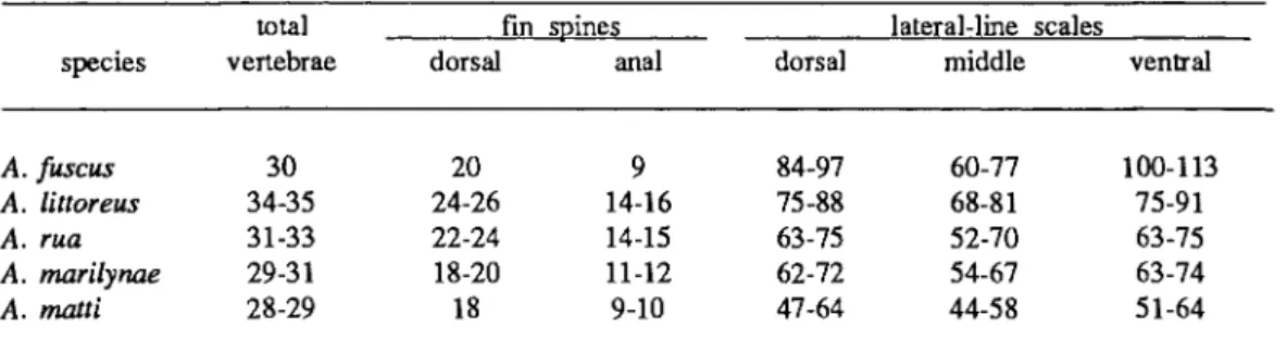
224 W. F. SMITH-VANIZ AND D. W. JOHNSON Table 2. Range of selected counts in species of Acanthoclinus1.
total vertebrae
fin spines lateral-line scales
species dorsal anal dorsal middle ventral
A. fuscus 30 20 9 84-97 60-77 100-113
A. littoreus 34-35 24-26 14-16 75-88 68-81 75-91
A. rua 31-33 22-24 14-15 63-75 52-70 63-75
A. marilynae 29-31 18-20 11-12 62-72 54-67 63-74
A. matti 28-29 18 9-10 47-64 44-58 51-64
'Data from Hardy (1985).
of Acanthoclinus, Whitley (1968) later gave Taumakoides full generic rank, and that "its allocation to a separate genus is justified." He listed three new characters that supposedly differentiate these two taxa. Two of these (position of the 5th branchiostegal ray relative to the epi/cera- tohyal joint; and the association of the an- terior anal-fin spine pterygiophores to the first three haemal spines) are virtually identical in our cleared and stained ma- terial of A. fuscus & littoreus, and the third character (slight difference in gill rakers) we consider to be of trivial signi- ficance. Furthermore, as discussed in the section "Acanthoclinine relationships," A.
littoreus and fuscus (the type species of Taumakoides and Acanthoclinus, respective- ly) are each others closest relatives; thus, recognition of Taumakoides either as a genus or subgenus cannot be justified.
Distribution.—Endemic to New Zealand.
Species comparisons.—The species of Acanthoclinus are contrasted in the preced- ing key, Table 2 and in the following spe- cies accounts.
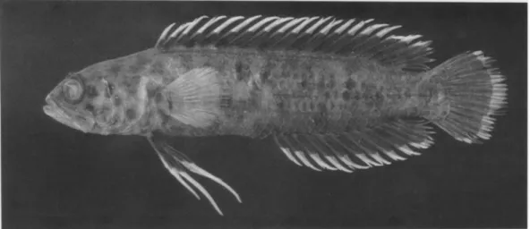
Acanthoclinus fuscus Jenyns Figs. 1A-2A, 5
Acanthoclinus fuscus Jenyns, 1842:92, pi.
18, fig. 2 (original descr.: Bay of Is., New Zealand); lectotype BMNH 1917.7.14:37); Hardy, 1985:360-364, fig. 1 (redescription; specimen photo- graph; synonymy; lectotype designa- tion).
Acanthoclinus taumaka Clarke, 1879:293, pi. 15 (original descr.: Jackson's Bay, New Zealand; holotype AIM).
Acanthoclinus quadridactylus [not Bloch &
Schneider]. Graham, 1953:320,326, pi on 321 (behavioral observations, in- cluding aspects of parental care of eggs); Jillett, 1968:1-7 (aspects of bio- logy: age, growth and food); Jillett, 1968a: 1-8 (aspects of biology: breeding
& development).
Blennius littoreus [not Forster]. Whitehead, 1969:pl. 30.
Diagnosis.—A species of Acanthoclinus with the following autapomorphies: 1) ven- tral lateral line with an accessory branch at the anal-fin origin (sometimes separate from the ventral lateral line) that continues anteriorly on either side of the ventral midline (Fig. 5), 2) the dorsal lateral line is unique in frequently having an anterior branch arising below base of 2nd dorsal-fin spine and directed obliquely onto predorsal area, but not joined with opposite member at the dorsal midline (often this accessory branch is present on one side only), and 3) haemal spine of second preural vertebra fused to vertebral centrum. Acanthoclinus fuscus also has the highest number of lateral-line scales, a unique combination of dorsal- and anal-fin spines (Table 2), and the predorsal area scaled (plesiomorphic character state present in no other species of Acanthoclininae).
Abbreviated description.—Dorsal fin XX, 4; anal fin IX, 4; pectoral fin 17-19;
vertebrae 12 + 18 = 30 total; tubed later-
al-line scales: dorsal 84-97, middle 60-77, ventral 100-113 (excluding accessory branch, which has 32-44 scales); lateral- line scales slightly to moderately larger than adjacent scales (Fig. 20A); posterior profiles of non-lateral-line scales somewhat ovate; anterodorsal angle of opercle scale- less; body, including predorsal area, en- tirely covered with cycloid scales; lower jaws anteriorly with 2 pairs of pores (Fig.
2A), each dentary with 5 pore positions (posterior three positions may each be oc- cupied by one to several pores); basihyal moderately broad and toothed; first pteryg- iophore of anal fin with a single super- numerary spine; haemal spine of second preural vertebra fused to vertebral centrum.
Color pattern (after Hardy (1985).—
When fresh, body and ventral half of dorsal fin olive-greenish, mottled with creamish blotches; broad creamish stripe medially on head and predorsal region; a similar broad stripe anteroventrally angled across cheek, and scattered creamish specks on remainder of head, lips, and opercular region; two or three small, creamish spots on pectoral-fin base; dark blotch posteriorly on dorsal fin over soft rays.
Remarks.—Unlike the other species of Acanthoclinus, our specimens of A. fuscus all have the same number of dorsal- and anal-fin rays and vertebral counts, the same as those reported by Hardy (1985).
Apparently these meristic characters have become genetically fixed in A. fuscus.
Maximum size.—Hardy (1985) reported the maximum size as 210 mm SL (250 mm TL). The largest specimens recorded by Jillet (1968) were 200 mm TL and thought to be 10 years old.
Material (all from New Zealand).—
ANSP 122745 (1:72), Bay of Islands, Uru- pukapuka Is.; ANSP 165085 (6:55.8-76, including 2:44.6-46.5 C&S), New Plymouth; USNM 83311 (1:92), "Balabac";
USNM 200547 (6:54.5-82.3), Kaikoura Peninsula; USNM 303548 (3:27.3-28.8), Wellington Harbor, Seaview; USNM 312932 (3:46-89), Auckland, Campbells Bay.
Acanthoclinus littoreus (Forster) Figs. 1B-2B
Blennius litoreus Forster in Bloch &
Schneider, 1801:177 (incorrect original spelling; name in synonymy).
Blennius quadridactylus Bloch & Schneider, 1801:177 (original descr.: [New Zealand] based on verbatim description of Blennius littoreus in J.R. Forster's unpublished manuscript).
Acanthoclinus trilineatus Griffin, 1933:330, 332-333, fig. 2, pi. 34 (original descr.:
Deep Water Cove, Bay of Islands, New Zealand).
Taumakoides trilineatus. Whitley, 1955:111 (type species of new subgenus of Acanthoclinus).
Taumakoides littoreus. Hardy, 1985:364- 367, fig. 5 (redescription; specimen photograph; synonymy); Mooi, 1990:463, fig. 7 (egg surface morph- ology).
Nomenclature.—Hardy (1985:359-60) gave a detailed discussion of the nomen- clatural confusion that has resulted from use of the name Acanthoclinus quadri- dactylus (Bloch & Schneider) by subse- quent authors. We agree with his opinion that the requirements of "first reviser" were fulfilled in essence by Cuvier & Valen- ciennes (1836), who tacitly implied that Foster's name had priority over that of Bloch & Schneider by including both names under the heading Clinus littoreus.
Diagnosis.—Acanthoclinus littoreus dif- fers from all other species of Acantho- clinus in having 34-35 (versus 28-33) total vertebrae; only it and A. fuscus have the first anal-fin pterygiophore with a single supernumerary spine and all body scales cycloid. The combination of high numbers of dorsal- and anal-fin spines and lateral- line scales (Table 2) is shared only with A. rua, and the latter counts scarcely over- lap.
Abbreviated description.—Dorsal fin XXIV-XXVI, 3-5; anal fin XIV-XVI, 3;
pectoral fin 18-20 (rarely 21); vertebrae 12 + 22-23 = 34-35 total; tubed lateral-line
226 W. F. SMITH-VANS AND D. W. JOHNSON scales: dorsal 75-88, middle 68-81, ventral
75-91; all lateral-line scales noticeably larger than adjacent body scales (Fig.
20B); posterior profiles of non-lateral-line scales appear somewhat ovate; anterodorsal angle of opercle with 1 or 2 partially embedded scales; body, except naked pre- dorsal area, entirely covered with cycloid scales; lower jaws anteriorly with 2 pairs of pores (Fig. 2B), each dentary with 5 pore positions; basihyal slender, toothless;
first pterygiophore of anal fin with a sin- gle supernumerary spine; haemal spine of second preural vertebra autogenous.
Color pattern.—Back and sides dark reddish brown. Opercle with dark brown blotch outlined by pale pinkish border.
Three dark lines, with pale borders, radi- ating from posterior margin of eye; the 1st from posterodorsal margin of orbital rim along upper surface of head, 2nd from middle of rim towards opercular blotch, sometimes continuous with it, 3rd from ventral margin of rim towards subopercle.
Maximum size.—Hardy (1985) reported the maximum size as 133 mm SL, based on examination of 122 specimens.
Material (all from New Zealand).—
ANSP 165089 (6:66.5-95.3, including 2:53- 69 C&S), Outer Chetwode Is.; ANSP 122742 (1:104), ANSP 122744 (1:74) and ANSP 122746 (5:27-116), Bay of Islands, vicinity Urupukapuka Is.; USNM 247331 (2:66.5-69), Wellington, Lyall Bay.
Acanthoclinus rua (Hardy) Figs. 1C-2C
Taumakoides rua Hardy, 1985:367-370, fig.
6 (original descr.: off Barrett's Reef, Wellington Harbour entrance, New Zealand; holotype NMNZ P.13852).
Diagnosis.—Acanthoclinus rua differs from all other species of Acanthoclinus in having the lower jaws anteriorly with one pair of pores (Fig. 2C), each dentary with 4 pore positions.
Abbreviated description.—Dorsal fin XXIV-XXVI, 3-4; anal fin XIV-XVI, 3-4;
pectoral fin 18-21; vertebrae 11 + 20-22 =
31-33 total; tubed lateral-line scales: dorsal 63-75, middle, 52-70, ventral 63-75;
lateral-line scales slightly larger than adja- cent body scales; posterior profiles of non- lateral-line scales somewhat ovate (Fig.
20C); anterodorsal angle of opercle with 2- 4 partially embedded scales; body, except naked predorsal area, covered anteriorly with cycloid scales, that become ctenoid under base of dorsal-fin soft rays and on caudal peduncle, cycloid scales also present basally on caudal fin; lower jaws anteriorly with one pair of pores, each dentary with 4 pore positions; basihyal slender, tooth- less; first pterygiophore of anal fin with 2 supernumerary spines; haemal spine of sec- ond preural vertebra autogenous.
Color pattern (after Hardy (1985).—
When fresh, "Body and head uniformly brown, a faintly pale-edged, darker blotch on operculum; white band medially on dorsal surface of snout and predorsal region (may also include 1st dorsal fin spine); dorsal fin webbing almost trans- parent pale grey, with fleshy appendages on spine tips pale to bright orange; dorsal fin soft rays tipped orange, a very narrow orange margin around caudal fin; anal fin similar to dorsal fin, but appendages on spine tips white; proximal half of pelvic fins greyish, distal half bright orange."
Maximum size.—The smallest species of Acanthoclinus; the largest of 37 types of A. rua examined by Hardy (1985) is 64 mm SL.
Material (all from New Zealand).—
ANSP 165086 (2:44-44.8), Stewart Is.;
ANSP 165087 (3:34.3-41.5, 46.3 C&S), Percy Is.
Acanthoclinus marilynae (Hardy) Taumakoides marilynae Hardy, 1985:370-
373, fig. 7 (original descr.: Scorching Bay, Wellington, New Zealand; holo- type NMNZ P. 14134).
Diagnosis.—A species of Acanthoclinus distinguished by the following autapo- morphic character state: the lateral lines on opposite sides of the body adjacent to the
dorsal- or anal-fin bases united behind these fins and continuing to caudal-fin base as a single series of tubed scales along the dorsal or ventral ridge of ped- uncle, respectively.
Abbreviated description.—Dorsal fin XIX-XX, 3-5; anal fin XI-XII, 3-5;
pectoral fin 18-21; vertebrae 11 + 18-20 = 29-31 total; tubed lateral-line scales: dorsal 62-72, middle 54-67, ventral 63-74; scales in dorsal and ventral lateral-line series moderately larger than adjacent scales, those in middle series subequal; posterior profiles of non-lateral-line scales broadly elliptical; anterodorsal angle of opercle with 5-6 small scales; body, except naked predorsal area, covered anteriorly with cycloid scales, that become ctenoid dorsal- ly and on sides from behind level of pectoral-fin base, ctenoid scales also basal- ly on caudal fin; lower jaws anteriorly with 2 pairs of pores, each dentary with 5 pore positions; basihyal slender, toothed;
first pterygiophore of anal fin with 2 supernumerary spines; haemal spine of sec- ond preural vertebra autogenous.
Color pattern (after Hardy (1985).—
When fresh (based on several specimens),
"body varying from mottled light and dark brown to deep chocolate-brown throughout;
head mottled medium brown and dark olive-green to deep chocolate brown (also predorsal and opercular region); dorsal and anal fin webbing dark greyish-brown to black; dorsal and anal fin spine tips pink- ish-brown to deep orange; outer margin of dorsal and anal fin soft rays, and caudal fin rays, narrowly lined with bright yel- lowish-orange; proximal half of pelvic fins medium brown to greyish, distal half me- dium to deep orange."
Remarks.—Hardy (1985:390) reported the basihyal toothless in A. marilynae; all our specimens have this bone distinctly toothed.
Maximum size.—The largest of 70 types of A. marilynae examined by Hardy (1985) is 134 mm SL.
Material (all from New Zealand).—
ANSP 122743 (3:24.9-60.8) and ANSP 122752 (1:58.2), Bay of Islands, NW of
Urupukapuka Is.; ANSP 122747 (2:60.2- 65.8), ANSP 122801 (1:49.8), and ANSP 134947 (2:62.8-66.6 C&S), Bay of Islands, SW of Cape Brett.
Acanthoclinus matti (Hardy) Taumakoides matti Hardy, 1985:373-375,
fig. 8 (original descr.: off Sentinel Rock, outer Marlborough Sounds, New Zealand; holotype NMNZ P. 15332).
Diagnosis.—Acanthoclinus matti differs from all other species of Acanthoclinus in its distinctive color pattern (see below), including the absence of dark blotch on opercle; it also has the largest body scales (Fig. 20D) and the lowest number of lateral-line scales (Table 2).
Abbreviated description.—Dorsal fin XVIII, 4-5; anal fin IX-X, 3-5; pectoral fin 19-20; vertebrae 11-12 + 17 = 28-29 total;
tubed lateral-line scales: dorsal 47-64, mid- dle 44-58, ventral 51-64; scales in dorsal and ventral lateral lines slightly larger than adjacent body scales, those of middle lateral line subequal or smaller than adja- cent scales (Fig. 20D); posterior profiles of non-lateral-line scales broadly elliptical;
anterodorsal angle of opercle with about 6 scales; body, except naked predorsal area, covered anteriorly with cycloid scales, that become ctenoid dorsally and on sides from about posterior margin of pectoral-fin base, ctenoid scales also on peduncle and basally on caudal fin; lower jaws anterior- ly with 2 pairs of pores, each dentary with 5 pore positions; basihyal slender, toothed;
first pterygiophore of anal fin with 2 supernumerary spines; haemal spine of second preural vertebra autogenous.
Color pattern.—Head dark chocolate brown, covered with conspicuous, small, white spots; body scales deep maroon with white upper and lower corners, resulting in a series of about 10 narrow white stripes on sides against a dark background; belly with 7 similar white stripes.
Maximum size.—Attains at least 125 mm SL.
Material.-- ANSP 165088 (1:51.0),
228 W. F. SMITH-VANE AND D. W. JOHNSON
Fiordland, mouth of Cunaris Sound, New Zealand.
Genus Belonepterygion McCulloch Belonepterygion McCulloch, 1915:51 (type
species Acanthoclinus fasciolatus Ogilby, 1889, by original designation and monotypy).
Ernogrammoid.es Chen and Liang, 1948:32 (type species Ernogrammoides fasciatus Chen and Liang, 1948, = Beloneptery- gion fasciolatum (Ogilby), by original designation and monotypy).
Calliblennius Aoyagi, 1954:213 (37) (type species Calliblennius rubescens Aoyagi, 1954, = Belonepterygion fasciolatum (Ogilby), by original designation and monotypy, preoccupied in fishes by Calliblennius Barbour 1912).
Description.—A monotypic acantho- clinine genus (maximum size 50 mm SL), with the following combination of char- acters: dorsal fin XVII-XX, 4-5; anal fin X-XII, 3-5; pectoral fin 17-19; total verte- brae 10-11 + 17-18 = 27-29 (rarely 29) total; caudal-fin rays (dorsal/ventral): pro- current 3/3, segmented rays 8/8, middle 14 branched, total elements 22. Body with three unbranched lateral lines: dorsal lateral line originates from upper angle of oper- cle, middle extends from near posterior margin of pectoral fin to caudal-fin base, and ventral (variable) typically extends from just anterior to pelvic fin or from above middle of belly; both the dorsal and ventral lateral lines terminate posteriorly at end of dorsal and anal fins, respectively.
Tubed lateral-line scales: dorsal 36-46, middle 23-34, ventral 17-44 (intermediate scales in latter two series may occasionally lack tubes); lateral-line scales no larger than adjacent body scales, some with a pair of vertically aligned superficial neuro- masts (not shown in Fig. 20E); posterior profiles of all scales broadly elliptical.
Head entirely naked; body and caudal-fin completely scaled, except predorsal area naked; anterior scales cycloid, becoming ctenoid dorsally and on sides from about
level of 5th dorsal-fin spine; ctenoid scales also on peduncle and basally on caudal fin. Sensory pores on head (Fig. ID) vari- able in number but infraorbital series typic- ally in a single series; lower jaws anterior- ly with 1 pair of pores Fig. 2D), each dentary with 4 pore positions; gill mem- branes fused in ventral midline to form a broad free fold across isthmus. Infraorbital bones 5 (one of four specimens checked for this character with 6 infraorbitals on left side only), 3rd infraorbital with a well-developed suborbital shelf (Fig. 13C).
Palatine toothless; metapterygoid with a very slight lateral flange; metapterygoid- quadrate joint not interdigitated; pre- opercular canal open posterolaterally; pri- mary opercular spine broadly rounded and platelike, no secondary spine present; max- illa very broadly rounded posteriorly (Fig.
14C), supramaxilla absent. Supracleithrum with well developed supracleithral sensory canal that is continuous anterodorsally with posttemporal canal and posteriorly with lateral-line scales; pectoral girdle with scapulo-coracoid joint smooth, posterolateral arm of coracoid relatively slender (Fig.
19B) and with radial formula 2-1-1; hyoid arch with ceratohyal-epihyal suturing medi- ally only. Infrapharyngobranchials 2-4 toothed; interarcual cartilage relatively long (Fig. 15B); basihyal slender, toothless.
Neural spine of first vertebra autogenous;
first pterygiophore of anal fin with 2 supernumerary spines; all but anteriormost pterygiophore of segmented dorsal- and anal-fin rays consisting of autogenous prox- imal, middle and distal radials; haemal spine of second preural vertebra fused to vertebral centrum. Caudal skeleton with 3 separate epurals, hypural 5 moderately long, and hypurapophysis very long and slender (Fig. 17B). Adductor mandibulae with Al section completely covering A2 section laterally, and Al fibers bundlelike anterodorsally (Fig. 22C).
Distribution.—Australia (east and west coasts), Lord Howe Island, New Caledonia, Philippines, Taiwan and Ryukyu Islands.
Belonepterygion fasciolatum (Ogilby) Figs. 1D-2D
Acanthoclinus fasciolatus Ogilby, 1889:63, pi. 3, fig. 3 (original descr.: Lord Howe Is.; lectotype AMS 1.1876).
Ernogrammoides fasciatus Chen & Liang, 1948: 32, fig. 1 (original descr.:
Keelung, Formosa; type species of new genus Ernogrammoides); Masuda et al. 1984:141, pi. 126, figs. P,Q (brief description; color photographs).
Calliblennius rubescens Aoyagi, 1954: 213 (37), fig. 1 (original descr.: Kowan,
"Okinawa-Honto"; holotype probably destroyed; type species of new genus Calliblennius).
Belonepterygion fasciolatum. Hardy, 1985:375-378, fig. 9 (redescription;
specimen figure; synonymy; lectotype designation; distribution); Mooi, 1990:463 (egg surface morphology).
Diagnosis.—Belonepterygion fasciolatum differs from all other acanthoclinines in having the combination of three lateral lines and the gill membranes fused in ventral midline to form a broad free fold across isthmus; it also is unique in lacking palatine teeth, in the bundlelike arrange- ment of adductor mandibulae section Al anterodorsally, and (in adults) having 9-16 narrow dark bands on body.
Description.—See generic description above.
Color pattern.—Body pale to darkish brown with 9-16 narrow dark brown bands on sides; opercle with an oblong, brown- ish-black blotch; throat and cheeks with pinkish-red suffusion; lower jaw, snout and upper part of head medium brown, the sharply delimited lower margin of this coloring on a line from inner angle of jaws, extending just above lower margin of eye to opercular blotch; dorsal and anal fins mostly red to dark brown, with fleshy distal tabs on spines white or very pale orange.
Geographic variation.—Hardy (1985) presented frequency tables of selected meristic characters by locality for B. fas-
ciolatum and documented geographic varia- tion, especially in number of lateral-line scales. He also noted that the number of bands on the sides varied considerably within and between populations but gave no frequency data.
Maximum size.—Hardy (1985) recorded 50 mm SL as the maximum size of 260 specimens examined.
Material.—ANSP 122316 (1:41.8) and ANSP 142690 (2:39.7 & 30.6 C&S), Queensland, Australia; USNM 257883 (6:33.5-40.6) and USNM 273813 (3:32-36), Taiwan.
Genus Beliops Hardy
Beliops Hardy, 1985:378 (type species Beliops xanthokrossos Hardy, 1985, by original designation and monotypy).
Description.—A genus of acanthoclinine fishes with two known species (maximum size 26 mm SL), with the following com- bination of characters: dorsal fin XVIII- XX, 2-4; anal fin XXI, 2-3; pectoral fin 15-18; pelvic fin 1,2 with innermost ray unbranched; vertebrae 10 + 16-17 = 26 or 27 total; caudal fin (dorsal/ventral): pro- current rays 2-4/2-3, segmented rays 7-8/7- 8, middle 12 or 14 branched, total ele- ments 18-22 . Body with two unbranched lateral lines: dorsal lateral line extends from upper angle of opercle to below a vertical from near base of last dorsal-fin spine, and middle (= ventral) extends from slightly to well behind posterior margin of pectoral fin to caudal-fin base; tubed lateral-line scales: dorsal 24-35, middle 9- 20; lateral-line scales no larger than adja- cent body scales; posterior profiles of scales broadly elliptical (Fig. 20F) or dis- tinctly lanceolate with membranous flaps (Fig. 20G). Head with 1-2 cycloid scales in anterodorsal angle of opercle (B. xantho- krossos) or entirely naked (B. batanensis);
body and caudal-fin base completely scaled, except predorsal and prepelvic areas naked (pectoral-fin base also naked in B.
batanensis); anterior scales cycloid, becom- ing strongly ctenoid posterior to pectoral
230 W. F. SMITH-VANE AND D. W. JOHNSON fin (B. xanthokrossos) or scales mostly
cycloid, some with a few weak ctenii (B.
batanensis). Sensory pores on head (Figs.
1E-F) variable in number and position but mandibulo-preopercular pores in a single series; lower jaws anteriorly with a single pair of pores, each dentary with 4 pore positions; gill membranes fused in ventral midline to form a broad free fold across isthmus. Infraorbital bones 5, the 3rd infra- orbital with a well-developed suborbital shelf (Figs. 13D-E). Palatine teeth present;
metapterygoid with a well-developed lateral flange; metapterygoid-quadrate joint inter- digitated; preopercular canal tubular, closed posterolaterally; primary opercular spine pungent, not platelike, no secondary spine present; maxilla rather slender posteriorly (Figs. 14D-E), supramaxilla present or absent. Supracleithrum with well developed supracleithral sensory canal that is continu- ous anterodorsally with posttemporal canal and posteriorly with lateral-line scales;
pectoral girdle with scapulo-coracoid joint interdigitated, posterolateral arm of corac- oid moderately slender (Fig. 19C) and with radial formula 2-1-1; hyoid arch with cera- tohyal-epihyal suturing medially only, infra- pharyngobranchials 3-4 toothed; interarcual cartilage relatively short, conical (Figs.
15C-D); basihyal moderately broad and toothed (Figs. 23A-B). Neural spine of first vertebra fused to vertebral centrum;
first pterygiophore of anal fin with 1 supernumerary spine; all pterygiophores of segmented dorsal- and anal-fin rays with fused proximal and middle radials; haemal spine of second preural vertebra fused to vertebral centrum. Caudal skeleton with epurals 2-3 fused distally, hypural 5 moderately long, and hypurapophysis rela- tively short (Fig. 17C). Adductor mandib- ulae with Al and A2 sections both visible laterally.
Distribution.—The two species of Beli- ops have allopatric distributions (Fig. 3);
B. xanthokrossos is an Australian endemic, and B. batanensis is known only from the Batan Islands, northern Philippines.
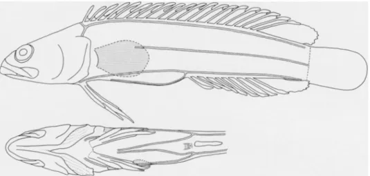
Beliops xanthokrossos Hardy Figs. 1E-2E, 6, 8
Beliops xanthokrossos Hardy, 1985:378-381, fig. 10 (original descr.: Kingston Reef, Rottnest Is., Western Australia; holo- type WAM P.26617-008).
Diagnosis.—The combination of two lateral lines, anterior dorsal-fin spines rela- tively slender and elongate with deeply incised interradial membranes, and scales posterior to the pectoral fin ctenoid and with elliptical profiles readily distinguishes Beliops xanthokrossos from all other acan- thoclinines. All the character states of B.
xanthokrossos that are not shared with its congener and that can be polarized with confidence appear to be plesiomorphic (refer to discussion in species account of B. batanensis).
Description (see also preceding generic description).—Dorsal fin XVIII-XIX, 2-4;
anal fin X, 2-3; pectoral fin 16-18; ver- tebrae 10 + 16-17; caudal-fin rays (dor- sal/ventral): procurrent 2-4/2-3; segmented 8/8, middle 14 branched, total elements 20- 22 ; gill rakers on first arch (epibranchial + ceratobranchial) 1-2 + 2-3; tubed lateral- line scales: dorsal 24-31, middle 9-18;
body scales elliptical and ctenoid posterior to pectoral fin; sensory pores on head rela- tively numerous on dorsum and with pore positions in the infraorbital series mostly bi-pored (Fig. IE); dorsal- and anal-fin spines with interradial membranes strongly incised, anterior dorsal spines incised to about one-half to two-thirds anteriorly, de- creasing thereafter; dorsal- and anal-fin spines relatively long and slender (Fig.8), with elongate fleshy tassels distally; longest dorsal spine 13.5-15.2 percent SL; terminal dorsal- and anal-fin rays without membran- ous attachment to caudal peduncle. Small supramaxilla present (Fig. 14D). Teeth in both jaws small and conical, curved back- ward, largest anteromedially; upper jaw with double row of teeth along sides, those in outer row larger; lower jaw similar but only a single row of teeth posteriorly.
Fig. 6. Beliops xanthokrossos, ANSP 165557, 25.7 mm SL, Western Australia, Duke of Orleans Bay.
Fig. 7. Beliops batanensis, USNM 288976, holotype, 21.0 mm SL, Philippines, Batanes Providence, Batari Island. (Drawn by Penelope K. Hollingsworth.)
Color pattern.—Hardy (1985:379) reported that B. xanthokrossos has two color forms, which may indicate sexual dimorphism. He illustrated the yellow form (holotype) and our Fig. 6 shows the mottled morph. His description of the two forms is as follows:
"Yellow form — head and body yel- lowish-brown to brownish-yellow, with variable number of small, darker brown spots on sides, each covering 1 or 2 scales; 3 or 4 smaller, brown spots on head, roughly following posterior curve of orbit. Dorsal, anal, pectoral, and caudal fin webbing simi- lar to body, fleshy appendages of dor-
sal and anal fin spines bright yellow.
Pelvic fin yellowish-brown to bright yellow, several dark, brown flecks on proximal half.
"Mottled form — head and body medium brown, densely covered with darker brown blotches, each blotch several scales in extent, with vertical, dark brown band on posterior of caudal peduncle; several smaller, dark brown spots on head and operculum.
Dorsal, anal, pectoral, and caudal fin webbing similar to body, a series of dark brown blotches extending length of dorsal and anal fins, close to base.
Fleshy appendages of dorsal and anal
232 W. F. SMITH-VANIZ AND D. W. JOHNSON fins bright yellow. Proximal half of
pelvic fin speckled with brown, distal half pale yellow."
Distribution.—Endemic to Australia (south and west coasts) and collected in depths of 3-15 m.
Maximum size.—Attains at least 25.9 mm SL.
Material.—ANSP 165557 (2:25.7 &
25.9 C&S), Western Australia, Duke of Orleans Bay.
Beliops batanensis, new species Figs. 1F-2F, 7-8
Diagnosis.—A species of Beliops dis- tinctive in having the posterior body scales with membranous flaps (Fig. 20G) and ctenii, if present, very reduced (Fig. 21G), short and robust dorsal-and anal-fin spines (Fig. 8), and the interradial membranes of the spinous dorsal fin very weakly incised.
Description (see also preceding generic description).—The following counts and measurements are given for the holotype first, with values for the paratype, if dif- ferent, in parentheses. Dorsal fin XX, 2;
anal fin XI, 2; pectoral fin 15/15 (16/16);
vertebrae 10 + 17; caudal-fin rays (dor- sal/ventral): procurrent 2/2; segmented 7/7, middle 12 branched, total elements 18; gill rakers on first arch (epibranchial + cerato- branchial) i,2 + 7,i; tubed lateral-line scales (L/R): dorsal 35/35 (34/33), middle 20/17 (20/20); head, nape, prepelvic area and pectoral-fin base naked, remainder of body including caudal-fin base covered with cycloid scales (some posterior scales with very reduced ctenii); scales lanceolate, those on half of body with membranous flaps; sensory pores on head relatively sparse with all pore positions occupied by single pores (Fig.IF), each dentary with 4 pore positions; dorsal- and anal-fin spines with interradial membranes very weakly incised, and with the spines relatively short and robust (Fig. 8), with distal fleshy tabs poorly developed; terminal dorsal- and anal-fin rays with basal third of ray mem- branously attached to caudal peduncle.
Supramaxilla absent (Fig. 14E). Teeth in both jaws mostly small and conical, curved backward, largest anteromedially; upper jaw anteriorly with three rows of teeth flanked by several much larger teeth, only a single row posteriorly; lower jaw similar but in- nermost row of large conical teeth not slanted backward.
Measurements as percent standard length: head length 27.6 (29.4); snout tip to dorsal-fin origin 30.5 (31.3); snout tip to anal-fin origin 58.0 (59.2); dorsal-fin base 68.6 (69.0); anal-fin base 38.0 (37.4);
pectoral fin 14.5 (13.7); longest pelvic-fin ray 13.8 (14.2); caudal fin 19.5 (20.1);
longest dorsal-fin spine 9.3 (10.2); longest anal-fin spine 10.5 (10.4); body depth at dorsal-fin origin 20.7 (19.9); body depth at anal-fin origin 17.1 (17.5); orbit diameter 5.7 (6.2); upper jaw length 15.0 (15.6);
postorbital head length 17.6 (19.2).
Color pattern (in alcohol).—Head and body mostly dark, slightly paler on opercle; posterior half of upper jaw and exposed branchiostegal membranes densely covered with small melanophores; dorsum with a poorly defined, pale stripe that extends from origin of dorsal fin across snout and onto upper and lower lips where it widens slightly; dorsal and anal fins mostly uniformly dark with scattered darker flecks, except tips of spines and rays and adjacent interradial membranes pale distal- ly; caudal fin dark except for conspicuous, narrow, white, distal margin; pelvic fin also dark, except spine lip pale.
In life (based on field observations of the freshly collected specimens), the darkly pigmented areas of the body and fins are black, the pale areas white.
Species comparison.—Beliops batanensis exhibits a surprisingly large number of apomorphies not present in its closest known relative as follows: 1) distinctive scale type (see diagnosis); 2) reduction in number of total caudal-fin rays (18 vs. 20- 22); 3) concomitant reduction in branched caudal-fin rays (12 vs. 14); 4) terminal dorsal- and anal-fin rays membranously attached to caudal peduncle; 5) interradial membranes of dorsal- and anal-fin spines